Method of selective excitation for improving contrast in magnetic resonance imaging

Inactive Publication Date: 2004-04-01
RAMOT AT TEL AVIV UNIV LTD
View PDF0 Cites 0 Cited by
  • Summary
  • Abstract
  • Description
  • Claims
  • Application Information

AI Technical Summary

Benefits of technology

[0098] Typically, a 90.degree. pulse is known to flip the magnetization in such manner. However, a 90.degree. pulse enforces a magnetization flip to all the non-zero spin nuclei, without distinction between various components such as solvent components and non-solvent components. It should be appreciated that the advantage of the present invention is in the elimination of the contribution of the solvent to the signal that may be observed at the end of the first radiofrequency pulse sequence. By the time the second radiofrequency pulse sequence starts, the solvent is in a state of zero transverse magnetization, hence the at least one type of molecules is exclusively flipped to the transverse plane.
[0099] Referring now again to FIG. 1, the fourth pulse with phase .phi..sub.4 restores the irreducible spherical tensor T.sub.1,0. The duration .theta. of the fourth pulse with phase .phi..sub.4 is selected so as to optimize the analysis performed in the acquisition step. A presently preferred value for .theta. is 90.degree..
[0100] Once the longitudinal magnetization of the at least one type of molecules is restored, a process of magnetization transfer from the at least one type of molecules to the solvent initiates. The duration of the process is set to be a magnetization transfer period denoted t.sub.LM (FIG. 1). At any time within t.sub.LM, the longitudinal magnetization value of the at least one type of molecules is proportional to that of the solvent. In accordance with the presently preferred embodiment of the invention, by measuring the solvent magnetization, a unique value of the desired magnetization of the at least one type of molecules can be calculated.
[0101] In the currently available conventional magnetic resonance spectrometers, the signal-to-noise ratio that may be obtained by direct measurement of longitudinal magnetization is extremely small. In the presently preferred embodiment of the invention, in order to receive an optimized signal from the solvent, a radiofrequency pulse subsequence (PS1 in FIG. 1) is further applied, so as to select a coherence of -1, which corresponds to a magnetization substantially engaging the transverse plane.
[0102] PS1 can be any subsequence known in the art, depending on the desired implementation of the present invention. For spectroscopic studies, PS1 can be a hard pulse or any pulse sequence suitable for localized spectroscopy, such as ISIS [Paul T. Callaghan, Principles of Nuclear Magnetic Resonance Microscopy, Clarendon Press-Oxford 1993, p. 287]. Alternatively, PS1 can be a slice selective pulse sequence, or other imaging sequence, such as but not limited to the GEFI sequence. [Paul T. Callaghan, Principles of Nuclear Magnetic Resonance Microscopy, Clarendon Press-Oxford 1993, p. 134].
[0103] The principles of the present invention may also be used to improve contrasts in slice images of tissues, such as a connective tissue.

Problems solved by technology

Connective tissues, such as ligaments, tendons and cartilage appear in standard magnetic resonance (MR) images with low signal-to-noise (S / N) ratio (SNR) due to the water short T.sub.2 relaxation times. Images performed with short echo time (TE), result in a significant loss of contrast.
However, when these systems are analyzed simultaneously, the signal from the protein spread out over the entire image and will not be visible.
Secondly, the long time scale in MTC prevents independent measurement of the protein relaxation time T.sub.2, and of intramolecular processes within the proteins, such as spin diffusion.
While these approaches do increase the MR signal of connective tissues and the contrast between connective and adjacent tissues, the results are not yet optimal for diagnostic purposes.
In all of the presently known MTC methods, the suppression of the water excitations is insufficient for differentiating between direct excitation of the water molecules and magnetization that was transferred from the protein to the water.

Method used


Image

  • Method of selective excitation for improving contrast in magnetic resonance imaging
  • Method of selective excitation for improving contrast in magnetic resonance imaging
  • Method of selective excitation for improving contrast in magnetic resonance imaging

Examples


example 1

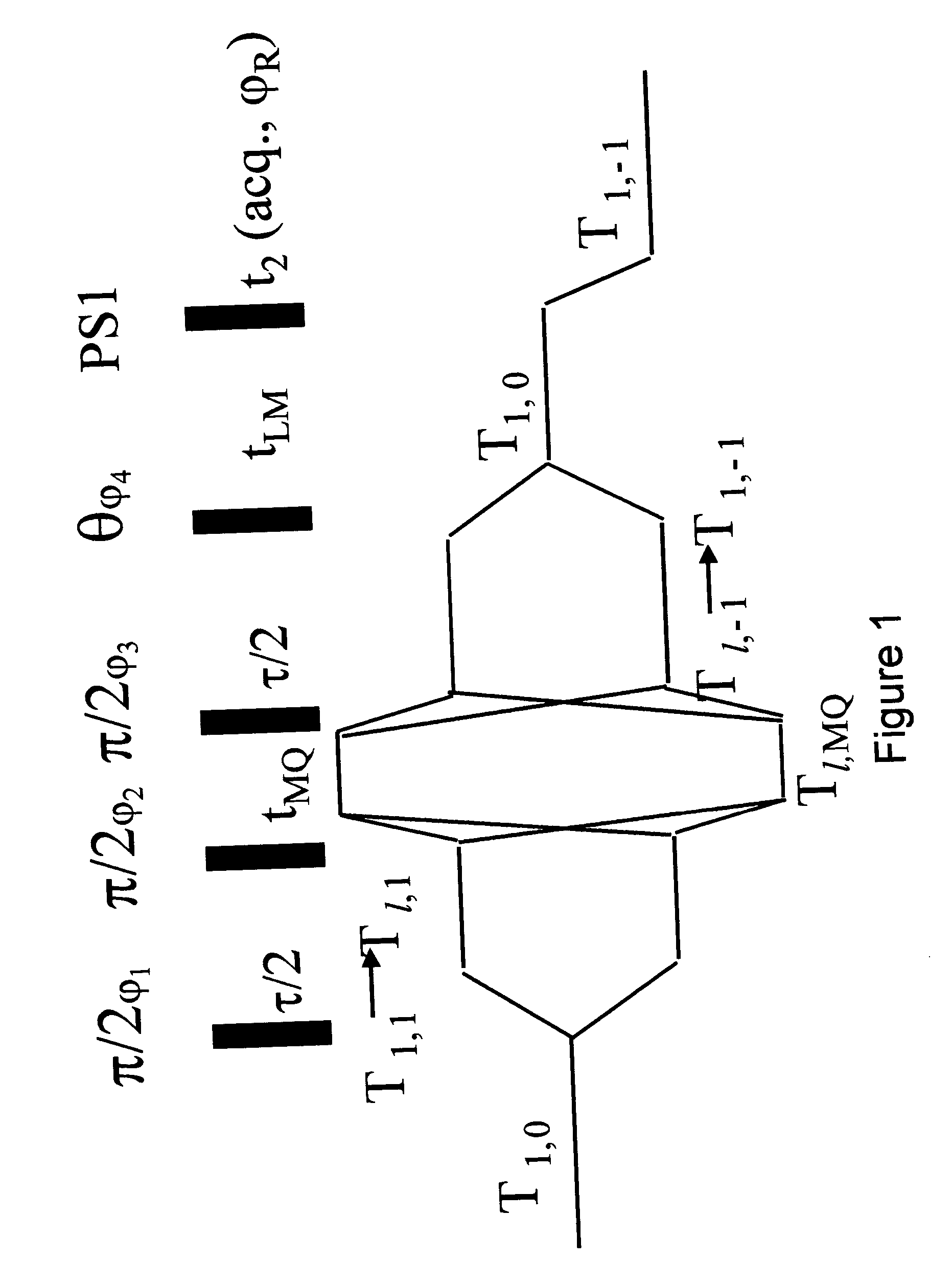
Magnetization Transfer from Protein to Water

[0116] The spectra of the water and protein signals as a function of the magnetization transfer period t.sub.LM, according to the present invention, are shown in FIG. 3. The simultaneous rise of the water signal and the decline of the broad protein signal represent the process of magnetization transfer from the protein to the water, within a short period of the order of tens of milliseconds. The effect is further exemplified in FIG. 4 illustrating graphically the dependence of the integrals of the water and protein spectra on the magnetization transfer period t.sub.LM.

[0117] Following the fast magnetization transfer process, both the water and the residual protein signals decay with a rate constant, which is a weighted average of the longitudinal relaxation rates of the water and the protein protons.

example 2

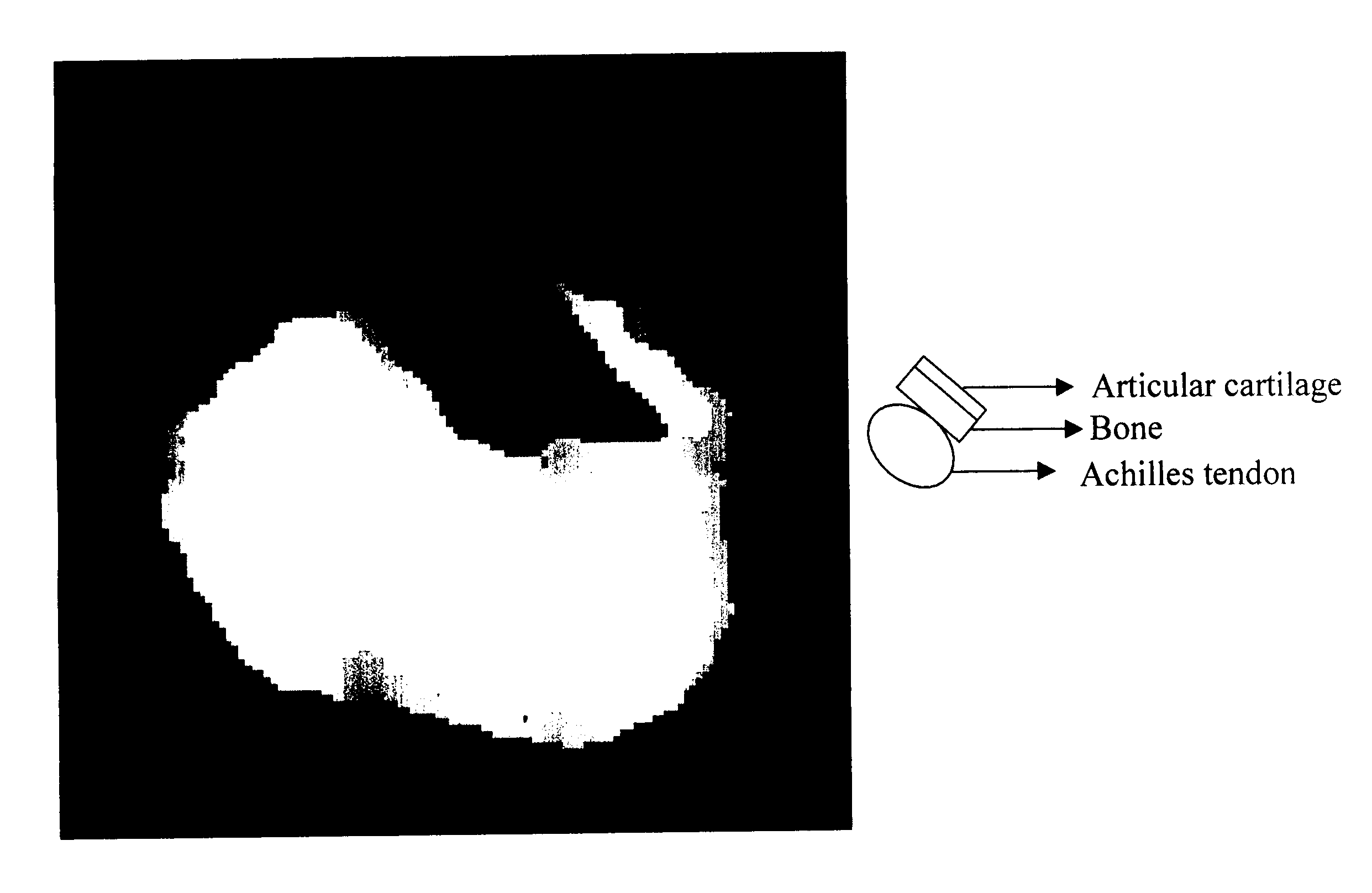
Imaging of an Achilles Tendon and an Articular Cartilage

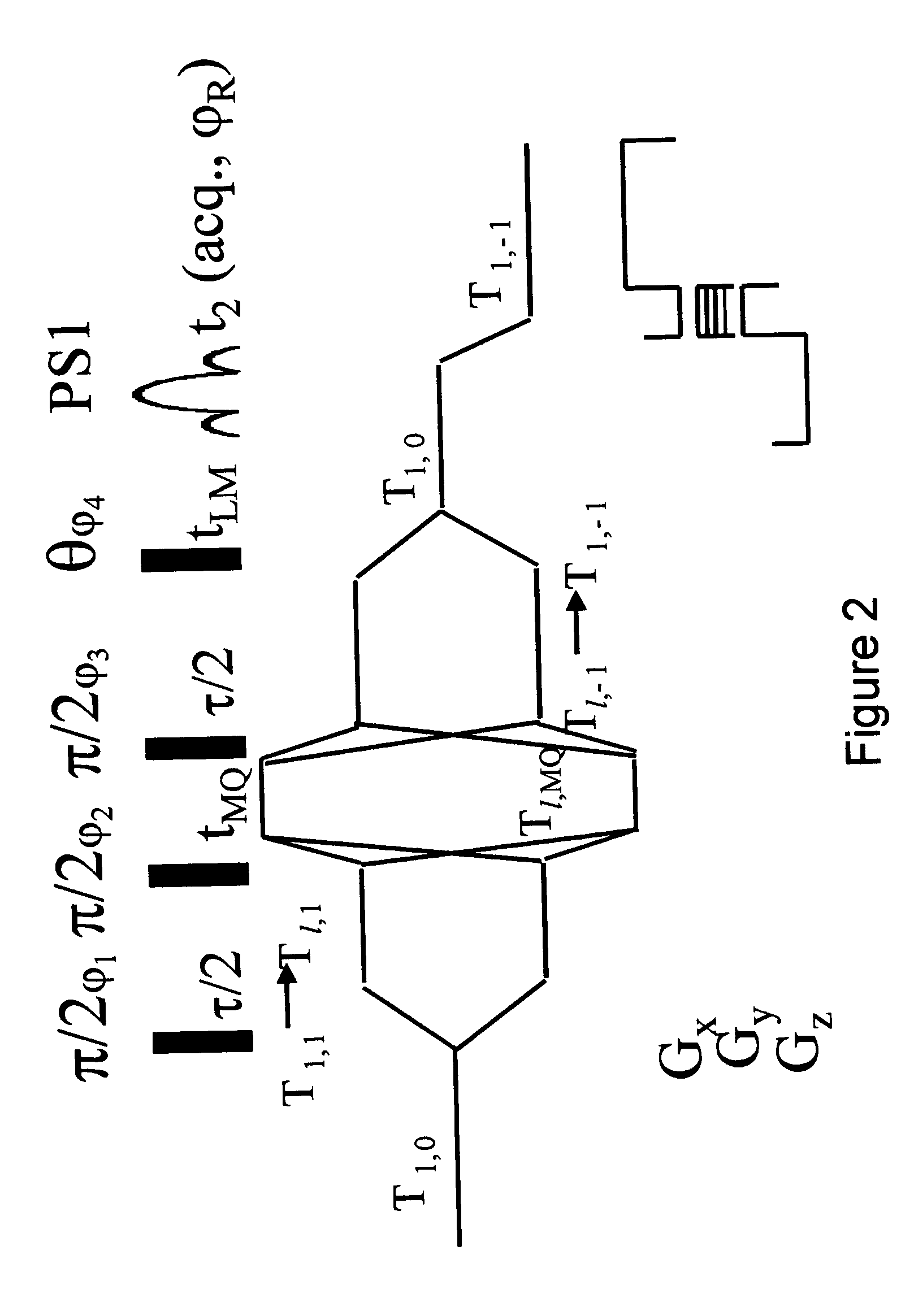
[0118] For illustrating the contrast obtained by the DQF-MTC imaging method a sample consisted of two types of tissues, bovine Achilles tendon and articular cartilage attached to one another.

[0119] Imaging was conducted on a 360 MHz Bruker WB spectrometer, using the DQF-MTC MRI pulse sequence shown in FIG. 2 with .tau. and t.sub.LM set as 7 .mu.s and 5 .mu.s respectively. The obtained image is shown in FIG. 5. The signals originating from the tendon, which gives in standard MRI a weaker signals than those of the cartilage, become comparable with the latter when imaged in accordance to the teachings of the the present invention.

example 3

Imaging of Mouse Brain

[0120] For illustrating the contrast obtained by the DQF-MTC imaging method a sample tube containing an excised mouse brain was prepared.

[0121] Imaging was conducted on a 360 MHz Brukers Avance spectrometer, using the DQF-MTC MRI pulse sequence shown in FIG. 2.

[0122] FIGS. 6a-d shows the imaging of a mouse brain using two different techniques. FIGS. 6a and 6b are respectively images of a transverse cross section and sagital cross section of a mouse brain, obtained by single quantum gradient echo imaging method. The obtained images provide very little information as the contrast there between the various parts of the brian is low. FIGS. 6c and 6d are the same images obtained using the DQF-MTC MRI method. Contrasts between various areas of the sample are clearly observed.

[0123] It is appreciated that certain features of the invention, which are, for clarity, described in the context of separate embodiments, may also be provided in combination in a single embodime...


PUM


Abstract

A method of magnetic resonance analysis of at least one type of molecules present in a solvent, the method comprises (a) applying a first radiofrequency pulse sequence selected so as to initially select a coherence of an order n for the at least one type of molecules in the solvent, wherein n is zero, a positive or a negative integer other than ±1, and thereafter to select an exclusive excitation of the at least one type of molecules in the solvent; (b) measuring at least one magnetic resonance parameter of the solvent; and (c) using the at least one magnetic resonance parameter of the solvent for calculating at least one magnetic resonance parameter of the at least one molecule.

Description

FIELD AND BACKGROUND OF THE INVENTION[0001] The present invention relates to Magnetic Resonance Imaging (MRI) and, more particularly, to a method of selective excitation so as to improve contrast in Magnetic Resonance Imaging of, for example, connective and other tissues.[0002] Magnetic Resonance Imaging (MRI) is a method to obtain an image representing the chemical and physical microscopic properties of materials, by utilizing a quantum mechanical phenomenon, named Nuclear Magnetic Resonance (NMR), in which a system of spins, placed in a magnetic field resonantly absorb energy, when applied with a certain frequency.[0003] A nucleus can experience NMR only if its nuclear spin I does not vanish, i.e., the nucleus has at least one unpaired nucleon. Examples of non-zero spin nuclei frequently used in MRI include .sup.1H (I=1 / 2), .sup.2H (I=1), .sup.23Na (I={fraction (3 / 2)}), etc. When placed in a magnetic field, a nucleus having a spin I is allowed to be in a discrete set of energy lev...

Claims


Application Information

Patent Timeline
IPC IPC(8): G01R33/46G01R33/465
CPCG01R33/4608Y10T436/24G01R33/465
Inventor ELIAV, UZINAVON, GILNEUFELD, ARNON
Owner RAMOT AT TEL AVIV UNIV LTD
Why Eureka
Try Eureka