
Inhibition of restenosis using a DNA-coated stent

a dna-coated stent and restraint technology, applied in the direction of prosthesis, blood vessels, genetic material ingredients, etc., can solve the problems of gradual narrowing of the vessel lumen, high chance of restnosis recurrence, and the possibility of recurrence very high

Inactive Publication Date: 2004-04-15
EPSTEIN STEPHEN E +1
View PDF0 Cites 46 Cited by
  • Summary
  • Abstract
  • Description
  • Claims
  • Application Information

AI Technical Summary

Benefits of technology

[0049] Another advantage of the invention is that the composition of the coating material can be tailored to preserve and support the DNA or cells to be incorporated into the coating. The material should, of course, not degrade the incorporated DNA. However, the design and formulation of the coating material is nevertheless simplified because it does not have to accommodate a wide variety of proteins and / or small molecules.
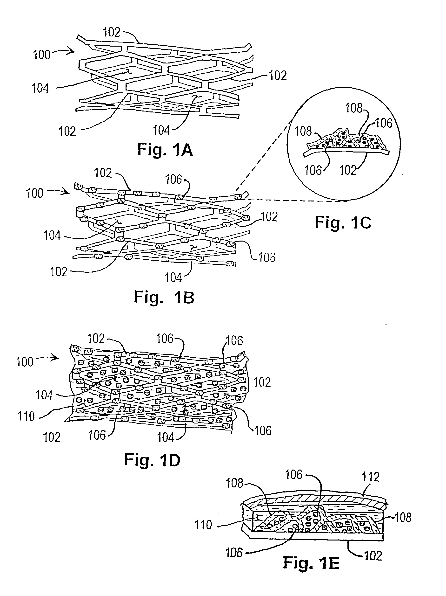
[0050] The stent used in the first embodiment of the invention is illustrated in FIGS. 1A-1E of the drawings. These figures illustrate a stent coated with DNA by incorporating plasmid DNA or a viral vector into a coating material that adheres to the stent (with or without a collagen gel) and into which DNA (as plasmid or viral vector) can be incorporated. The coated stent facilitates DNA delivery to, and transfection of, cells within the injured vessel wall, or cells that are migrating from the media and / or adventitia to form the neointima. The genes within the stent coating will be selected or created to encode gene products with anti-restentosis activities.
[0051] FIG. 1A illustrates the bare stent 100 without coating and without DNA or viral vectors. The stent comprises struts 102 having interstices or openings 104 between them.
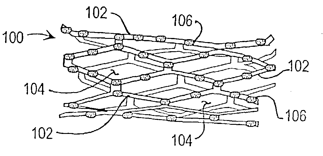
[0052] FIG. 1B illustrates the stent 100 with a coating that has plasmid DNA or viral vectors 106 incorporated into it. The coating and its contained genes cover the metal struts 102 but not the intervening spaces 104 FIG. 1C is a greatly enlarged view of a cross-section of a portion of the stent 100 of FIG. 1B, as indicated by the guidelines, showing the coated struts 102 with associated DNA 106. The lower portion of the figure shows a cross-section of a strut 102 of the stent 100. The irregular lattice work of hooplike structures 108 represents the polymer of the stent coating, which has plasmid DNA 106 (small dots) incorporated therein.
[0053] FIG. 1D illustrates the stent 100 of FIG. 1A provided with a coating of collagen 110 containing plasmid DNA or viral vectors 106. The stent 100, with its lattice-work of polymer hoops 108, serves as a scaffold for supporting the collagen gel 110 that has plasmid DNA or viral vectors 106 incorporated into it. The coating of the collagen gel 108 with contained genes 106 supported by the stent 100 covers not only the metal struts 102 (which cover only 15-20% of the arterial wall over which the stent extends), but also the intervening spaces 104, providing total coverage of the arterial wall.
[0054] FIG. 1E is a greatly enlarged cross-sectional side view of the stent 100 shown in FIG. 1D. It can be seen that the stent 100 incorporating a collagen gel layer 110 provides a "DNA / collagen barrier" to cells migrating from the media or adventitia of the arterial wall 112 on their way to form the expanding neointima. These cells, as they pass through the DNA / collagen barrier 110, will transiently reside in a perfect anatomic milieu for efficient DNA transduction. The collagen gel 110 is held in place by the lattice-work of polymer hoops 108.

Problems solved by technology

Importantly, when restenosis occurs within a stent, the chance that restenosis will recur is very high.
Thus, the problem of restenosis is still formidable, despite recent advances in reducing its incidence.
First, recoil of the vessel wall (negative remodeling) leads to gradual narrowing of the vessel lumen.
Second, an exaggerated healing response of medial and / or adventitial smooth muscle cells (SMCs) to vascular injury, which involves the excessive proliferation of SMCs and the migration of SMCs to the subintima, where they continue to proliferate and begin to secrete extracellular matrix.
Ultimately the expanding lesion narrows the vessel, increases resistance to blood flow, and causes ischemic symptoms.
Given these pathophysiologic mechanisms, the problem of controlling restenosis occurring with stent deployment becomes largely the problem of controlling the development of the neointimal mass.
Although many have been reported to be successful in inhibiting neointima development in various experimental models, almost invariably their translation to clinical interventions has been without success.
It would be very unlikely that such high concentrations could be achieved by any other approach than local delivery.
Unfortunately, despite years of development and testing, the consensus is that catheter delivery systems are too inefficient to provide a high probability of success.
Although this strategy may ultimately prove to be successful with specific drugs, one of the possible problems is that proteins and small molecules have short therapeutic half-lives.
They may undergo degradation such that proper concentrations at the target area will not be achieved, or not be achieved for a long enough time to attain anti-restenosis activity.
This raises an important practical problem inherent with most current coatings, i.e., existing polymers must be tailored to each protein or small molecule that is being tested for anti-restenosic activity, thereby making it extremely difficult and labor-intensive to design appropriate coatings for each different candidate drug.
Another problem with existing coating polymers is that they may degrade any DNA (genes) incorporated into them.
An additional problem is that if the coating is to contain transfected cells expressing anti-restenosis gene products, existing polymers may be toxic to such cells.
This poses what could be a formidable problem; it means that 80-85% of the vessel wall to which the stent is apposed will not directly contact the therapeutic agent, or the cell expressing a-potentially therapeutic gene product.
Thus, a number of problems can be foreseen in such attempts to deliver drugs and the like by means of coated stents and a number of problems can be foreseen in attempts to deliver DNA or cell-based delivery of agents using existing stent coatings.
Existing stent coatings may degrade any incorporated DNA.
Existing stent coatings may be toxic to incorporated transfected cells.

Method used


Image

  • Inhibition of restenosis using a DNA-coated stent
  • Inhibition of restenosis using a DNA-coated stent
  • Inhibition of restenosis using a DNA-coated stent

Examples


first embodiment

[0039] In a first embodiment or strategy of the invention, plasmid DNA or viral vector is incorporated into a stent coating, which comprises a substance that adheres to the stent and incorporates the DNA or viral vector, or transformed cells, without damaging them. Thereby the coating facilitates DNA delivery to, and transfection of, cells within the injured vessel wall, or cells that are migrating from the media and / or adventitia to form the neointima. The genes within the stent coating will encode gene products with anti-restenosis activities. The coating can be formed from any material that can cover the surface of the stent and that has the above characteristics. One such candidate coating has been created by the Photolink.RTM. process of the SurModics company (Eden Prairie, Minn.).

[0040] Within the first embodiment or strategy of the invention, two alternatives may be used:

[0041] 1. DNA is incorporated in the stent coating, covering stent struts but not intervening spaces.

[0042...

second embodiment

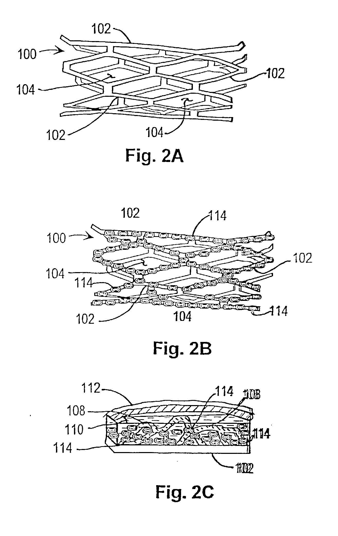
[0055] In a second embodiment or therapeutic strategy of the invention, progenitor endothelial cells transduced with therapeutic transgenes are incorporated into a stent coating. The coating comprises a substance that adheres to the stent and incorporates the cells without damaging them. The implanted endothelial cells will have been transfected (or infected) ex vivo, with vectors containing transgenes encoding gene products with anti-restenosis activities. This anatomic platform facilitates exposure of cells within the injured vessel wall (or cells that are migrating from the media and / or adventitia to form the neointima) to the therapeutic gene product expressed by the endothelial cells.

[0056] As with the first invention strategy or embodiment, this variant of the invention can employ any coating that can be attached to a stent and that has the above characteristics. One such candidate coating has been created by the Photolink.RTM. process of the SurModics Company (Eden Prairie, M...


PUM

PropertyMeasurementUnit
timeaaaaaaaaaa
massaaaaaaaaaa
resistanceaaaaaaaaaa

Abstract

Restenosis of arteries after angioplasty is inhibited by implanting in the treated artery a stent incorporating genes that encode gene products having anti-restenotic activity. The genes may be incorporated into a coating on the stent structure or in cells that are affixed to the stent. The genes or cells containing them may be adhered to the struts of the stent or incorporated in a collagen matrix that forms a coating covering the struts and interstices of the stent

Description

RELATIONSHIP TO OTHER APPLICATIONS[0001] This application claims the benefit of the priority of copending U.S. Provisional Patent Application No. 60 / 251,579, filed Dec. 8, 2000.[0002] 1. Field of the Invention[0003] This invention relates to preventing restenosis of arteries after angioplasty and more particularly to use of a stent platform to deliver gene products through DNA or transfected cells that have been incorporated into a coating applied to the stent, the gene products of which will prevent such restenosis[0004] 2. Brief Description of the Prior Art[0005] Coronary angioplasty has become an important method of treating narrowed (stenotic) arteries supplying the heart or the legs. Although the initial success rate of coronary angioplasty for opening obstructed coronary arteries exceeds 95%, restenosis occurs at the site of angioplasty in 25-50% of patients within six months, regardless of the type of angioplasty procedure used. Although the use of stents has appreciably redu...

Claims


Application Information

Patent Timeline
Patent Type & Authority Applications(United States)
IPC IPC(8): A61K47/48A61K48/00A61L31/00A61L31/10A61L31/16C07K14/52C07K14/78C12N9/68
CPCA61K47/48992C12Y304/21007A61K48/0025A61L31/005A61L31/10A61L31/16A61L2300/258A61L2300/416A61L2300/64C07K14/52C07K14/78C12N9/6435A61K48/00C08L89/06A61K47/6957
Inventor EPSTEIN, STEPHEN E.FUCHS, SHMUEL
Owner EPSTEIN STEPHEN E
Learn More