
Method for loading lipid like vesicles with drugs or other chemicals

Inactive Publication Date: 2004-10-21
RGT UNIV OF CALIFORNIA
View PDF27 Cites 14 Cited by
  • Summary
  • Abstract
  • Description
  • Claims
  • Application Information

AI Technical Summary

Benefits of technology

[0012] In accordance with a third aspect of the present invention, a pharmaceutical preparation for administration in vivo to an animal is provided by the method of encapsulation described above. The chemical species in this instance is a drug. The osmolarity of the buffer within the vesicles is within the physiological range of the animal, the vesicles are suspended for administration in the bulk solution, and the pH of the bulk solution is physiologically benign.
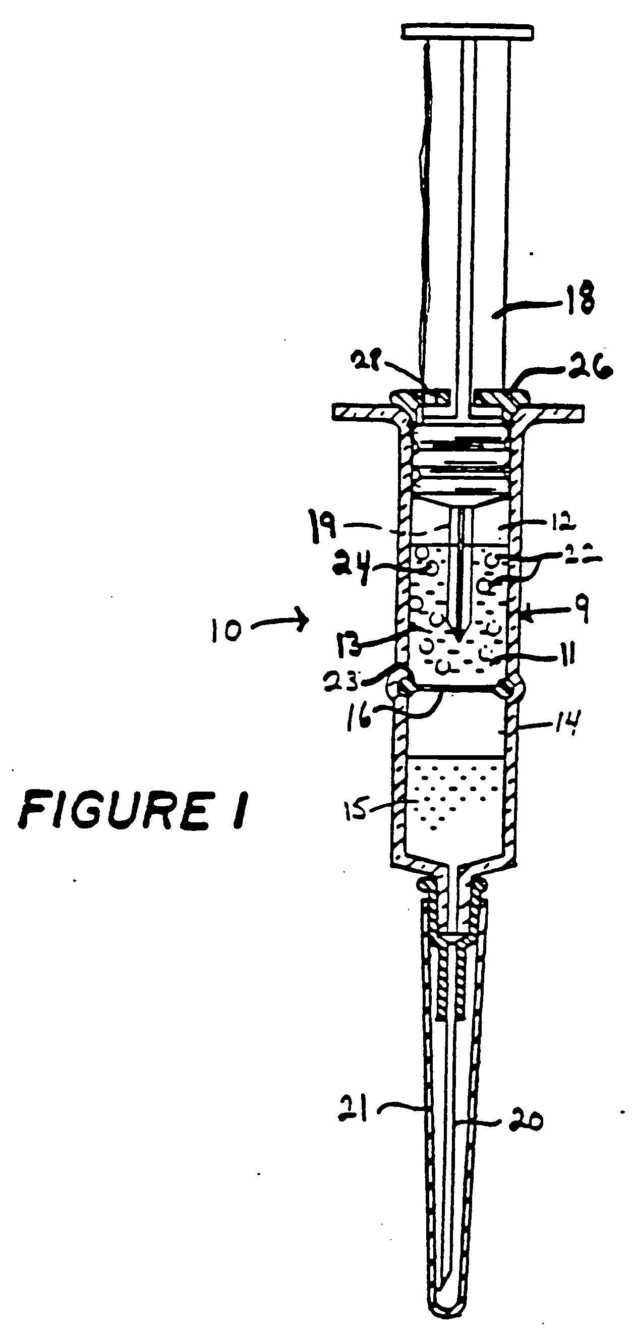
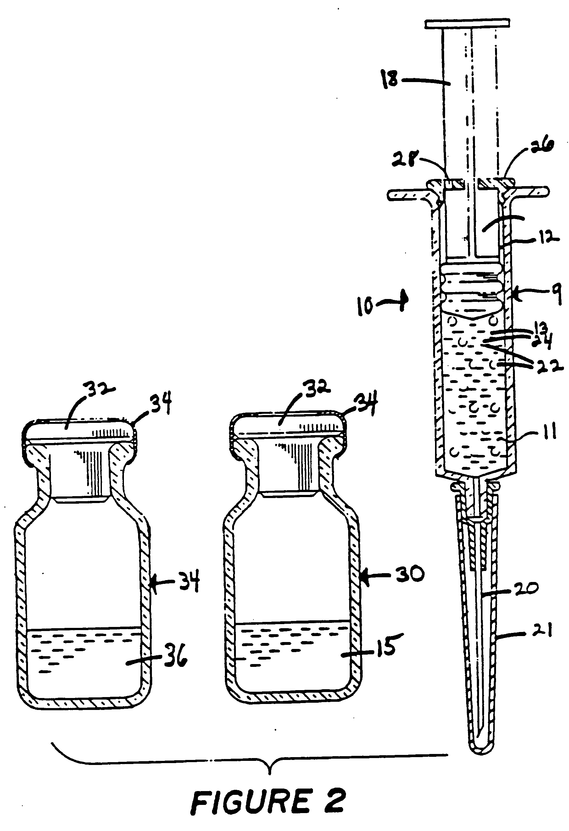
[0036] The syringe (10) also contains the chemical (11) to be loaded. The chemical (11) is present in the solution which affords it the greatest stability and may therefore be in either the first or second solution (13, 15) depending upon the properties of the chemical and the solutions. For purposes of illustration, the chemical (11) in FIG. 1 is located in the first solution (13). The second solution (15) is an acid if the buffer (24) inside the vesicles (22) is a base and a base if the buffer (24) inside the vesicles (22) is acidic. The second solution (15) has a pH such that a mixture of said first and second solutions (13, 15) will have a pH, respectively, of at least about 0.5, 0.3 or 0.2 pH units higher than the buffer (24) if the buffer (24) is acidic and the chemical (11) has respectively one, two or three or more basic pH responsive groups and at least about 0.5, 0.3, or 0.2 pH units lower than the pH of the buffer (24) if the buffer (24) is basic and the chemical (11) has respectively, on, two or three or more acid pH responsive groups. To encapsulate the chemical (11) within the vesicles (22), the syringe (10) is turned with the needle (20) facing upwards (the opposite direction from FIG. 1) and the plunger (18) is forced upward just enough to create enough pressure to break the barrier (16). The barrier may also be broken by a sharp implement (19) attached to the plunger (18). In this instance the implement (19) is prevented from damaging the barrel (9) by a stop (23). The volume of the second solution (15) in the second compartment (14) should be such that when the barrier (16) is broken only a minimal amount of solution will enter the needle (20). Once the barrier (16) is broken the plunger (18) is withdrawn to around its initial position at the top of the first compartment (12). The syringe (10) is then agitated and the two solutions are allowed to mix for an appropriate period of time. If the resulting pH of the mixture of the first and second solutions (13, 15) is physiologically benign the entire mixture including the vesicles (22) containing the now encapsulated chemical species may be injected directly into an animal. For facilitation in operating the plunger (18) the syringe may have a block (26) at the bottom of the barrel (9) with an air hole (28).

Problems solved by technology

This method of encapsulation, however, only allows for oral administration of the encapsulated material, since the droplets of lipids enclosing the encapsulated material are too large to be delivered parenterally.
All of the methods described, either employ laborious procedures requiring skill and training or the use of sophisticated and expensive equipment, have a low efficiency of encapsulation or low encapsulation rate or involve encapsulating the drug simultaneously with the preparation of the vesicle, thereby invoking possible leakage of the encapsulated chemical.
Also these methods leave a substantial portion of the substance sequestered outside of the vesicle since at best only 50% enclosed volumes of the encapsulated material relative to total volumes of the vesicles have been reported.
These methods therefore require that expensive drugs used for encapsulation be recovered from the drug solution in which the vesicles were prepared.
The prior art field of encapsulation methods thus has a number of very serious problems

Method used


Image

  • Method for loading lipid like vesicles with drugs or other chemicals
  • Method for loading lipid like vesicles with drugs or other chemicals

Examples


example 2

[0039] Liposomes were prepared by sonicating 0.5 grams of asolectin in 10 mls of 100 mM sodium citrate buffer, pH 4. An amount of 542 microliters of five normal sodium hydroxide was added. This raised the pH of the bulk solution containing the liposomes to 7.4. An intravenous catheter system consisting of a 27-gauge needle, connected to a 1.0 ml syringe by 4 inches of PE20 (polyethylene) plastic tubing was used for the infusion of the liposome suspension into the lateral tail veins of two female Wistar rats, 250 grams each. The liposome suspension was infused into the rats at a rate of about 0.2 mls per minute until a total volume of 0.7 mls had been infused. The rats appeared somewhat disoriented upon completion of the infusion, and release from the restraining cones, but otherwise none the worse from the experience. One hour later the animals were examined and were completely normal in appearance, and after one week's observation, no long-term effects of the infusion could be dete...

example 3

[0040] Lipid vesicles, containing 15 mg / ml of Sigma Type II-S phosphatidyl choline were prepared by sonication in a 120 mM lysine / phosphate buffer (chloride-free) at pH 10.5. The total sonication time was three minutes, with intermittent cooling. The vesicles were incubated for two minutes with 20 .mu.M of a spin-labeled carboxylic acid, prepared by reacting 1M succinic anhydride with one equivalent of Tempamine in choloroform, in the presence of a sufficient amount of a 100 mM citric acid to lower the external pH to 6 (approximately 1 volume equivalent). Analysis of the intravesicular concentration of the spin-labeled acid by ESR spectroscopy revealed that a more than 1,000-fold increase had occurred in response to the imposed pH gradient.

[0041] The vesicles were then transferred into a piece of dialysis tubing that had been spread into a flattened geometry to minimize the diffusion path of internal molecules to its surface. When the dialysis tubing was placed in a large volumes of...

example 5

[0044] Liposomes are prepared according to Example 1 or 2 and are concentrated by means of a standard filtration concentration to a concentration of approximately 50 mg asolectin per 1 ml of 100 mM sodium citrate. The resulting lipid-like solution is injected in mice as described in Example 2 such that the final infusion is approximately 1% of total fluid body volume of asolectin. This example indicates that large volumes of liposomes having substantial pH gradients can be injected into animals without serious adverse effects.


PUM


Abstract

A method for accumulating drugs or other chemicals within synthetic, lipid-like vesicles by means of a pH gradient imposed on the vesicles just prior to use is described. The method is suited for accumulating molecules with basic or acid moieties which are permeable to the vessels membranes in their uncharged form and for molecules that contain charge moieties that are hydrophobic ions and can therefore cross the vesicle membranes in their charged form. The method is advantageous over prior art methods for encapsulating biologically active materials within vesicles in that it achieves very high degrees of loading with simple procedures that are economical and require little technical expertise, furthermore kits which can be stored for prolonged periods prior to use without impairment of the capacity to achieve drug accumulation are described. A related application of the method consists of using this technology to detoxify animals that have been exposed to poisons with basic, weak acid or hydrophobic charge groups within their molecular structures.

Description

[0002] The invention relates to a method for loading lipid-like vesicles with a drug or other chemical species by establishing a preimposed pH gradient.[0003] The use of membranous vesicles such as liposomes and the like as adjuvants and carriers for drugs, other chemicals and biologically active compounds such as antigens and antibodies is well known in the field (U.S. Pat. Nos. 4,053,585; 4,397,846; 4,411,894; 4,427,649).[0004] Also, many methods exist to encapsulate the various drugs or other chemicals within the vesicles. U.S. Pat. No. 4,241,046, discloses a method for encapsulating biologically active materials within liposomes by providing a combination of lipids in an organic solvent and an aqueous mixture of the material for encapsulation, emulsifying the provided mixture, removing the organic solvent, and suspending the resulting gel in water. The biologically active material is encapsulated by being processed with the liposome during preparation of the liposome.[0005] U.S....

Claims


Application Information

Patent Timeline
IPC IPC(8): A61K9/127
CPCA61K9/1278
Inventor MEHLHORN, ROLF JOACHIM
Owner RGT UNIV OF CALIFORNIA
Learn More