Internal marker device for identification of biological substances

Inactive Publication Date: 2005-01-06
RGT UNIV OF MICHIGAN +1
View PDF14 Cites 23 Cited by
  • Summary
  • Abstract
  • Description
  • Claims
  • Application Information

AI Technical Summary

Benefits of technology

[0016] In one aspect, the present invention is directed to a device for visualizing structure located in the interior of a substance that is visually opaque. As used herein, the term “visually opaque” is taken to mean obscure or not apparent using characteristic imaging modalities. The present invention provides a method and apparatus based on removably positionable internal fiducial markers as a targeting aid or a landmarking or verification device for various imaging techniques. The device is capable of being removably inserted into a suitable lumen or cavity in the subject to be scanned or imaged

Problems solved by technology

No effective internally positionable devices have been developed.
Malignant cells, such as those found in cancerous tumor tissue, generally exhibit elevated energy requirements resulting in elevated levels of glucose consumption or other functional processes.
While both PET and SPECT allow distinction of tumor from normal tissues, there are instances in which PET and SPECT are difficult to use as a single imaging modality.
One drawback of such radiopharmaceutically assisted imaging techniques is that the visualized area of increased tracer accumulation is best localized in comparison to known anatomical references in order to be precisely characterized and located in the subject's body.
This requirement becomes problematic in regions of greater anatomical variation, and in regions which have little radiotracer uptake on scan.
This problem becomes more pronounced in situations where imaging data generated from PET or SPECT scans are to be integrated with imaging data derived from other methods such as, i.e., MRI, CT, or ultrasound.
The methods and devices described in the Wahl and Thomton references present difficulties when employed to visualize regions where greater patient-to-patient anatomical variation is encountered.
Such regions often lack internal landmarks or accurate correlation with the positioning of external fiducial markers.
However, external devices such as the Mount

Method used


Image

  • Internal marker device for identification of biological substances
  • Internal marker device for identification of biological substances
  • Internal marker device for identification of biological substances

Examples


example i

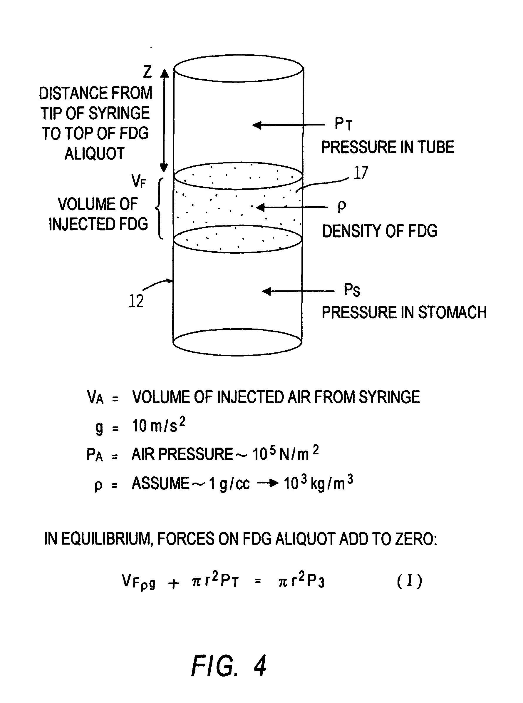
[0098] Studies were conducted in order to determine the potential uptake by a surrounding substance of imaging materials contained in the device of the present invention. A 10-French tube having a length of 109 cm, composed of polyurethane (TECOTHANE), was charged with a 2% hypaque saline solution. X-ray studies of the device thus prepared demonstrated fluid placement without air pockets in the fluid column. A copy of this x-ray is presented in FIG. 6.

[0099] This study indicated that an introduced solution would remain in a single volume in an elongated tube without intervening air bubbles or the like.

example ii

[0100] Three devices of the present invention were prepared according to the disclosure of the present invention. The devices were each 10-French polyurethane tubes of 109 cm2. Each device was charged with 1.6 ml of 2-deoxy-2-[18F]fluoro-D-glucose in suspension prepared to have a total activity of approximately 44.6 microcuries to yield an apparent tracer activity on PET greater than that of normal soft tissue.

[0101] Each device so prepared was fastened at its proximal end and allowed to suspend with a distal end in an Erlenmeyer flask filled with water. Each flask was mechanically moved to simulate body activity. At the end of one hour, radioactive counts of the flask water were taken and did not exceed total background radiation. From this data, it can be concluded that radioactive infiltration into the surrounding water from the device of the present invention did not occur. It could be concluded from these results that material introduced into the device of the present inventio...

example iii

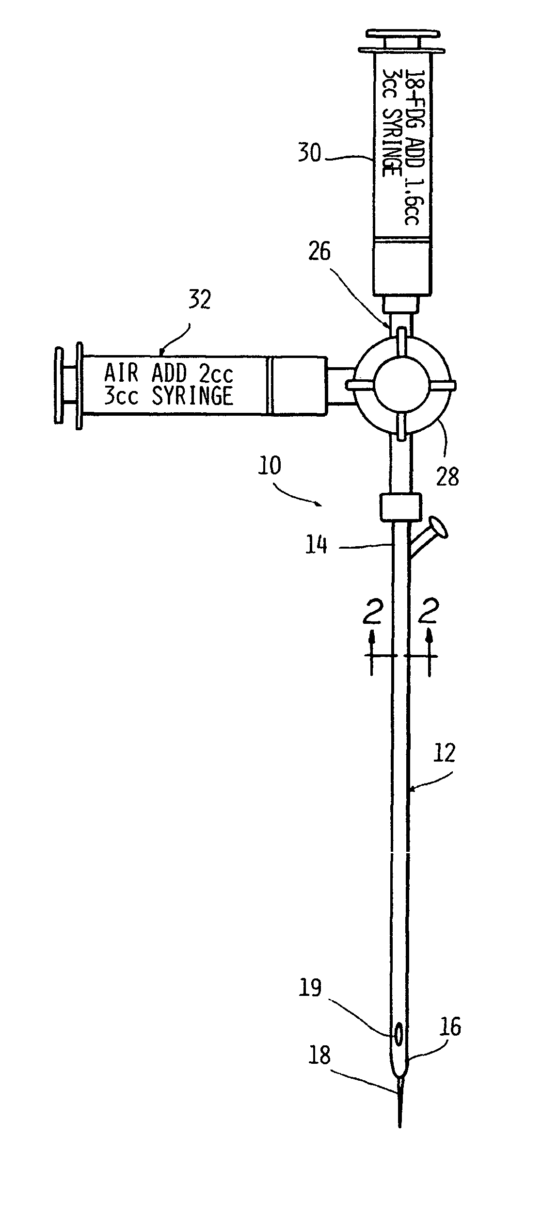
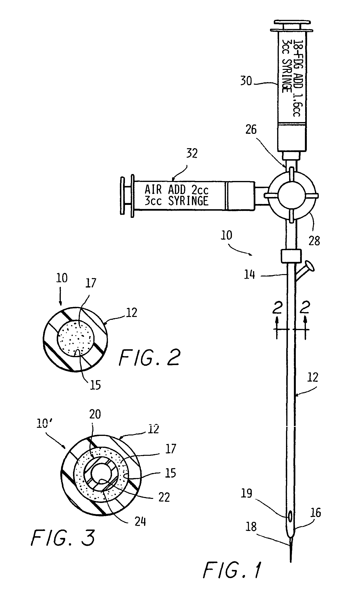
[0102] In order to simulate use, the device of the present invention on a test subject, the procedure was simulated in a phantom. A 109 cm 10-French enteral feeding tube was positioned at a location simulating a subject's esophagus. The associated stylet was removed from the tube and an AP chest x-ray of the simulation dummy (phantom) was taken to confirm proper placement of the tube.

[0103] After proper placement was ascertained, the access port cap was cut off and replaced with a suitable three-way stopcock with a male luer lock. The existing flushing port of the tube was capped. Two 3-ml luer-lock syringes were affixed to the two open ports of the stopcock. One syringe was charged with 1.6 ml of FDG prepared in the manner described in Example II. The other syringe was charged with 2 ml of air.

[0104] In order to charge the lumen, the stopcock handle was turned to block flow from the air-filled syringe and to open a channel to the FDG-filled syringe. Injection of 1.6 cubic centime...


PUM


Abstract

A device for visualizing structure located on the interior of a biological substance. The device includes a marker member that may be a solid cylinder or lumen having an interior volume having a distal end removably insertable in the biological substance relative to the interior structure to be visualized. An image-enhancing material is contained relative to the marker member in a manner such that the imaging material does not directly contact the biological substance. The imaging material of choice is one capable of producing an emission or signal detectable external to the biological substance by suitable imaging instrumentation. Also disclosed is a method for visualizing critical structures or radiation therapy targets in imaging processes such as positron emission tomography and/or single photon emission computerized tomography, MRI, or ultrasound either used alone or in combination or in registration with anatomical imaging processes such as computed tomography or mammography.

Description

CROSS-REFERENCE TO RELATED APPLICATIONS [0001] This application is a continuation-in-part of U.S. Ser. No. 09 / 539,002 filed Mar. 30, 2000.STATEMENT OF GOVERNMENT SUPPORT [0002] This invention was made in the couse o research partially supported by a grant from the National Institute of Health (NIH CA 52880). The U.S. government has certain rights in the invention.FIELD OF THE INVENTION [0003] The present invention is related to identifying landmarks that assist in visualizing internal structures during diagnostic and / or therapeutic procedures, including medical procedures. BACKGROUND OF THE INVENTION [0004] Targeting aids or landmarking devices have been employed in diagnostic imaging techniques such as computed tomography (CT), magnetic resonance imaging (MRI) and ultrasound. These devices, commonly referred to as fiducial markers, generally occur as two types: internally occurring markers that are inherent in the subject's anatomy; and externally positionable imaging aids that can...

Claims


Application Information

Patent Timeline
IPC IPC(8): A61B19/00
CPCA61B19/54A61B6/037A61B2019/5454A61B90/39A61B2090/3954
Inventor THOMAS, CHERRY T.WAHL, RICHARD L.FISHER, SUSAN J.
Owner RGT UNIV OF MICHIGAN
Why Eureka
Try Eureka