Paramagnetic polymerized protein microspheres and methods of preparation thereof

a polymerized protein and microsphere technology, applied in the field of contrast agents, can solve the problems of reducing the efficacy of contrast agents, restricting the use of particular contrast agents to one imaging mode, complex interaction between encapsulated microbubble contrast agents and ultrasound, etc., and achieve the effect of accurate simulation

Inactive Publication Date: 2005-01-13
THE BOARD OF TRUSTEES OF THE UNIV OF ILLINOIS
View PDF19 Cites 1 Cited by
  • Summary
  • Abstract
  • Description
  • Claims
  • Application Information

AI Technical Summary

Benefits of technology

Ultrasound pressure waves of current B-mode imaging machines typically destroy microbubbles. Fixed concentrations of the contrast agent are stabilized in cellulose dialysis tubes to determine the characteristics of unmodified and surface-modified GOAM in a commercial diagnostic ultrasonic field. The two different ultrasound transducers described above are used to acquire the RF data and B-mode images. The acoustic power of the ultrasound machine as reflected by the mechanical index (“MI”) provided on the machine is set in 0.1 MI steps, from 0.2 to 0.8 MI. The tube contains fresh contrast agents for each MI level test for each transducer. The backscatter coefficient for each step for each contrast agent is determined.
Different suspension conditions affect the properties of ultrasound contrast agents. The effects are tested by changing different air concentrations, diluting the contrast agent, and utilizing different carrier media.
The effects of air concentration are assessed for both unmodified and surface-modified GOAM at fixed concentrations using the methods described in Sboros. (Sboros et al., “An In Vitro Comparison of Ultrasonic Contrast Agents in Solutions with Varying Air Levels,” Ultrasound in Med. & Biol., 26:807-18 (2000) which is incorporated by reference herein). Sterile water is used as the suspension medium. A sterile bag filled with sterile water is infused with helium or air to achieve partial oxygen pressures (pO2) of 1.5 or 24.7 kPa, respectively. These suspensions are injected slowly in the cellulose dialysis tubing. The imaging data is gathered under these conditions using the Aloka 5500 PHD RF machine to acquire the RF data and B-mode images. Microbubble concentration and size are determined for the suspensions. Normalized ultrasonic backscatter vs. concentration is examined.
Ultrasound wave propagation in a synthetically generated medium was simulated. The simulation consisted of a bubble with an outer diameter of 250 μm and inner diameter of 225 μm. The bubble was surrounded by an albumin shell of thickness 12.5 μm. FIGS. 5a-5d illustrate a sequence of snapshots of the absolute value of the scattered wave. The simulation shows the progress of an ultrasound wave toward a single spherical target. FIGS. 5a-5d illustrate that the ultrasonic wave hitting the sphere, as well as the wave being reflected (the backscattered wave), is accurately simulated.

Problems solved by technology

The differences in the imaging techniques involved with various modalities, however, have thus far generally restricted the use of any particular contrast agent to one imaging modality.
The interaction of encapsulated microbubble contrast agents with ultrasound is complex.
A significant problem with the use of microbubble contrast agents result from the machinery associated with the imaging process.
This acoustic pressure range can destroy some microbubble contrast agents during the imaging process, thus reducing the efficacy of the contrast agent and also reducing the effective imaging time (half-life) of the contrast agent.
The major drawbacks associated with use of Albunex® as a contrast agent for ultrasound are its short plasma half-life and its acoustic instability relative to pressure changes.
Moreover, albumin microbubbles cannot by used with other modalities such as magnetic resonance imaging or computed tomography because the microbubbles do not have the functional characteristics required for such modalities.
Work by de Jong showed large differences in non-linear behavior between ideal and Albunex® microspheres due to the additional restoring force and friction inside the shell that surrounds the Albunex® microsphere.
Thus, although there are a number of ultrasonic contrast agents now available commercially, and despite significant research directed to many of these agents, limitations still exist with these agents.
Furthermore, few ultrasonic contrast agents can be used with other imaging modalities.
With this method of preparation, residual GdCl3 is likely to remain in the Gd2O3 preparation, such that extreme toxicity from inadvertently incorporated free GdCl3 is possible.
With most chelated gadolinium contrast agents, only one gadolinium atom per molecule is present in commercially-available contrast media manufactured for use in MR imaging, so that the enhancement capabilities of the contrast agent are limited.
In addition, synthesis of albumin particles and also albumin microspheres tagged with gadolinium chelates on the surface would also be expected to have decreased MR sensitivity due to the limited number of sites for conjugation of the gadolinium chelate to the microsphere surface.
Because iron oxide is predominately a T2 relaxation agent, MAM has limited usefulness in conventional MR imaging.
Additionally, based on the lower density of iron oxide relative to other heavy metals, iron oxide, and thus MAM, has a very limited utility for other imaging modalities, such as computed tomography.
As with contrast agents for US, contrast agents for MR also have limitations, both when used with MR and if used with other imaging modalities.
A major drawback associated with using Gd-DTPA contrast agents for CT imaging is the fact that only one electron dense (gadolinium) atom per molecule is present in commercially-available contrast media.
Thus, presently available MR contrast agents provide sub-optimal CT enhancement and / or are not well-suited for use with other imaging modalities, such as CT and US.
To date, few contrast agents have been used for imaging studies utilizing multiple imaging modalities.
Magnevist® (Gd-DTPA) and a few other gadolinium-containing MR contrast agents have been used for this purpose, but limitations associated with the dosage and cost of commercially available MR contrast agents have prevented widespread use.
Further, these agents would confer no obvious benefit to US imaging due to their low compressibility and the high concentrations required in order to provide effective US imaging.
However, neither fluorine MR imaging nor signal void imaging have found widespread use in hospital or clinical practice, where T1 (and to a lesser extent, T2) imaging of protons is typical.
Despite the significance of contrast agents in medical diagnostics and the ever-present need for correlative studies, no single commercially-available contrast agent provides effective, cost-efficient image enhancement utilizing more than one imaging modality.

Method used


Image

  • Paramagnetic polymerized protein microspheres and methods of preparation thereof
  • Paramagnetic polymerized protein microspheres and methods of preparation thereof
  • Paramagnetic polymerized protein microspheres and methods of preparation thereof

Examples


example 1

Ultrasound Studies

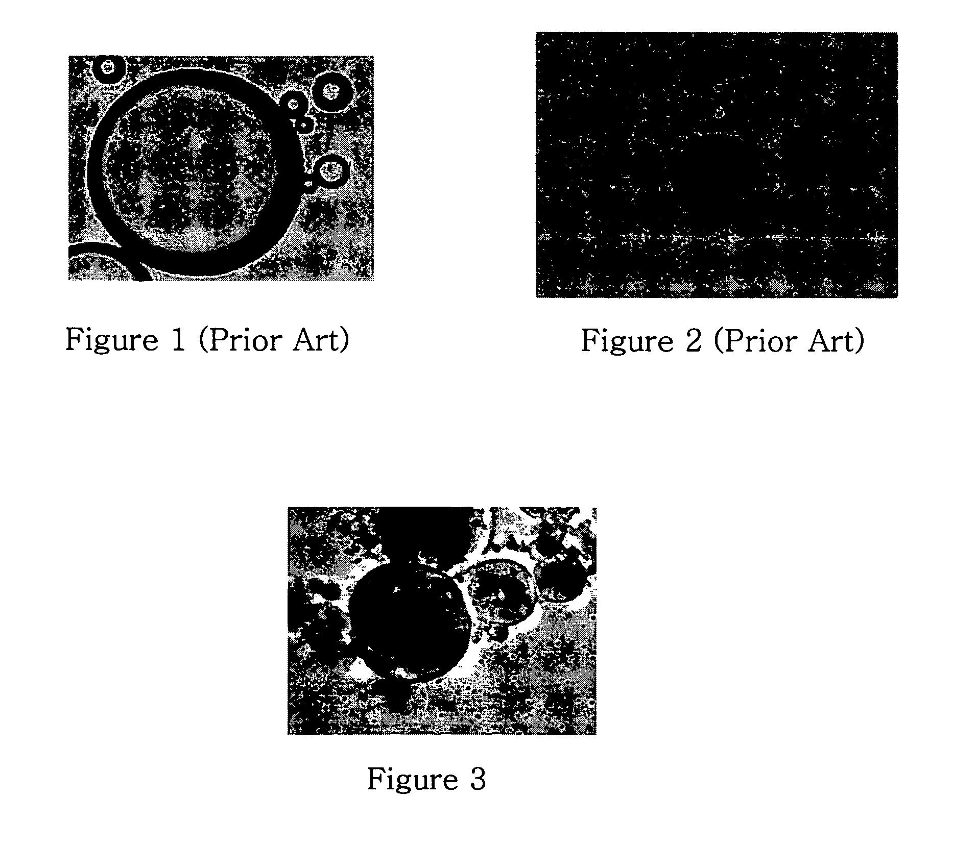
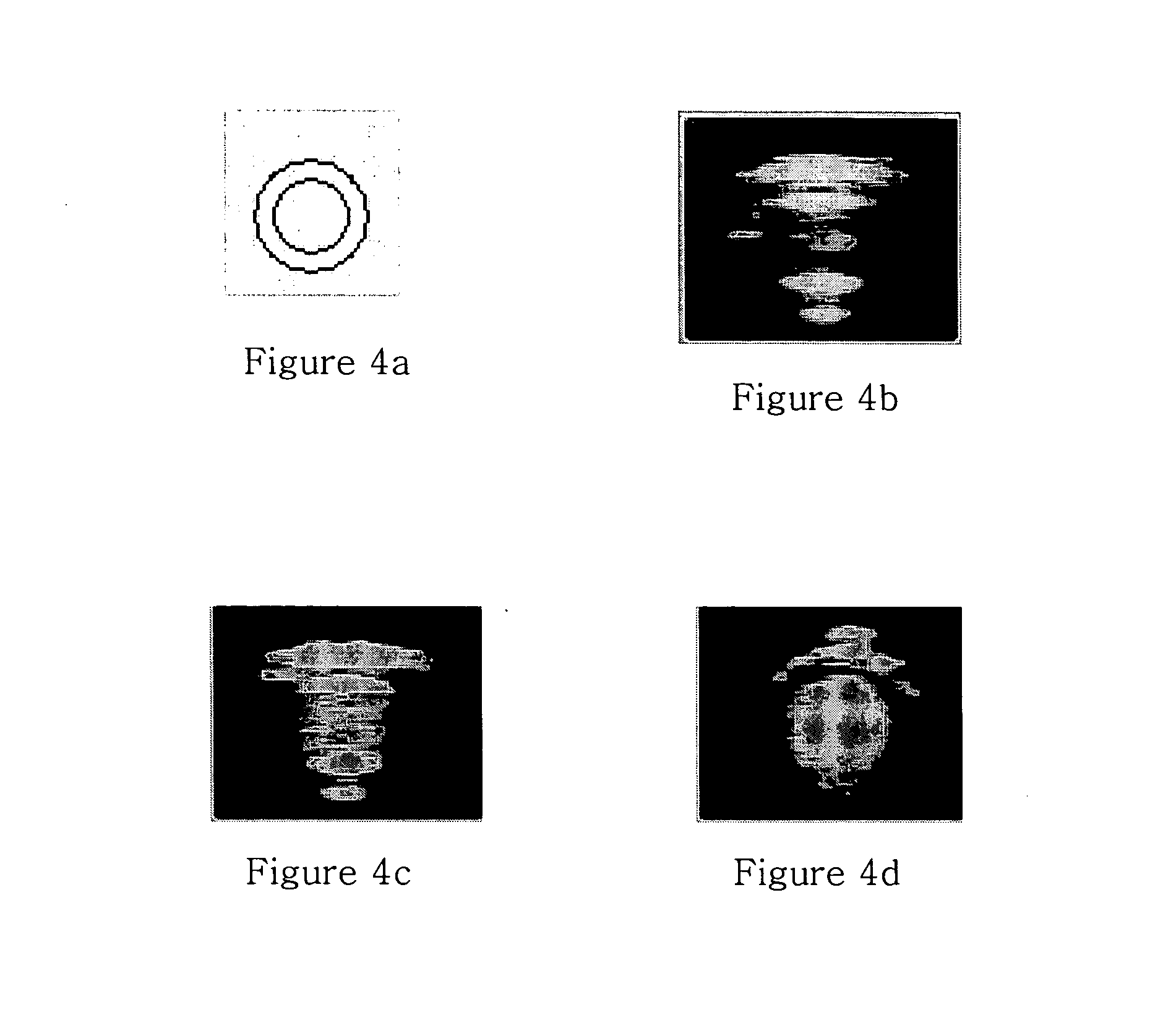
Preliminary imaging studies were conducted to compare the cross-sectional ultrasound images of oil (containing no contrast agents), air-filled albumin microspheres, and unmodified and surface-modified GOAM flowing through a tube. An Aloka SSD5500 PHD ultrasound machine with a linear transducer (UST 5539 10 MHz) was used to create traditional B-mode ultrasound images. In these experiments, the above solutions were injected into clear plastic Tygon tubing (OD=0.318 cm; ID=0.159 cm) (FIG. 4a) immersed in degassed water at room temperature. Cross-sectional images of the tube were captured using a personal computer, video frame grabber and real time video capture software (Capture©, Watkin, 1997). These images are shown in FIGS. 4b-4d.

These images clearly demonstrate the full circumference visualization capabilities of the various media—oil (no contrast media), air-filled albumin microspheres, and GOAM. Differences are clearly evident in the cross-sectional B-mode u...

example 2

Physical Characterization of Unmodified and Surface-Modified GOAM

The determination of the size distribution, concentration, and size fractionation of the synthesized GOAM is accomplished via Coulter counter analysis. In addition, optical microscopic images (Bausch & Lomb) are used to verify the size and conformation of GOAM.

example 3

Acoustic Simulations

Two different simulation approaches are required to describe the characteristics of the acoustic driving forces on the developed microsphere as well as the acoustic propagation of the scattered ultrasonic energy. One approach uses boundary element method (BEM) modeling to describe the acoustic behavior of the microsphere. Finite difference time domain modeling (FDTD) is used to examine the backscattering properties of the reflected acoustic pressure waves.


PUM

PropertyMeasurementUnit
diameteraaaaaaaaaa
molar concentrationaaaaaaaaaa
timeaaaaaaaaaa

Abstract

The present invention relates to a composition that includes gadolinium particles encapsulated in microsphere shells. The composition is suitable for use as a contrast agent with a plurality of imaging modalities, including, for example, ultrasound, magnetic resonance imaging, and computed temography. The compositions also are useful for therapeutic applications, including neutron capture therapy.

Description

BACKGROUND OF THE INVENTION 1. Field of the Invention The invention relates to contrast agents and methods of preparation thereof for use in various imaging modalities, and / or for use in therapy. 2. Description of Related Art Introduction to Imaging Modalities, Various in vivo imaging processes, including ultrasound, magnetic resonance and computed tomography, are used as medical diagnostic tools. The underlying principle of each imaging modality is generally that the differences in a particular property or properties (e.g., acoustic properties, proton density, etc.) of the organs, tissue and other substances within the body at a location to be examined are detected and then translated into an image. The various modalities, however, rely on very different principles to generate images. The effectiveness of any of these imaging processes, and the resolution of the resulting images, in a great part depends on the degree of contrast between the body parts that the imaging equipmen...

Claims


Application Information

Patent Timeline
Patent Type & Authority Applications(United States)
IPC IPC(8): A61K9/50A61K41/00A61K49/18A61K49/22
CPCA61K9/5052A61K41/009B82Y5/00A61K49/225A61K49/1821
Inventor MCDONALD, MICHAEL A.WATKIN, KENNETH L.
Owner THE BOARD OF TRUSTEES OF THE UNIV OF ILLINOIS
Why Eureka
Try Eureka