
Method for combined parallel agent delivery and electroporation for cell structures and thereof

a cell structure and electroporation technology, applied in the field of combined parallel agent delivery and electroporation for cell structures, can solve the problems of difficult transfer of all these compounds and particles

Inactive Publication Date: 2005-02-03
CELLECTRICON
View PDF6 Cites 29 Cited by
  • Summary
  • Abstract
  • Description
  • Claims
  • Application Information

AI Technical Summary

Benefits of technology

"The present invention is a method for quickly and easily delivering multiple agents to cells using a technique called electroporation. This method allows for the simultaneous or sequential delivery of agents to cells using two or more electrodes. The technique can be used for screening drugs and identifying proteins inside cells, as well as for studying the function of intracellular proteins and metabolic pathways. The method can also be used to detect and identify receptor ligands and enzymes substrates. The invention also includes a method for altering the biochemical content of cells by introducing agents into the cell's cytoplasm. Overall, the invention provides a faster and more efficient way to deliver agents to cells and study their effects."

Problems solved by technology

Although these probes and indicators are available, the main challenge so far in applying these probes and indicators is to introduce them into the cellular interior.
It is, however, difficult to transfer all these compounds and particles to the cytosol of a cell owing to the presence of a cell plasma membrane barrier, which acts as a physical boundary to the external solution that prevents the entrance of exogenous compounds and particles.

Method used


Image

  • Method for combined parallel agent delivery and electroporation for cell structures and thereof
  • Method for combined parallel agent delivery and electroporation for cell structures and thereof
  • Method for combined parallel agent delivery and electroporation for cell structures and thereof

Examples


second embodiment

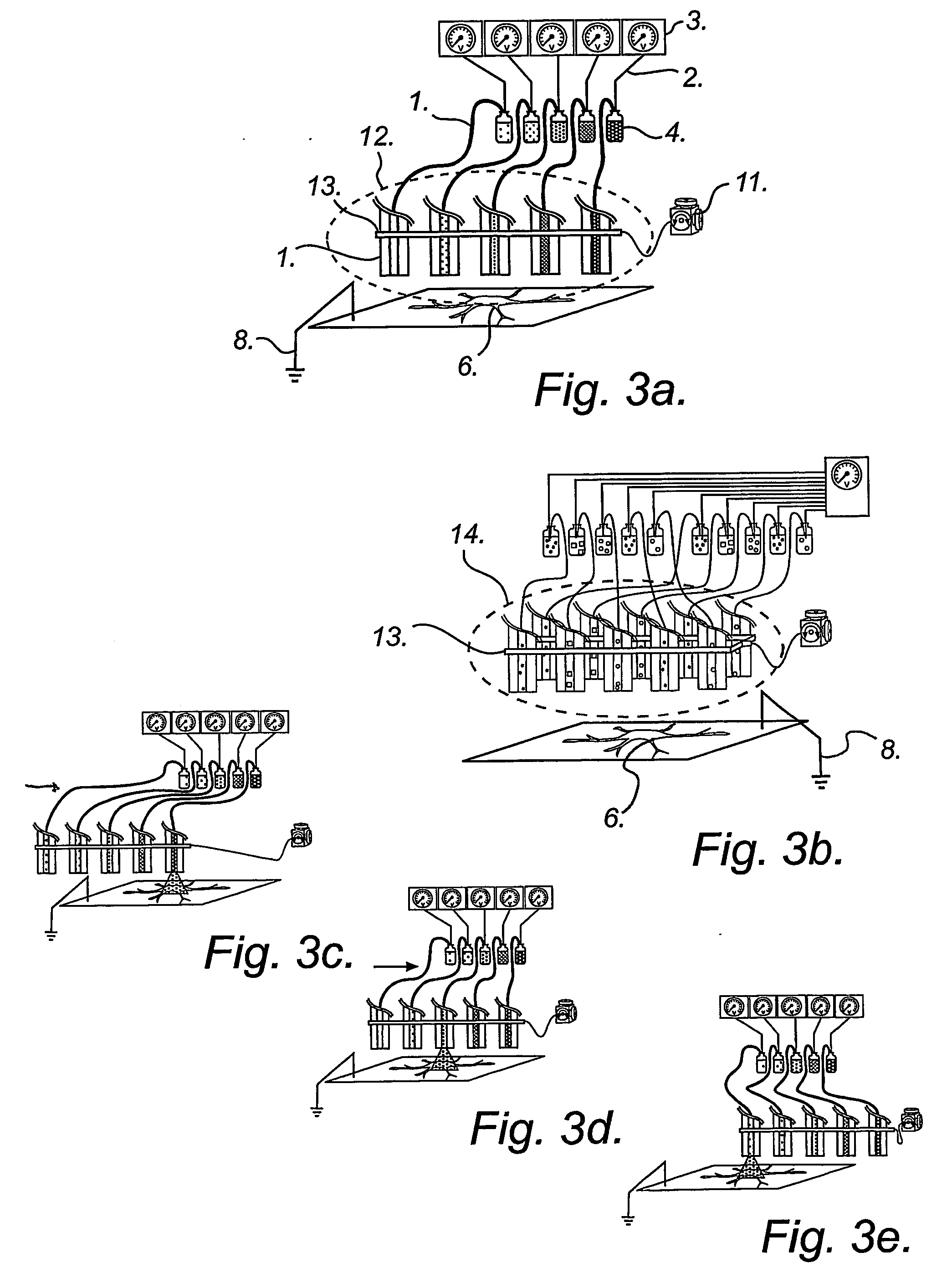
According to another embodiment, sequential delivery of multiple cell-loading loading agents is achieved by connecting a conventional EFC to a microfluidic switch for sample stacking. In this configuration a plurality of loading agents can be sequentially introduced into the EFC and subsequently be delivered to a single or several cellular targets. The cell-loading agents can be introduced in discrete zones in the EFC by any of the pumping modes discussed above. Either the EFC is preloaded with cell-loading agents before electroporation experiments or the EFC is loaded on-the fly, that is during electroporation experiments. An example of this second embodiment is illustrated in FIG. 5.

According to yet another embodiment, sequential delivery of multiple cell-loading agents is achieved by introducing a separation step while pumping the fluid through the EFC. When using electrophoretic pumping for delivery of reagents, all species present in the electrolyte solution is separated based...

example 1

Detection of the Intracellular Receptor Ryanodine Type II

Fluo-3 AM ester was from Molecular Probes (Leiden, The Netherlands). Cyclic ADP ribose and the chemicals used for buffer solutions, were all of analytical grade and purchased from Sigma (St. Louis, Mo., USA). All solutions were made in distilled water from a Milli-Q system (Millipore).

NG108-15 cells were plated on no. 1 cover slips or in a Petri dish and allowed to grow for 1-3 days. Cell dishes were mounted in a circular polycarboneat holder and transferred to the stage on the microscope. Prior to experiments the culture medium was replaced by a HEPES buffer (NaCl 140 mM, KCl 5.0 mM, MgCl2 1.0 mM, CaCl2 1.0 mM, D-glucose 10 mM, HEPES 10 mM, pH was adjusted to 7.4 with NaOH).

The cells were stained with fluo-3 AM ester by incubating the cells for 30 minutes in dye solution (10 μM fluo-3 AM ester in HEPES buffer) at room temperature. To remove excess uncaptured dye, the cells were washed three times in HEPES buffer and st...

example 2

Detection of Intracellular Enzymes I. Detection of Proteases

Casein BODIPY FL was obtained from Molecular Probes (Leiden, The Netherlands). The chemicals used for buffer solutions were all of analytical grade and purchased from Sigma (St. Louis, Mo., USA). All solutions were made in distilled water from a Milli-Q system (Millipore).

Cell culturing and preparations were made according to methods used in example 1 above, and apparatus and instrumentation was the same as in example 1.

Electroporation was performed as in example 1 and an EFC (30 cm long, 50 μm id., 375 μm o.d.) was used. Casein BODIPY FL was used in a concentration of 100 μg / ml and electroporated into cells using a 10 second pulse at 10 kV.

The results of detection of intracellular proteases using casein-BODIPY-FL is shown in FIG. 9. Specifically, the intracellular protease activity was investigated using a protein, casein, which was heavily loaded with the green-fluorescent molecule BODIPY FL, as enzyme substrate....


PUM

PropertyMeasurementUnit
voltageaaaaaaaaaa
voltageaaaaaaaaaa
close distanceaaaaaaaaaa

Abstract

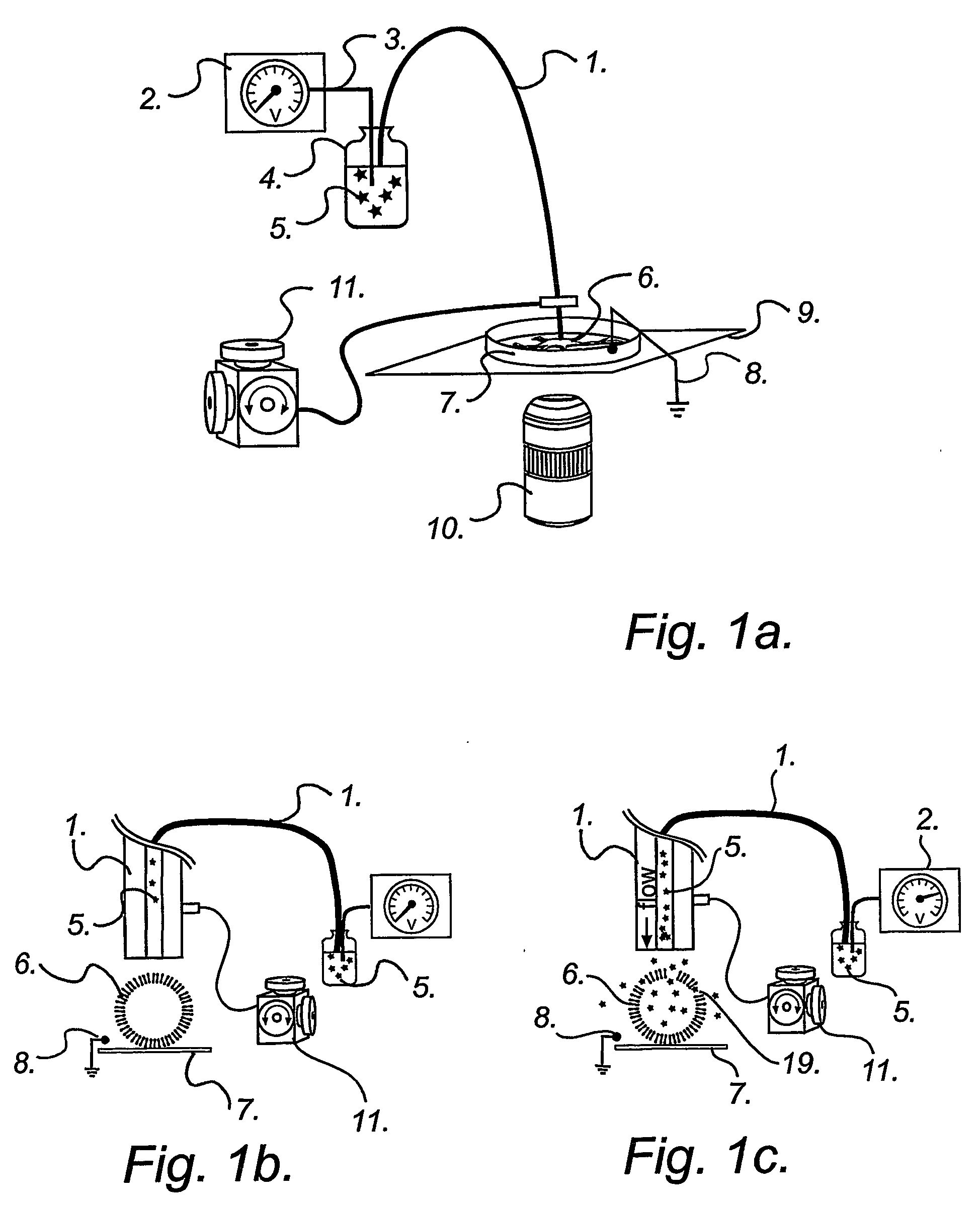
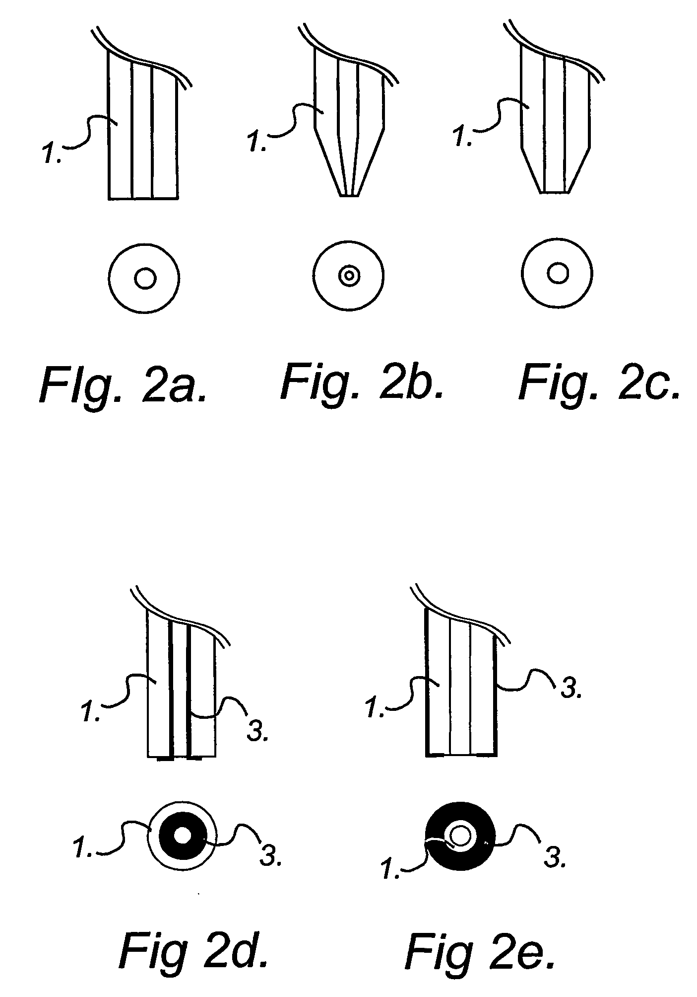
Disclosed is a method for parallel delivery of agents to and / or into a cell structure, wherein at least two electrolyte-filled tubes are provided together with a counter electrode, the tubes being connected to a voltage or current generator, said agents being introduced into the electrolyte solution contained in the tubes, which are placed close to the cell structure, whereupon the agents are transported through the tubes to said cell structure and into the said structure through pores which have been formed by application of an electric field focused on the cell structure, resulting in electroporation of the cell structure. Also different applications of the method is disclosed, e.g. use of the method in order to transfer cell-impermeant solutes, such as drugs or genes, into the cell structure or out of the cell structure.

Description

FIELD OF THE INVENTION The present invention relates to a highly spatially resolved method for the electroporation and parallel delivery of one or several different compounds into cell structures, such as cells, cell-like structures, or a population of cells. Preferably, at least two electrolyte-filled capillaries (EFCs), a linear array of EFCs, or a two-dimensional matrix array of EFCs coupled to a voltage generator are used as a combined electroporation and delivery tool of, for example, multiple dyes, drugs, DNA, RNA, antisense oligonucleotides and biomolecules into the cytoplasm of single cells or populations of cells in a sequential or parallel manner or combinations thereof. The invention also relates to the use of these methods. In particular, it relates to methods for rapid screening of drugs that affect intracellular chemistry, and for the identification and detection of intracellular proteins. BACKGROUND OF THE INVENTION Fast and reliable methods for investigating drug a...

Claims


Application Information

Patent Timeline
Patent Type & Authority Applications(United States)
IPC IPC(8): G01N33/50A61K31/74A61K33/00A61K35/12A61K38/00A61K38/43A61K39/00A61K39/395A61K45/00A61K48/00A61N1/30A61P43/00C12N13/00C12N15/09C12N15/87G01N21/78G01N33/15G01N33/487G01N33/86G01N35/10
CPCA61N1/30G01N2035/1039G01N35/1074C12N15/87A61P43/00
Inventor ORMAR, OWEKARLSSON, MATTIASNOLKRANTZ, KERSTINFARRE, CECILIA
Owner CELLECTRICON
Learn More