Devices and methods for disruption and removal of luminal occlusions

Inactive Publication Date: 2005-02-10
UNIV OF FLORIDA RES FOUNDATION INC
View PDF65 Cites 106 Cited by
  • Summary
  • Abstract
  • Description
  • Claims
  • Application Information

AI Technical Summary

Benefits of technology

[0015] The subject invention concerns a device and methods for disrupting and/or removing occlusive material from lumens, such as biological lumens within the body. Although the subject invention is particularly useful for the disruption and/or removal of thrombus from the vasculature, it is also applicable in other lumens of the body, such as the ureter, urethra, fallopian tubes, bile duct, intestines, and the like. The subject invention provides for effective disruption and/or removal of the occlusive material

Problems solved by technology

This is particularly desirable in treatment of the vasculature,

Method used


Image

  • Devices and methods for disruption and removal of luminal occlusions
  • Devices and methods for disruption and removal of luminal occlusions
  • Devices and methods for disruption and removal of luminal occlusions

Examples


example 1

Construction of a Thrombectomy Device

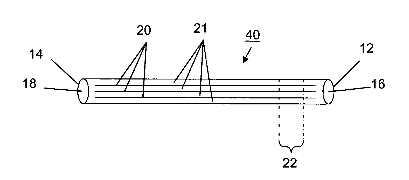
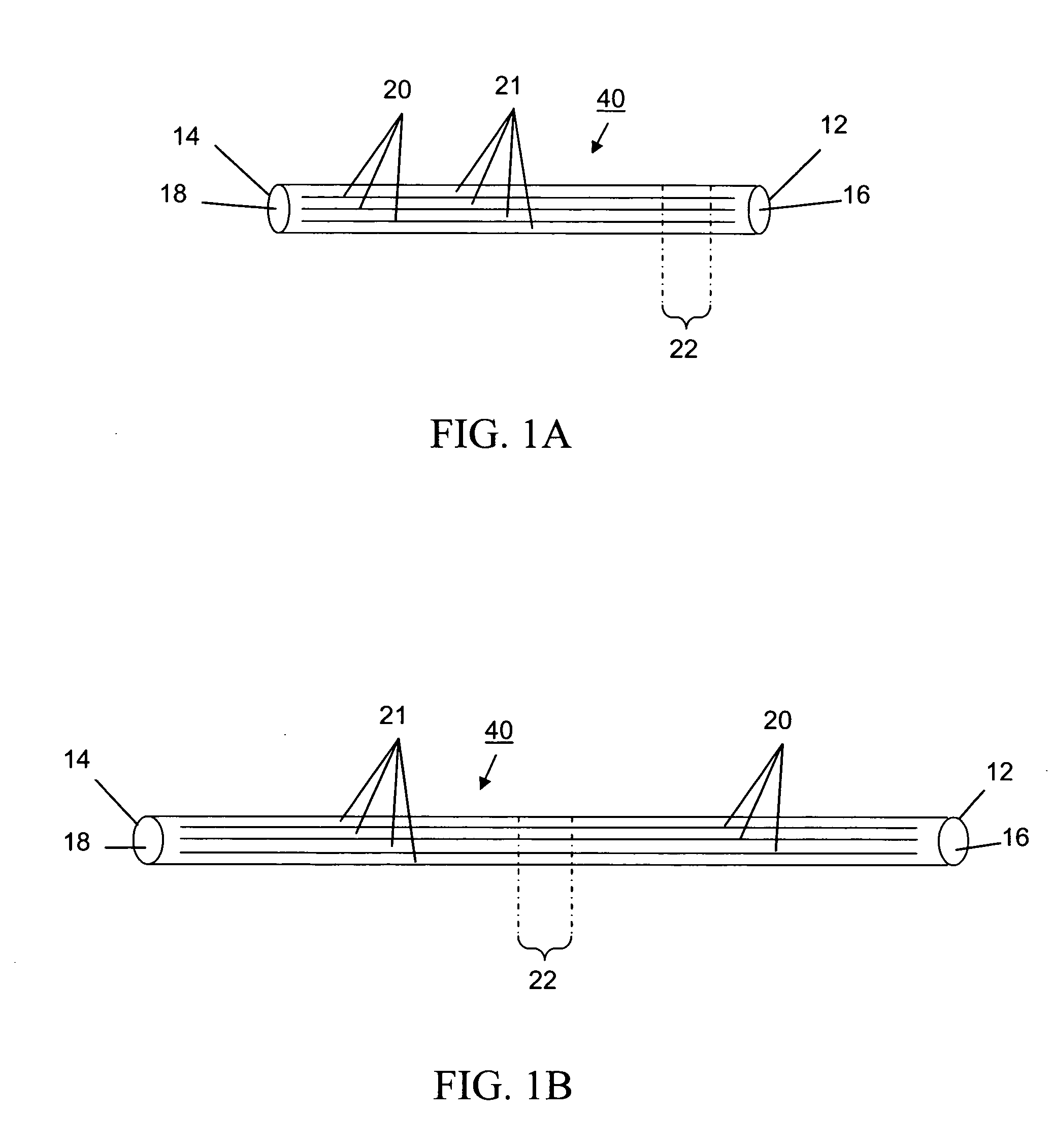
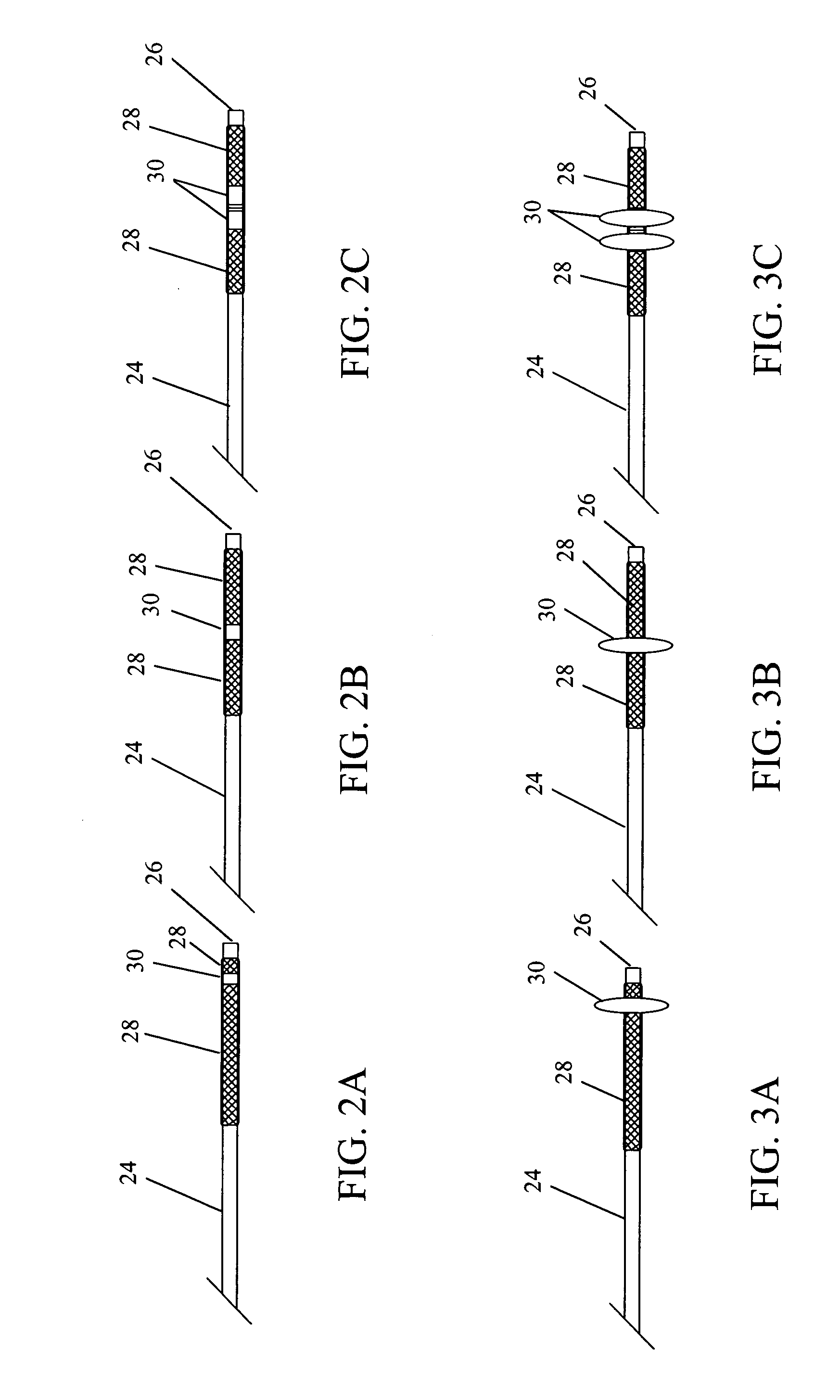
[0074] A thrombectomy device of the subject invention has been made in a modular fashion. The device consists of an elastomeric polymer tube of an appropriate size and a balloon catheter.

[0075] Tubes were made from CARBOSIL 40 90A (The Polymer Technology Group, Inc., Berkeley, Calif.), a solution grade elastomeric, tear-resistant silicone-polyurethane thermoplastic copolymer, by the dip-coating method. Several 21 (0.80 mm outer diameter, OD) gauge injection needles (MONOJECT from Sherwood Medical, St. Louis, Mo.) were used as mold substrates. Each needle was 1.5 inches long and the size (OD) of the needle represents approximately the inner diameter (ID) of the resulting tube. Two times dipping of each needle in 15 wt% polymer solution in tetrahydrofuran (THF) at about a 4 mm / sec withdrawal rate and a 30 minute interval between two successive dippings formed an approximately 90 μm thick polymer coating. Coated needles were then vacuum-dried. Coa...

example 2

Mechanical Testing of a Modified Balloon Under Applied Saline Pressure

[0080] To determine the inflated balloon size with respect to the applied saline pressure, the device was installed on a micromanipulator. Using a digital inflation device and fluid dispensing syringe (MONARCH 25, MERIT MEDICAL SYSTEMS, Inc., South Jordan, Utah) the saline pressure was applied. The micromanipulator read the inflated balloon diameter quite accurately at different saline pressures. The digital inflation device showed the applied saline pressure directly. A plot (calibration curve) of balloon diameter versus saline pressure was constructed as shown in FIG. 6. A control experiment with a bare balloon was performed and compared with the device. As expected, higher pressure was required to inflate the modified balloon in the device. This type of calibration curve will be helpful for both in vitro and in vivo device operation.

example 3

In Vitro Performance Testing of the Thrombectomy Device in Silicone Tubing

[0081] 40 cm long silicone tubing (ID 2.5 mm) was selected for the preliminary in vitro performance testing of the device 60. Two syringes were placed at both ends of the tube 61. Due to the flexible nature of the tube 61, an approximately tortuous shape could be given without kinking, as shown in FIG. 7A. Rabbit blood clots were used as models for the experiment. 1 ml of blood was allowed to coagulate in a 7 ml sterile blood collection tube for a period of 1 hour. The tube 61 was then opened and the bulk clot was removed and cut in half. Each section was weighed and recorded. The silicone tube 61 was prepared by flushing it with saline solution to remove any air bubbles from the tube 61. The blood clot was placed in a 3ml syringe and injected into the silicone tube 61. Saline was then injected into the silicone tube 61 until the clot reached approximately 20 cm from the proximal end (first end, 62) of the tu...


PUM


Abstract

The subject invention pertains to an elastic sheath, device, and methods for disrupting and/or removing occlusive material from lumens, particularly biological lumens, such as the vasculature, ureter, urethra, fallopian tubes, bile duct, intestines, and the like. The subject invention provides for effective disruption and removal of occlusive material, such as a thrombus, from the body lumen with minimal risk of injury to the lumen wall. Advantageously, the invention can be used to achieve a high degree of removal while minimizing the amount of occlusive material that is released into the body lumen. The subject invention further pertains to methods for disrupting and removing occlusive material from a biological lumen. In another aspect, the present invention concerns a device useful as an in vitro model of luminal occlusion and methods for using the device to test the efficacy of devices and methods for treating luminal occlusions.

Description

CROSS-REFERENCE TO RELATED APPLICATION [0001] The present application claims the benefit of U.S. Provisional Application Ser. No. 60 / 470,067, filed May 12, 2003, which is hereby incorporated by reference herein in its entirety, including any figures, tables, nucleic acid sequences, amino acid sequences, or drawings.BACKGROUND OF THE INVENTION [0002] The blockage of human arteries can lead to a variety of serious medical complications. Arterial blockages reduce blood flow through the affected artery and may result in damage to the tissue that is relying upon the blood's supply of oxygen (ischemia). For example, if the blockage is in an artery that supplies blood to the heart itself, a heart attack may result. [0003] Thrombosis and atherosclerosis are common ailments that result from the deposition of thrombus on the walls of blood vessels. When such deposits harden, they are commonly referred to as plaque. These plaque deposits occur commonly in blood vessels that feed the brain, hea...

Claims


Application Information

Patent Timeline
IPC IPC(8): A61BA61B17/22
CPCA61B17/221A61B2017/2212A61B17/320725
Inventor SANTRA, SWADESHMUKULMERICLE, ROBERT A.BATICH, CHRISTOPHER D.STANLEY, JESSIE T.ESKIOGLU, ERIC
Owner UNIV OF FLORIDA RES FOUNDATION INC
Why Eureka
Try Eureka