Fluorescence polarization imaging devices and methods

a fluorescence polarization and imaging device technology, applied in the field of real-time methods for imaging organic tissue, can solve the problems of difficult excision, time-consuming and laborious procedures, and visual detection of tumor borders, and achieve the effect of increasing the number of surgical procedures and improving standard cancer surgery

Inactive Publication Date: 2005-05-05
THE GENERAL HOSPITAL CORP
View PDF31 Cites 38 Cited by
  • Summary
  • Abstract
  • Description
  • Claims
  • Application Information

AI Technical Summary

Benefits of technology

[0032] The present invention is directed to meeting the aforementioned needs and addressing the deficiencies particularly discussed above and generally in the prior art. The methods, system and apparatus constructed in accordance with the present invention are particularly useful for real-time intraoperative delineation o

Problems solved by technology

These cancers are a major cause of morbidity in the Caucasian population.
Unfortunately, most of these tumors have poorly defined boundaries, which makes visual detection of the tumor borders and, consequently, precise excision a challenging problem.
While precise and accurate, MMS is also a time-consuming and staff-intensive procedure.
However, all these techniques lack one or more elements necessary for their practical use in a clinical setting.
The major disadvantage of in vivo confocal microscopy for the assessment of skin tumor margins is the small field of view (0.25 mm to 0.3 mm).
This process takes time and the resulting image may be distorted by patient's motion.
Thus, this method includes an inability to use spectral information encoded in white light image for lesion characterization and comparatively poor contrast of the nonmelanoma cancer lesion in the image.
Therefore, the method suggested in the paper may be applied for nonmelanoma cancer detection, but cannot be used for precise tumor demarcation during surgery.
The predictive capability of Pp IX fluorescence imaging and its ability to demarcate lateral extent of the tumor are still questionable.
In many cases the differences of optical signals from normal and diseased tissues are subtle, therefore a lot of effort is devoted to the development and evaluation of novel

Method used


Image

  • Fluorescence polarization imaging devices and methods
  • Fluorescence polarization imaging devices and methods
  • Fluorescence polarization imaging devices and methods

Examples


examples

[0062] The present invention will be further described by referring to FIGS. 5-9, which depict images of tissues acquired by the PLI method. As will be readily apparent, applying contrast agents, such as MB and TB, greatly improves the contrast of the tumor within the image.

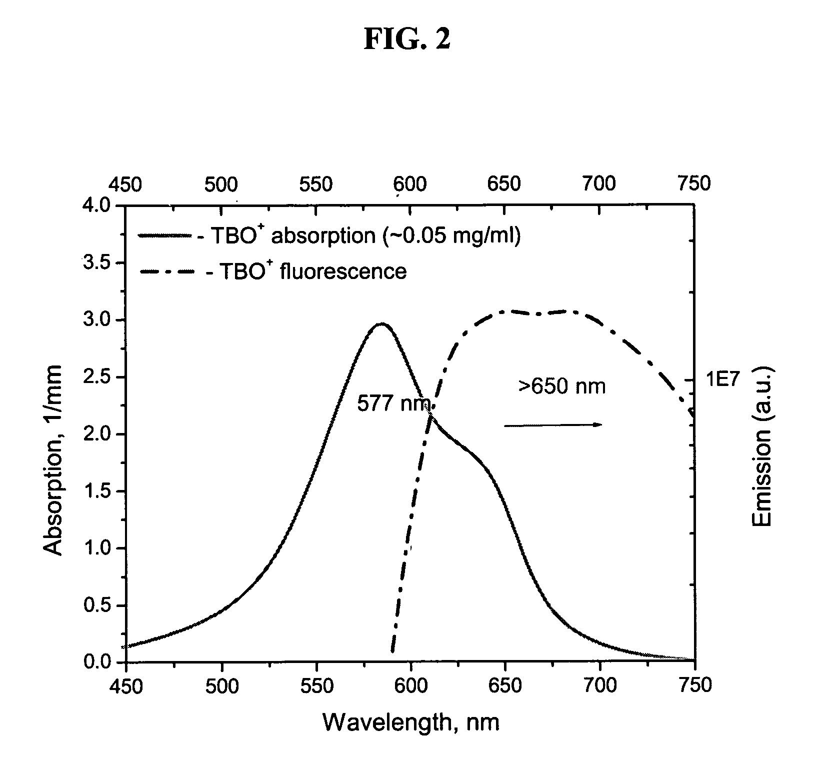
[0063] Image of tissue acquired by the FPLI method will be further described by referring to FIGS. 2-6. As will be readily apparent, applying fluorescent contrast agents, such as TBO and TCN, greatly improves the contrast of the tumor within the image.

[0064] TCN is a highly fluorescent antibiotic. The fluorescence of TBO can be readily detected also. The absorption and fluorescence emission spectra of TBO and TCN are shown in FIGS. 2 and 3, respectively. Commercially available TBO (TB 1% AQ, LC26165-1, Fischer Scientific Company, USA) and tetracycline were used to stain the tissue.

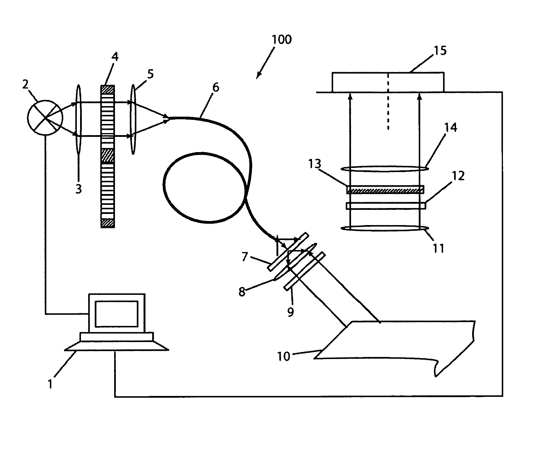
[0065] An additional filter was introduced in front of the CCD 15 of imaging apparatus 100 to cut-off the excitation light. Excitat...


PUM


Abstract

The present invention is directed to a novel multi-spectral exogenous fluorescence polarization imaging technique that enables rapid imaging of large tissue fields. The imaging device includes a tunable monochromatic light source and a CCD camera. Linear polarizers are placed into both the incident and collected light pathways in order to obtain fluorescence polarization or/and anisotropy image. To acquire exogenous fluorescence image, fluorescent contrast agents are delivered to a target tissue.

Description

CROSS-REFERENCE TO RELATED APPLICATION(S) [0001] The subject application claims the benefit of priority to U.S. Provisional Patent Application No. 60 / 504,513 filed Sep. 19, 2003, the disclosure of which is herein incorporated by reference in its entirety.BACKGROUND OF THE INVENTION [0002] 1. Field of the Invention [0003] The present invention is directed to a real-time method for imaging organic tissue. The method employs a polarization-enhanced fluorescence imaging system in order to obtain images of the organic tissue. The obtained images can be used in the demarcation of, among other things, nonmelanoma skin cancers. [0004] 2. Background of the Related Art [0005] Nonmelanoma skin cancers are the most common forms of human cancer. About 75% of all skin cancers are basal cell carcinomas (BCC) and about 20% are squamous cell carcinomas (SCC). These cancers are a major cause of morbidity in the Caucasian population. They commonly appear on sun-exposed areas of the body such as the he...

Claims


Application Information

Patent Timeline
IPC IPC(8): A61BA61B5/00G01N21/64
CPCA61B5/0059A61B5/0071A61B5/444G01J3/02G01J3/0224G01N2021/6423G01J3/4406G01J2003/1217G01N21/6428G01N21/6445G01N21/6456G01J3/10
Inventor YAROSLAVSKY, ANNA N.ANDERSON, RICHARD R.
Owner THE GENERAL HOSPITAL CORP
Why Eureka
Try Eureka