Methods of sterilizing biological mixtures using stabilizer mixtures

a technology of biological mixtures and stabilizers, applied in the direction of biocide, plant growth regulators, biochemistry apparatus and processes, etc., can solve the problems of not always reliable, use may contain unwanted and potentially dangerous biological contaminants or pathogens, and is not always reliabl

Inactive Publication Date: 2005-05-19
CLEARANT
View PDF2 Cites 33 Cited by
  • Summary
  • Abstract
  • Description
  • Claims
  • Application Information

AI Technical Summary

Benefits of technology

[0025] The present invention also provides a biological composition comprising at least one biological material and at least one stabilizer mixture in which the residual solvent content has been reduced

Problems solved by technology

Many biological materials that are prepared for human, veterinary, diagnostic and/or experimental use may contain unwanted and potentially dangerous biological contaminants or pathogens, such as viruses, bacteria (including inter- and intracellular bacteria, such as mycoplasmas, ureaplasmas, nanobacteria, chlamydia, rickettsias), yeasts, molds, fungi, single or multicellular parasites, and/or prions or similar agents responsible, alone or in combination, for TSEs.
Such procedures, however, are not always reliable and are not able to detect the presence of certain viruses, particularly in very low numbers.
This reduces the value or certainty of the test in view of the consequences associated with a false negative result.
False negative results can be life threatening in certain cases, for example in the case of Acquired Immune Deficiency Syndrome (AIDS).
Furthermore, in some instances it can take weeks, if not months, to determine whether or not the material is contaminated.
Moreover, to date, there is no reliable test or assay for identifying prions within a biological material that is suitable for screening out potential donors or infected material.
Thus the products of unicellular natural or recombinant organisms or tissues carry a risk of pathogen contamination.
In addition to the risk that the producing cells or cell cultures may be infected, the processing of these and other biological materials creates opportunities for environmental contamination.
Interestingly, even products from species as different from humans as transgenic plants carry risks, both due to processing contamination as described above, and from environmental contamination in the growing facilities, which may be contaminated by pathogens from the environment or infected organisms that co-inhabit the fac

Method used


Image

  • Methods of sterilizing biological mixtures using stabilizer mixtures
  • Methods of sterilizing biological mixtures using stabilizer mixtures
  • Methods of sterilizing biological mixtures using stabilizer mixtures

Examples


example 1

[0093] In this experiment, the protective effect of the combination of ascorbate (20 mM), urate (1.5 mM) and trolox (200 FM) on gamma irradiated freeze-dried anti-insulin monoclonal immunoglobulin supplemented with 1% bovine serum albumin (BSA) was evaluated.

Methods

[0094] Samples were freeze-dried for approximately 64 hours, stoppered under vacuum, and sealed with an aluminum, crimped seal. Samples were irradiated at a dose rate of 1.83-1.88 kGy / hr to a total dose of 45.1-46.2 kGy at 4° C.

[0095] Monoclonal immunoglobulin activity was determined by a standard ELISA protocol. Maxisorp plates were coated with human recombinant insulin at 2.5 μg / ml overnight at 4° C. The plate was blocked with 200μl of blocking buffer (PBS, pH 7.4, 2% BSA) for two hours at 37° C., and then washed six times with wash buffer (TBS, pH 7, 0.05% TWEEN 20). Samples were re-suspended in 500 μl of high purity water (100 ng / μl), diluted to 5 μg / ml in a 300 μl U-bottomed plate coated for either overnight or f...

example 2

[0097] In this experiment, the protective effect of the combination of 200 :M Trolox, 1.5 mM urate, and 20 mM ascorbate on freeze-dried anti-insulin monoclonal immunoglobulin supplemented with 1% human serum albumin (HSA) and, optionally, 5% sucrose, irradiated at a high dose rate was evaluated.

Method

[0098] Samples were freeze-dried for approximately 64 hours, stoppered under vacuum, and sealed with an aluminum, crimped seal. Samples were irradiated at a dose rate of approximately 1.85 kGy / hr to a total dose of 45 kGy at 4° C.

[0099] Monoclonal immunoglobulin activity was determined by a standard ELISA protocol. Maxisorp plates were coated with human recombinant insulin at 2.5 μg / ml overnight at 4° C. The plate was blocked with 200 μl of blocking buffer (PBS, pH 7.4, 2% BSA) for two hours at 37° C., and then washed six times with wash buffer (TBS, pH 7, 0.05% TWEEN 20). Samples were re-suspended in 500 μl of high purity water (100 ng / μl), and diluted to 5 μg / ml in a 300 μl U-bott...

example 3

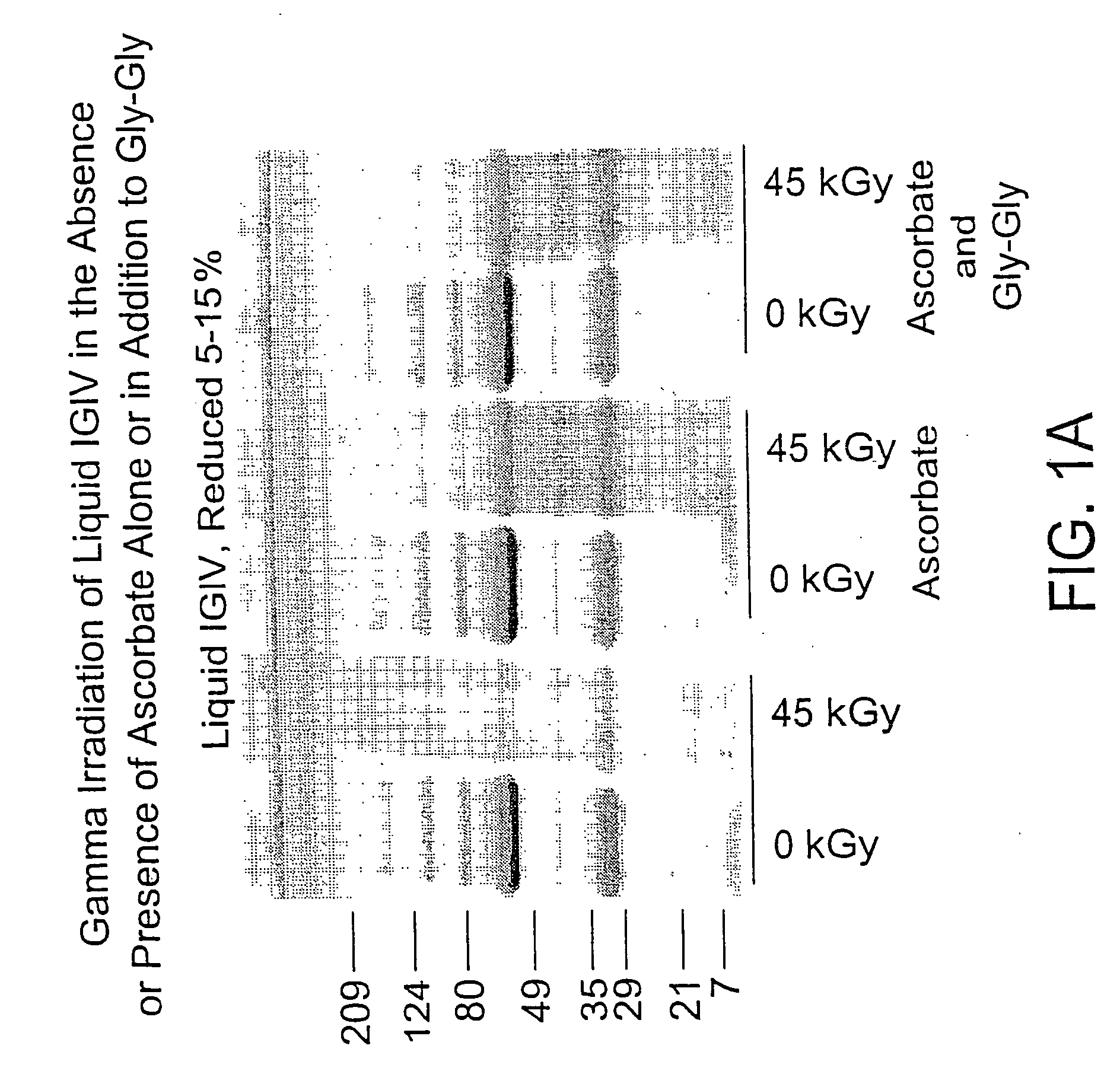
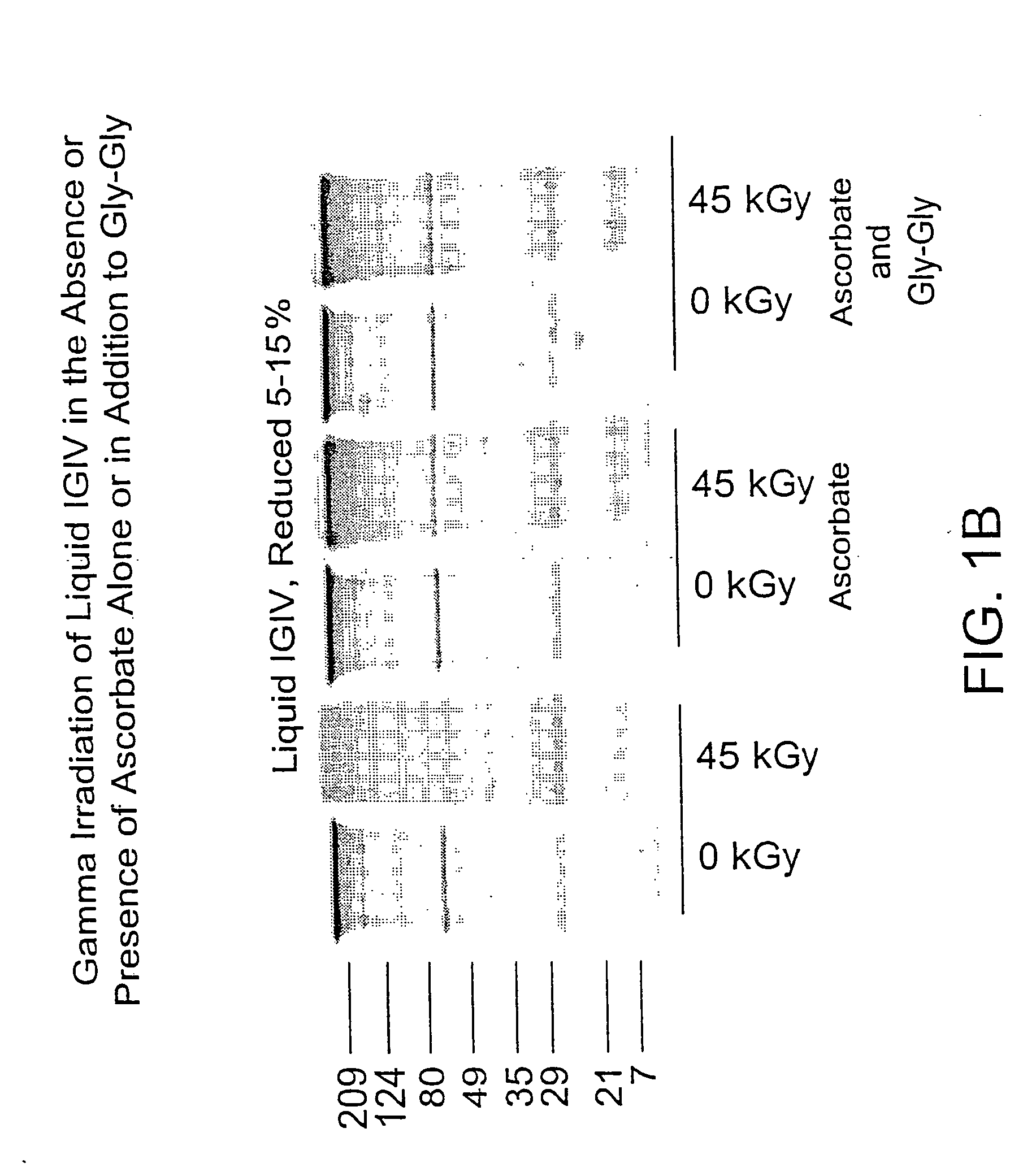
[0102] In this experiment, the protective effect of ascorbate (200 mM), alone or in combination with Gly-Gly (200 mM), on a liquid polyclonal antibody preparation was evaluated.

Method

[0103] In 2 ml glass vials, samples of IGIV (50 mg / ml) were prepared with either no stabilizer or the stabilizer of interest. Samples were irradiated with gamma radiation (45 kGy total dose, dose rate 1.8 kGy / hr, temperature 4° C.) and then assayed for functional activity and structural integrity.

[0104] Functional activity of independent duplicate samples was determined by measuring binding activity for rubella, mumps and CMV using the appropriate commercial enzyme immunoassay (EIA) kit obtained from Sigma, viz., the Rubella IgG EIA kit, the Mumps IgG EIA kit and the CMV IgG EIA kit.

[0105] Structural integrity was determined by gel filtration (elution buffer: 50 mM NaPi, 100 mM NaCl, pH 6.7; flow rate: 1 ml / min; injection volume 50 μl) and SDS-PAGE (pre-cast tris-glycine 4-20% gradient gel from Nov...


PUM


Abstract

Methods are disclosed for sterilizing biological materials to reduce the level of one or more biological contaminants or pathogens therein, such as viruses, bacteria (including inter- and intracellular bacteria, such as mycoplasmas, ureaplasmas, nanobacteria, chlamydia, rickettsias), yeasts, molds, fungi, single or multicellular parasites, and/or prions or similar agents responsible, alone or in combination, for TSEs. These methods involve the use of stabilizer mixtures in methods of sterilizing biological materials with irradiation.

Description

BACKGROUND OF THE INVENTION [0001] 1. Field of the Invention [0002] The present invention relates to methods for sterilizing biological materials to reduce the level of one or more biological contaminants or pathogens therein, such as viruses, bacteria (including inter- and intracellular bacteria, such as mycoplasmas, ureaplasmas, nanobacteria, chlamydia, rickettsias), yeasts, molds, fungi, single or multicellular parasites, and / or prions or similar agents responsible, alone or in combination, for TSEs. The present invention particularly relates to the use of stabilizer mixtures in methods of sterilizing biological materials with irradiation: [0003] 2. Background of the Related Art [0004] Many biological materials that are prepared for human, veterinary, diagnostic and / or experimental use may contain unwanted and potentially dangerous biological contaminants or pathogens, such as viruses, bacteria (including inter- and intracellular bacteria, such as mycoplasmas, ureaplasmas, nanoba...

Claims


Application Information

Patent Timeline
IPC IPC(8): A01N1/02A01N25/32A61K39/395A61L2/00A61L2/08A61L2/10A61L2/16C07K16/26
CPCA01N1/02C07K16/26A01N25/32A61K39/39591A61L2/0011A61L2/0035A61L2/0041A61L2/0047A61L2/0052A61L2/0058A61L2/007A61L2/0082A61L2/10A61L2/16A01N1/0294
Inventor BURGESS, WILSONDROHAN, WILLIAM N.MACPHEE, MARTIN J.MANN, DAVID M.
Owner CLEARANT
Why Eureka
Try Eureka