
Methods for treating tumors and cancerous tissues

a tumor and cancerous tissue technology, applied in the field of immunotherapy, can solve the problems of single antigens not being sufficient for tumor clearance, affecting the efficacy of tumor treatment, and poor candidates for immunotherapy for immunosuppression patients with high tumor burden, and achieve the effect of increasing the bioavailability of prostate specific antigens

Inactive Publication Date: 2005-09-29
SANGRETECH BIOMEDICAL
View PDF2 Cites 154 Cited by
  • Summary
  • Abstract
  • Description
  • Claims
  • Application Information

AI Technical Summary

Benefits of technology

[0061] inducing necrosis or apoptosis against at least a portion of the tumor or cancerous tissue by selectively applying cryotherapy, heat ablation, chemotherapy, radiation therapy, ultrasound therapy, or a combination thereof against the tumor or cancerous tissue, thereby liberating cancer-specific antigens from the tumor or cancerous tissue and increasing the bioavailability of the cancer-specific antigens within and proximate to the tumor or cancerous tissue and within in the bloodstream;
[0067] inducing necrosis or apoptosis against the prostate cancer by selectively freezing at least a portion of the prostate cancer by using cryotherapy, thereby liberating prostate specific antigens from the prostate cancer and increasing the bioavailability of the prostate specific antigens within and proximate to prostate cancer and within in the bloodstream;

Problems solved by technology

These often very ill, immunosuppresed patients with high tumor burden are not good candidates for testing vaccine-based immunotherapy.
Moreover, single antigens do not suffice for effective clearance of tumors, which consist of polyclonal cells and express or lose a whole range of antigens.
Unfortunately, it is often difficult to verify whether this goal has been attained, and there is a risk of leaving out crucial antigenic components, thereby jeopardizing the efficacy of tumor treatment.
At temperatures between −20 and −40 degrees Celsius, cells may encounter osmotic distress (due to extracellular ice formation which results in water withdrawal from the cell), cell membrane rupture, and microthrombi formation (leading to hypoxia).
Any of these events may lead to lethal (i.e. necrosis, apoptosis) or sub-lethal (i.e. increased cell permeability, alterations in cellular pH) damage to the cell(s).

Method used


Image

  • Methods for treating tumors and cancerous tissues
  • Methods for treating tumors and cancerous tissues

Examples


example 1

Production and Testing of Autologous Dendritic Cells

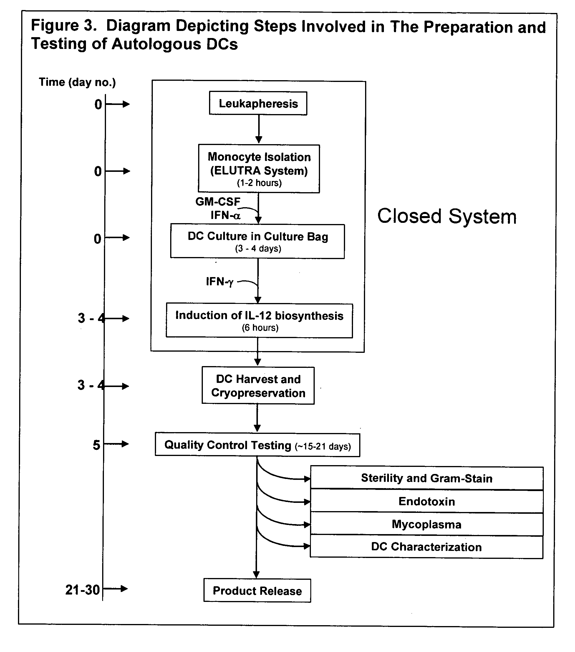
[0139]FIG. 3 is a flow diagram, illustrating the steps of the preparation and testing of autologous dendritic cells (DCs).

[0140] The production process of autologous DCs can be divided into 4 steps: (1) leukapheresis of patients, (2) isolation of DC (monocytes) using the Gambro ELUTRA™ system, (3) culture and maturation of DCs in a gas permeable bag, (4) harvest and cryopreservation of autologous DCs. Each of these steps is described below.

1. Leukapheresis of Patients

[0141] A single-stage White Blood Cell (WBC) Channel (or chamber) is used to collect the mononuclear cells. The anticoagulated whole blood enters the chamber through the inlet tubing. As it flows into the channel, it is separated into 3 blood components, the red blood cells (RBC), the WBCs, and the platelet-rich plasma. The separation of all 3 of these components is controlled by the specific gravity differences between the blood components and the pressure, densi...

example 2

Cryoablation and Intraprostatic DC Injection

[0152] The rationale for this protocol is based on the recognition that the liberation of tumor-associated antigen or prostate-associated antigen resultant from the cryoablation event allows the locally injected, autologous dendritic cells to uptake antigen, migrate to the lymphatic system, and affect a systemic immune response against tumor cells far removed from the prostate. Subtotal cryoablation (rather than total cryoablation) of the prostate is performed in order to allow for the creation of early necrotic prostatic tissue while minimizing the likelihood of freezing other, non-prostatic structures such as the neuro-vascular bundles, the anterior rectal wall, and other uninvolved bowel.

[0153] Immediately prior to the cryoablation procedure, four cryopreserved vials containing the patient's cultured dendritic cells are thawed to room temperature. The cell preparation should be thawed for a total of less than about 60 minutes prior to...

example 3

Treatment of Human Malignant Melanoma by Radiotherapy and Intratumoral Injection of DCs

[0164] Human malignant melanoma is often highly metastatic and radioresistant (Weichselbaum, et al., Proc. Natl. Acad. Sci. USA 82:4732-4735 (1985); Rubin, P. (1993) Clinical Oncology: A Multidisciplinary Approach for Physicians and Students 7Ed, Vol. 306 ,72 W. B. Saunders Philadelphia), however, ionizing radiation has shown therapeutic benefits. Ionizing radiation is a portion of the high energy electromagnetic radiation spectrum which can penetrate and be transmitted through tissues. A melanoma patient is subjected to ionizing radiation treatment, following standard protocol. The level of melanoma antigens, including Melan-A / MART-1, MAGE, NY-ESO-1, is monitored following irradiation. For a more detailed list of tumor antigens which may be additionally or alternatively monitored, see, e.g. Urban and Schreiber, Annu Rev. Immunol. 10:617-44 (1992), and Renkvist, et al., Cancer Immmunol. Immunothe...


PUM

PropertyMeasurementUnit
temperatureaaaaaaaaaa
temperaturesaaaaaaaaaa

Abstract

The invention disclosed herein relates generally to immunotherapy and, more specifically, to therapeutic methods for treating tumors and cancerous tissues by first inducing necrosis or apoptosis (e.g., cryotherapy, chemotherapy, radiation therapy, ultrasound therapy, or a combination thereof applied against at least a portion of the tumor or cancerous tissue), and then delivering one or more se doses of antigen presenting cells (e.g., autologous dendritic cells) intratumourally or proximate to the tumor or cancerous tissue, but only after a selected period of time sufficient for the bioavailablity of liberated cancer-specific antigens (monitored over the selected period of time) resulting from the necrosis or apoptosis to be at or near a maximum value. The present invention provides an alternative strategy to the ex vivo loading of target antigen to antigen presenting cells such as, for example, enriched autologous dendritic cells for purposes of enhancing an immune response.

Description

[0001] This application claims priority under 35 U.S.C 119(e) to provisional application Ser. No. 60 / 557,111, filed on Mar. 25, 2004, the entire disclosure of which is hereby expressly incorporated by reference.TECHNICAL FIELD [0002] The present invention relates generally to immunotherapy and, more specifically, to therapeutic methods for treating tumors and cancerous tissues by distressing tumor cells in order to liberate tumor antigen(s), and delivering antigen presenting cells, capable of exploiting the liberated antigen(s), to the tumor or cancerous tissue. BACKGROUND OF THE INVENTION Cancer Immunotherapvy [0003] Interest in cancer vaccination arose based on William Coley's early observation of cancer regression after streptococcus pyogenes infection (Coley, W. B., Clin. Orthop. 1991(262 ):3-3-11 (1893 )). Coley noted dramatic regression of some sarcoma lesions following experimental streptococcal infection of the skin (erysipelas). While discredited during his time, Coley's o...

Claims


Application Information

Patent Timeline
Patent Type & Authority Applications(United States)
IPC IPC(8): A61B18/02A61K39/00A61K48/00C12N5/0784
CPCA61B18/02A61B2017/00274A61B2018/00547A61K39/0011C12N2501/24A61K2039/5154C12N5/0639C12N2501/22A61K48/00A61P35/00A61P35/04A61P43/00A61K39/4615A61K2239/49A61K39/46449A61K39/464486A61K39/4622A61K2239/31A61K39/464482A61K39/464406A61K39/4644
Inventor CAVANAGH, WILLIAM ALOYSIUS IIITJOA, BENJAMIN ALANJACOB, CHARLES W. III
Owner SANGRETECH BIOMEDICAL
Learn More