
Methods for using pet measured metabolism to determine cognitive impairment

a cognitive impairment and metabolic technology, applied in the field of diagnostic imaging and radiology, can solve the problems of closed head injury and lack of quantitative diagnostic methods for concomitant cognitive impairment, and achieve the effects of enhancing and prolonging life, high sensitivity of cdi, and straightforward mathematical algorithms

Inactive Publication Date: 2005-09-29
THE BIOMEDICAL RES FOUND OF NORTHWEST LOUSNA
View PDF11 Cites 157 Cited by
  • Summary
  • Abstract
  • Description
  • Claims
  • Application Information

AI Technical Summary

Benefits of technology

[0052] This method enables the early detection of Mild Cognitive Impairment, a prodrome to Alzheimer's Disease. The methodological components include the specific location of the brain volumes of interest (VOIs) along with specific weighting factors derived from comparison of patient data and normal controls, and the creation of the normalized CDI from the mean weighted VOIs. Spatial normalization and filtering of a given image can be coded simply without having to use Matlab or SPM, and a one cm diameter spherical VOI at each coordinate can be sampled without the use of Marsbar, as these are fairly straightforward mathematical algorithms. Results of an exemplar study used to establish the preferred embodiment of the instant invention are presented here in a group comparison format, but the methodology taught is applicable to develop a CDI for and to evaluate a single patient. The preferred embodiment of the instant invention comprises a set of software routines that can independently provide a CDI value for an appropriately processed FDG PET scan of a patient's brain. In addition to these considerations, the high sensitivity of the CDI enables its use as a screening tool for the early detection of a variety of cognitive disorders.
[0053] Data from experiments performed to develop the instant invention show that analyses of groups of premorbid individuals can be discriminated from groups of normal controls To date, there is no reliable method of detecting MCI in the early stages, at the clinical level, in individual patients. The development of the methods taught in the instant invention are overdue for clinical use to put this early detection capability into the hands of clinicians. Detection of the degenerative processes in premorbid or very early stages of brain degenerative disease, drug abuse or immediately following brain injury would enable the early treatment with medication to enhance and prolong quality of life, to provide an answer to the patients' questions regarding their potential for cognitive decline, to help them plan and prepare for the future, and hopefully one day, to prevent these diseases altogether or provide increasingly effective rehabilitative measures for injury.

Problems solved by technology

Similarly, head trauma resulting in closed head injury with concomitant cognitive impairment lacks quantitative diagnostic methods.

Method used


Image

  • Methods for using pet measured metabolism to determine cognitive impairment
  • Methods for using pet measured metabolism to determine cognitive impairment
  • Methods for using pet measured metabolism to determine cognitive impairment

Examples


Embodiment Construction

[0075] The data, analysis, calculations and procedures forming the preferred embodiment were produced in studies of four groups of patients: [0076] 1. Control subjects from the normal control database, [0077] 2. Patients identified retrospectively with early cognitive decline who received negative PET workups, [0078] 3. Patients identified retrospectively with cognitive decline and with the pathognomonic changes of AD present on PET imaging, [0079] 4. Patients with MCI from the pilot study.

The data and statistics used as exemplars in the figures and tables are taken from these studies.

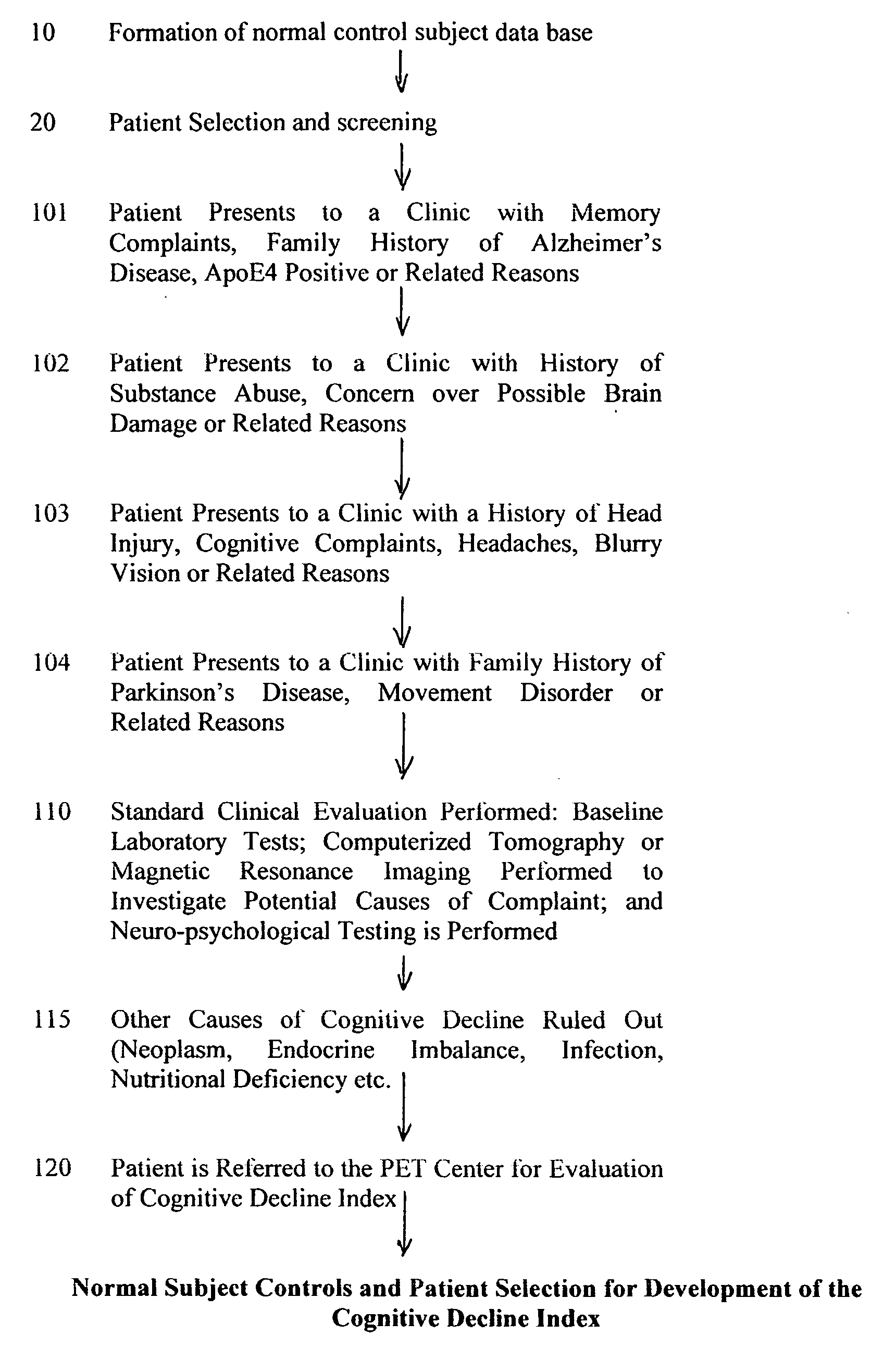
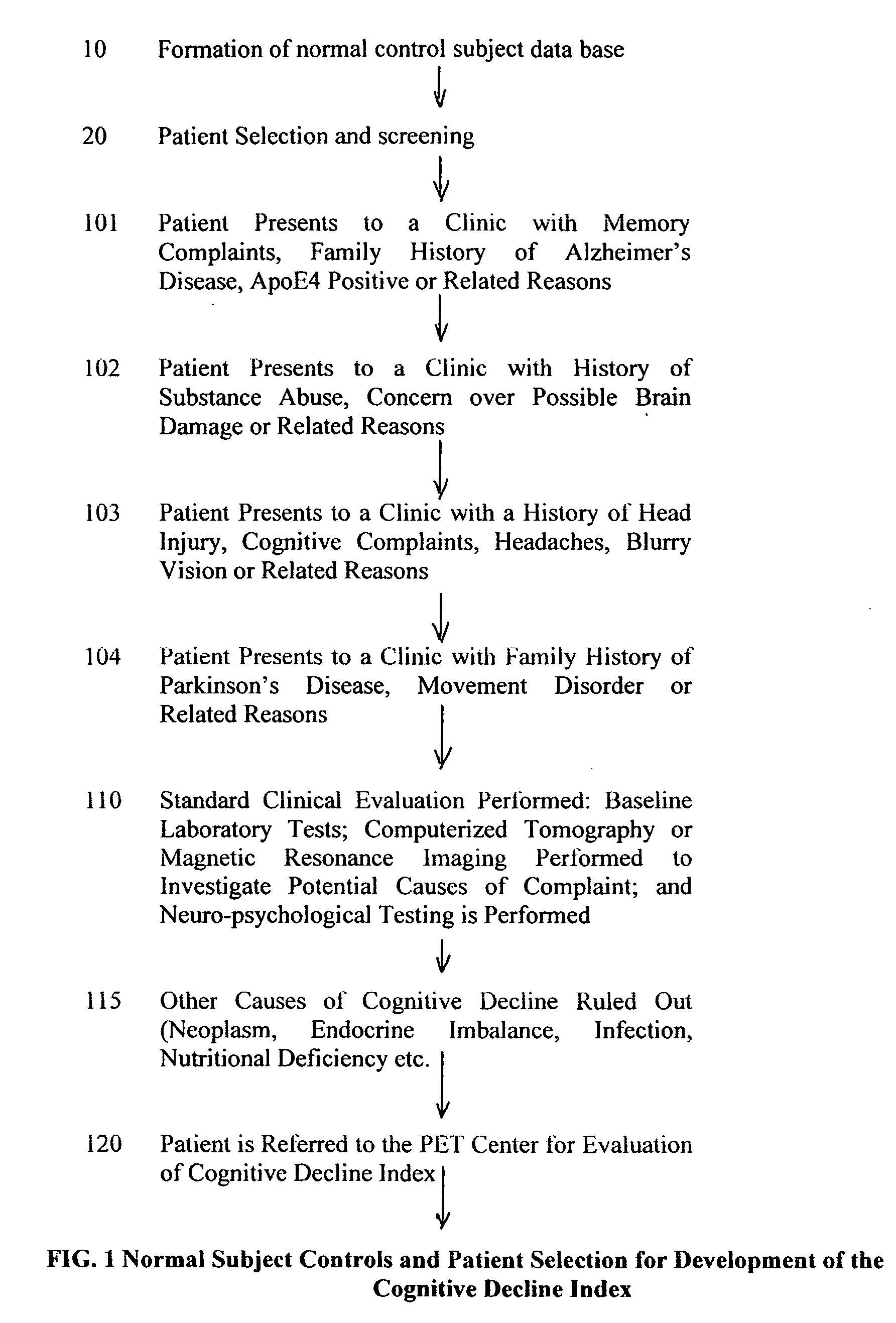
[0080] Referring first to FIG. 1, a database 10 is compiled for normal control subjects.

[0081] The database of FDG brain scans from healthy control subjects was created to enable the objective examination of a variety of patients who presented for clinical evaluation of cerebral pathology. These subjects are physically examined and screened for neurological and psychiatric illness. Entry into the d...


PUM


Abstract

A non-invasive, early stage method to obtain quantitative measures of mild cognitive impairment useful in diagnosing and following degenerative brain disease or closed head injuries by utilizing the image data from individual patient positron emission tomographic scans to construct a cognitive decline index that serves as a diagnostic and screening tool to reveal the onset of mild cognitive impairment and nervous system dysfunction which are sequelae of degenerative brain diseases and closed head injury. The method involves using weighted values of brain region intensities derived from comparing scans of normal subjects to a scan of the patient to calculate a cognitive decline index that is useful as a diagnostic tool for mild cognitive impairment. The weights for the intensity values for each region are derived from the differences of intensity values from regions of the brain of the patient selected by comparing the patient to normal control subjects.

Description

BACKGROUND OF THE INVENTION [0001] Diagnostic imaging and radiology began as a medical sub-specialty in the first decade of the 1900's after the publication in 1898 describing experiments on x-rays by Professor Wilhelm Roentgen. The development of radiology grew at a steady rate until World War II. Extensive use of x-ray imaging during the Second World War, and the advent of the digital computer and new imaging modalities like ultrasound, magnetic resonance imaging, single photon emission computed tomography and positron emission tomography have combined to create an explosion of diagnostic imaging techniques in the past 25 years. [0002] In general, radiological imaging can address two issues: structure and function. It is possible either to view structures in the body and image anatomy or view chemical processes and image biochemistry. Structural imaging techniques can image anatomy and include ultrasound, X-rays, computerized axial tomography (CAT) and magnetic resonance imaging (...

Claims


Application Information

Patent Timeline
IPC IPC(8): A61B5/05A61B6/00G06T7/00
CPCG06K9/00A61B6/037G06T7/0012G06K2209/05G06V2201/03
Inventor PATTERSON, JAMES C. II
Owner THE BIOMEDICAL RES FOUND OF NORTHWEST LOUSNA
Learn More