Methods and compositions for the detection of cervical disease

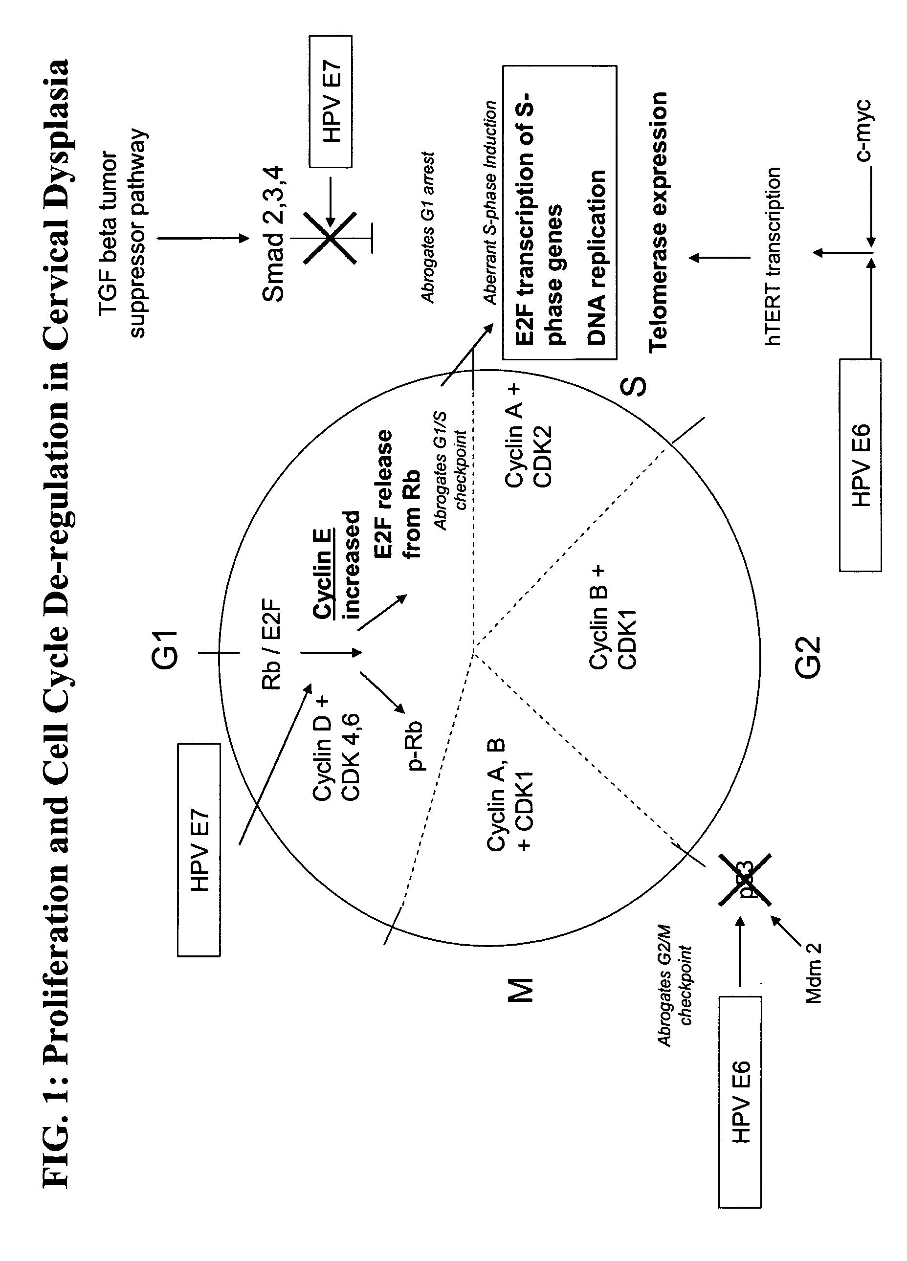
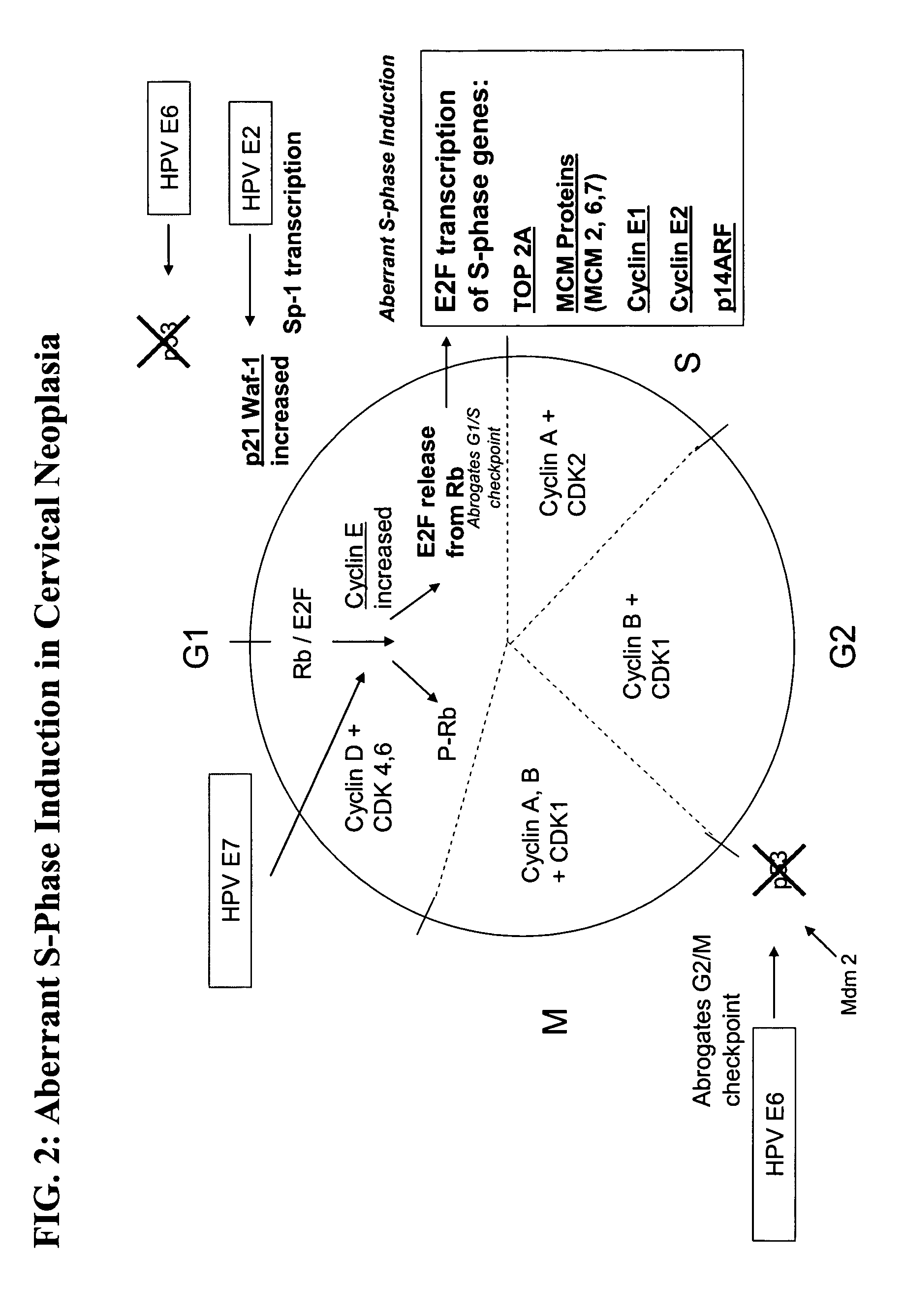
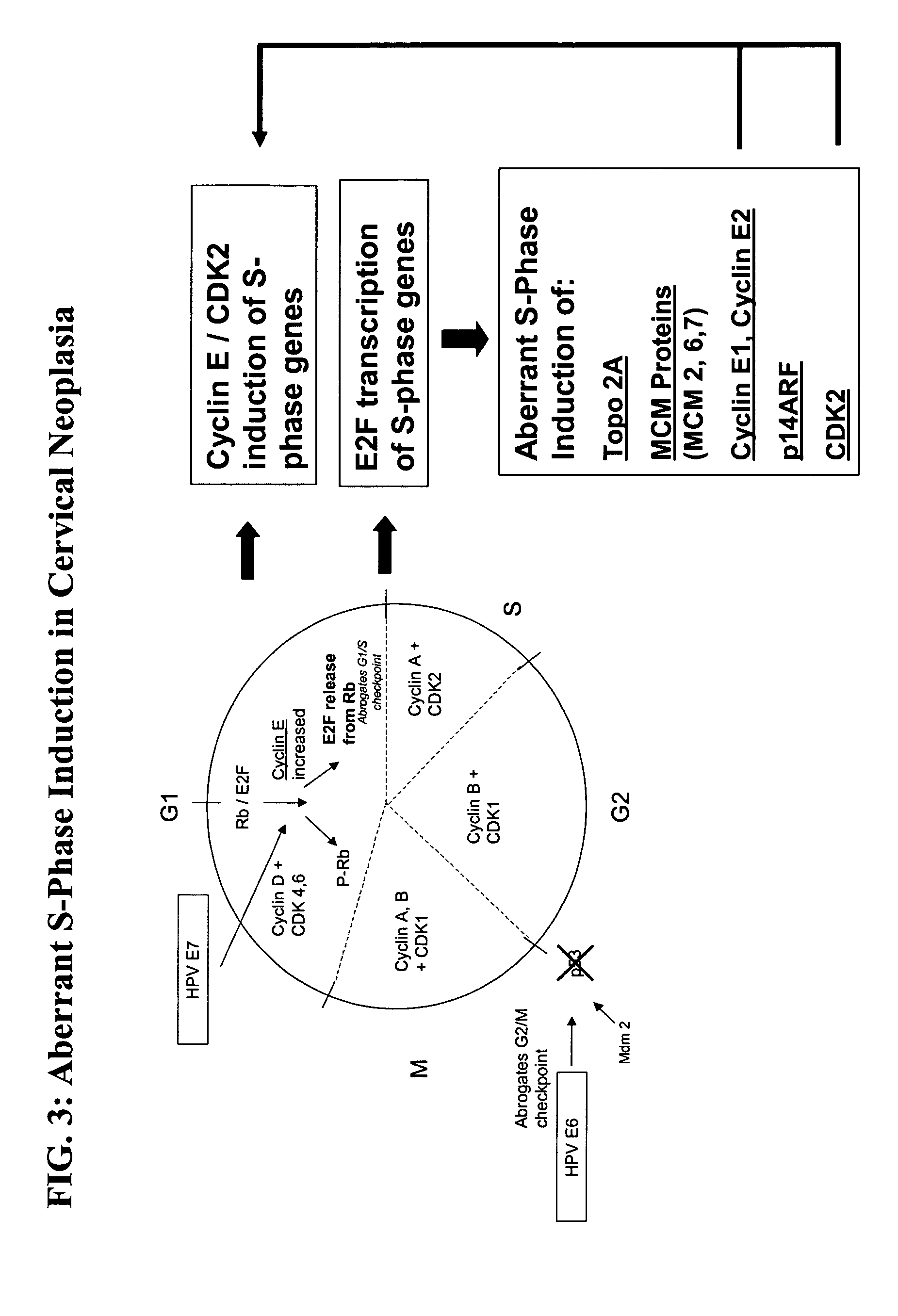
a technology of compositions and cervical disease, applied in the field of methods and compositions for the detection of high-grade cervical disease, can solve the problems of many false positives, hpv-positive women will ever develop high-grade cervical disease or cancer, and the clinical problem is more serious, so as to accelerate the cell proliferation and increase the level of c-my

Active Publication Date: 2005-11-24
TRIPATH IMAGING INC
View PDF8 Cites 125 Cited by
  • Summary
  • Abstract
  • Description
  • Claims
  • Application Information

AI Technical Summary

Benefits of technology

[0017]FIG. 4 provides a schematic representation of the role of c-myc in aberrant S-phase induction. C-myc is an important transcriptional activator in cellular proliferation. The gene encoding c-myc is located on the chromosome. This is the same site that HPV 18 integration has been...

Problems solved by technology

In many developing countries where mass screening programs are not available, the clinical problem is more serious.
However, HPV infection is very common, possibly occurring in 5-15% of women over the age of 30, but few HPV-positive women will ever develop high-grade cervical disease or cancer.
The presence of HPV alone is indicative only of infection, not of high-grade cervical disease, and, therefore, testing for HPV infection alone results in many false positives.
For those individuals who develop persistent infections with one or more oncogenic subtypes of HPV, there is a risk for the development of neoplasia in comparison to patients without an HPV infection.
Howev...

Method used


Image

  • Methods and compositions for the detection of cervical disease
  • Methods and compositions for the detection of cervical disease
  • Methods and compositions for the detection of cervical disease

Examples


example 1

Detection of Biomarker Overexpression Using Immunocytochemistry

Slide Preparation and Pretreatment

[0098] Patient cervical samples are collected and placed into a SurePath™ collection vial (TriPath Imaging, Inc.). Cervical cells are collected from the liquid medium and deposited in a thin layer on a glass slide using the PrepStain™ slide processor system (TriPath Imaging, Inc.). Prepared slides are immediately transferred to a pretreatment buffer (1% RAM) and heated for 45 minutes at 95° C. The slides are cooled to room temperature and rinsed three times (2 minutes per rinse) in TBS (tris buffered saline).

Manual Immunocytochemistry

[0099] To prevent non-specific background staining, slides are not permitted to dry out during the staining procedure. Furthermore, in order to block non-specific staining, hydrogen peroxide is applied to the slides for 5 minutes, followed by a TBS rinse. An antibody directed to MCM6 is applied to the slide for 1 hour at room temperature. Following inc...

example 2

Detection of Biomarkers in Clinical Samples

[0102] Approximately 180 cervical cytology patient samples representing various diagnoses were collected. The presence or absence of cancerous cells or lesions indicative of high-grade disease in these patients was previously confirmed by colposcopy. The following table indicates the number of samples within each diagnosis group analyzed in this study, as well as a description of the colposcopy findings (e.g., presence or absence of high-grade lesions).

TABLE 1Specimens analyzedDiagnosisCountDescriptionNIL72HPV NegativeASC-US2626 without lesion0 with lesion or high risk HPVLSIL4842 negative for high grade lesion6 positive for high grade lesionHSIL25Cancer10Squamous Cell Carcinoma andAdenocarcinoma

[0103] The samples were analyzed by immunocytochemistry methods to identify high-grade cervical disease. Antibodies were used to detect the overexpression of six biomarkers of interest: MCM2, MCM6, MCM 7, p21waf1, Cyclin E, and Topo2A. Assay cont...

example 3

Detection of Biomarkers in Clinical Samples Using Antibody Cocktails

[0123] Approximately 180 colposcopy-confirmed cervical cytology samples were analyzed by immunocytochemistry methods to identify high-grade cervical disease. Each sample was analyzed for the expression of multiple biomarkers of interest. Specifically, various combinations of antibodies directed to MCM2, MCM6, MCM 7, p21waf1, Cyclin E, and Topo2A were analyzed for their ability to detect high-grade cervical disease. These samples were evaluated for the expression of multiple biomarkers of interest using the immunocytochemistry methods and slide interpretation guidelines described in Example 2.

[0124] The immunocytochemistry results were compared with the results previously obtained by colposcopy. Each slide was then given a final result of true positive (TP), true negative (TN), false positive (FP), false negative (FN), or indeterminate. Sensitivity, specificity, positive predictive values, and negative predictive v...


PUM

PropertyMeasurementUnit
Fractionaaaaaaaaaa
Fractionaaaaaaaaaa
Fractionaaaaaaaaaa

Abstract

Methods and compositions for identifying high-grade cervical disease in a patient sample are provided. The methods of the invention comprise detecting overexpression of at least one biomarker in a body sample, wherein the biomarker is selectively overexpressed in high-grade cervical disease. In particular claims, the body sample is a cervical smear or monolayer of cervical cells. The biomarkers of the invention include genes and proteins that are involved in cell cycle regulation, signal transduction, and DNA replication and transcription. In particular claims, the biomarker is an S-phase gene. In some aspects of the invention, overexpression of a biomarker of interest is detected at the protein level using biomarker-specific antibodies or at the nucleic acid level using nucleic acid hybridization techniques. Kits for practicing the methods of the invention are further provided.

Description

CROSS REFERENCE TO RELATED APPLICATIONS [0001] This application claims the benefit of U.S. Provisional Application Ser. No. 60 / 556,495, filed Mar. 24, 2004, which is incorporated herein by reference in its entirety.FIELD OF THE INVENTION [0002] The present invention relates to methods and compositions for the detection of high-grade cervical disease. BACKGROUND OF THE INVENTION [0003] Carcinoma of the cervix is the second most common neoplasm in women, accounting for approximately 12% of all female cancers and causing approximately 250,000 deaths per year. Baldwin et al. (2003) Nature Reviews Cancer 3:1-10. In many developing countries where mass screening programs are not available, the clinical problem is more serious. Cervical cancer in these countries is the number one cause of cancer deaths in women. [0004] The majority of cases of cervical cancer represent squamous cell carcinoma, although adenocarcinoma is also seen. Cervical cancer can be prevented by population screening as...

Claims


Application Information

Patent Timeline
IPC IPC(8): C07K16/18C07K16/30C12P19/34C12Q1/70G01N33/53G01N33/574G01N33/68
CPCC07K16/3069C12Q1/6886C12Q2600/112G01N2333/025G01N33/57411G01N33/6875C12Q2600/158G01N33/53G01N33/68
Inventor FISCHER, TIMOTHYMALINOWSKI, DOUGLASTAYLOR, ADRIANNPARKER, MARGARET
Owner TRIPATH IMAGING INC
Why Eureka
Try Eureka