Maternal antibodies as fetal cell markers to identify and enrich fetal cells from maternal blood
a technology of maternal antibodies and fetal cells, applied in the field of methods of identifying and enriching can solve the problems of 1% increased risk of miscarriage following cvs, and difficulty in isolation of fetal cells from maternal blood, and achieve the effect of increasing the number of fetal cells
- Summary
- Abstract
- Description
- Claims
- Application Information
AI Technical Summary
Benefits of technology
Problems solved by technology
Method used
Image
Examples
example 1
Enrichment of Fetal Cells from Maternal Blood
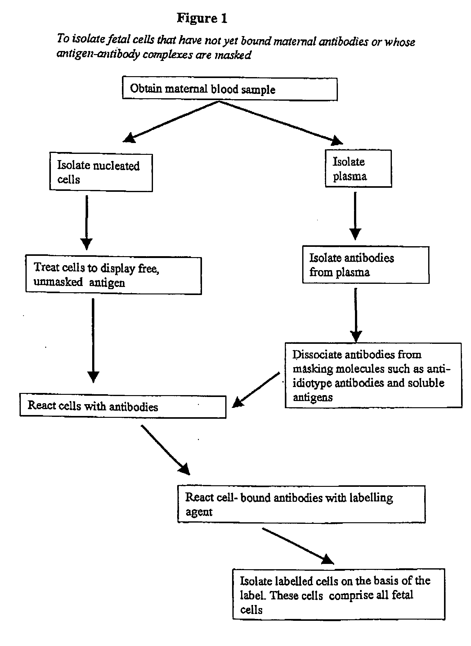
[0153] Blood samples (10 ml) from five pregnant women were obtained. Gestational ages ranged between 10 and 14 weeks, with unknown fetal gender and condition. Blood samples were processed as follows:
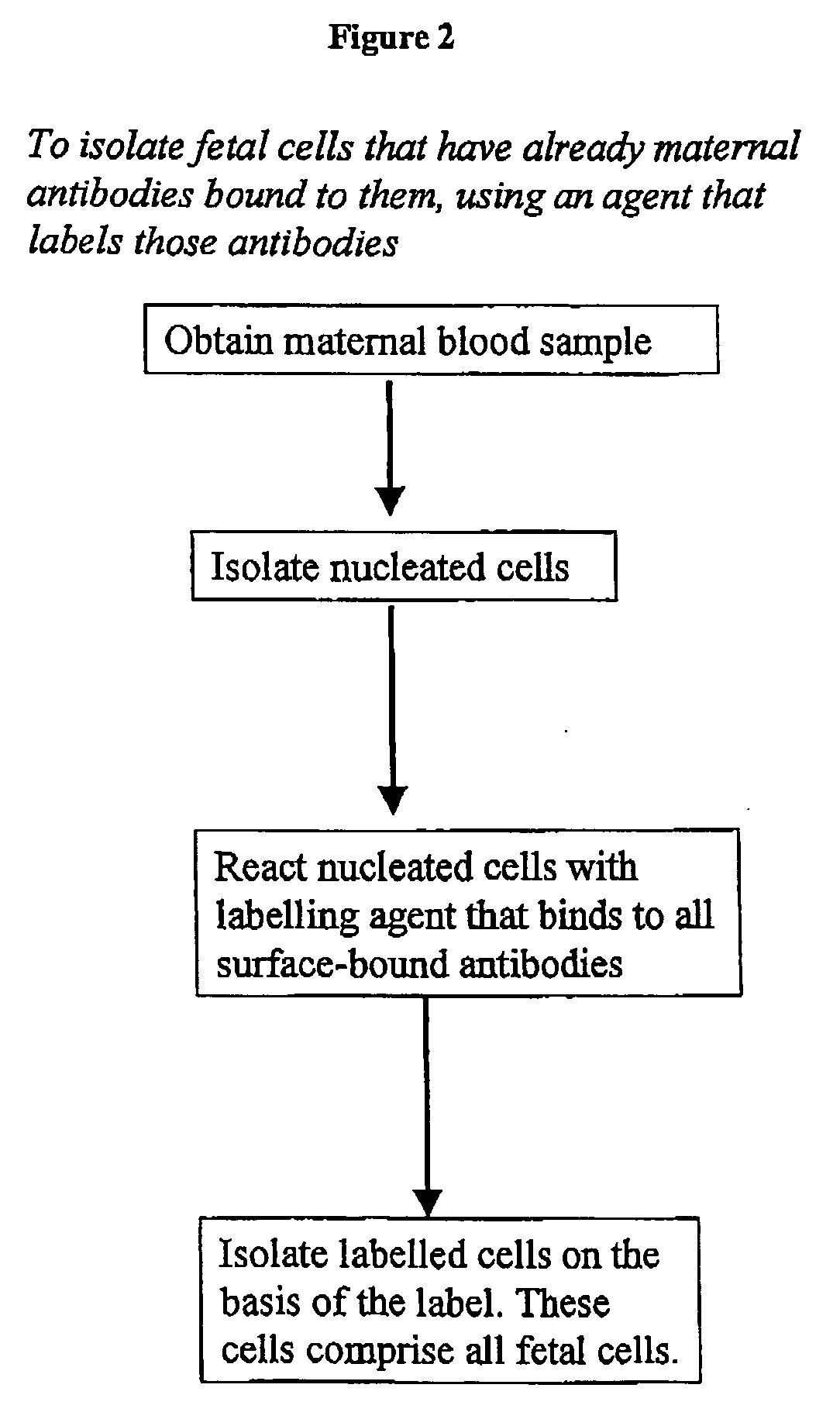
[0154] Nucleated cells were isolated using a density gradient. B lymphocytes and monocytes were removed using anti-CD19 and anti-CD14 magnetic beads. The remaining isolated cells were exposed for 30 min to a mixture of goat F(ab′)2 anti-human IgG and goat F(ab′)2 anti-human IgM, both of which were conjugated with the fluorescent dye phycoerythrin (PE). The cells were washed and suspended in PBS with 1% (v / v) formaldehyde, DNA-specific dye Hoechst 33342 and 0.05% (v / v) Triton X100.
[0155] The nucleated cells in the samples (5-10 million per sample) were subjected to fluorescence-activated cell sorting (FACS), to isolate Hoechst+ / PE+ cells (i.e. cells having normal DNA content and that bind to PE-conjugated antibody fragments). The threshold l...
example 2
Isolation of Reactive Maternal Antibodies from Plasma
Affinity Capture of IgG and IgM
[0159] Plasma isolated from maternal blood is first clarified (centrifuge at 10,000 g and filtered through 0.45 μm filter) and passed through a Protein-A or Protein-G column (Amersham Biosciences) to bind total human IgG. Simultaneously, the clarified maternal blood is passed through a Kaptiv-M™-Sepharose column (Interchim) or a mannin-binding protein (MBP)-Sepharose column (Nevens et al., J. Chromatogr. 597, 247-256, 1992; Pierce), to bind total IgM (Sisson and Castor, J. Immunol. Methods 127, 215-220, 1990). All steps are performed at about 4° C.
[0160] Once IgG and IgM factions are bound to the columns, the columns are washed with TBS, and the columns are uncoupled.
[0161] Initially, the IgG is eluted using 3M magnesium chloride / 25% ethylene glycol / HEPES pH 8.0 as the mobile phase to maintain the individual IgG fractions as unbound components. Next the Protein-A or Protein-G column is uncoupled...
example 3
Dissociation of Soluble HLA Antigens from Anti HLA Antibodies
[0200] Plasma isolated from maternal blood is first treated with caprylic acid to precipitate the bulk of plasma proteins without affecting Ig and some α2 macroglobulins. Following this initial precipitation, the supernatant is taken off and then the Ig fraction is precipitated with ammonium sulfate (Tsang and Wilkins, J. Immunol. Meth. 138, 291-299, 1991).
[0201] Following isolation, the Ig fraction is redissolved in HEPES pH 7.2 and then made up to 3M magnesium chloride / 25% ethylene glycol / HEPES pH 7.2. This solution is then rapidly desalted from the magnesium chloride and peptide HLA antigens by batch-wise column desalting on PD-10 columns or FAST-desalting columns (Amersham Biosciences) using Tris-buffered saline as the mobile phase (note PBS cannot be used at this stage as magnesium chloride will precipitate phosphate based buffers). The high-molecular weight fraction obtained is essentially free of peptide HLA antig...
PUM
 Abstract
Description
Claims
Application Information
 - R&D
- Intellectual Property
- Life Sciences
- Materials
- Tech Scout
- Unparalleled Data Quality
- Higher Quality Content
- 60% Fewer Hallucinations
Browse by: Latest US Patents, China's latest patents, Technical Efficacy Thesaurus, Application Domain, Technology Topic, Popular Technical Reports.
© 2025 PatSnap. All rights reserved.Legal|Privacy policy|Modern Slavery Act Transparency Statement|Sitemap|About US| Contact US: help@patsnap.com


