GLUT1 transporters expressed in cancer cells

Inactive Publication Date: 2006-01-05
XENOPORT
View PDF1 Cites 1 Cited by
  • Summary
  • Abstract
  • Description
  • Claims
  • Application Information

AI Technical Summary

Benefits of technology

[0009] Some methods further comprise determining that the agent, conjugate or conjugate moiety is transported by at least one efflux transporter. Additional methods further comprise modifying the agent, conjugate or conjugate moiety; establishing that the modified agent, conjugate or conjugate moiety retains GLUT1 substrate activity; and comparing the ratio of GLUT1 substrate activity to the ratio of efflux substrate activity for the agent, conjugate or conjugate moiety and the

Problems solved by technology

Disruption of cell division not only slows growth but can also kill tumor cells by triggering cell death.
Unfortunately, these drugs also kill normal populations of proliferating cells such a

Method used


Image

  • GLUT1 transporters expressed in cancer cells
  • GLUT1 transporters expressed in cancer cells
  • GLUT1 transporters expressed in cancer cells

Examples


Example

Example 1

Quantitative PCR Detection of GLUT Transporters Expression in Tumor Cells

[0082] To measure the level of GLUT transporter expression in human tumors, quantitative PCR was performed on human tumor mRNA purchased from Ardais Corporation. For comparison with normal colon, human colon mucosal tissue was obtained from endoscopy procedures. Table 3 shows high levels of GLUT1 mRNA in human tumors.

[0083] Intestinal biopsy samples were obtained, with patient consent, from routine endoscopies or colonoscopies. Biopsies were taken from healthy sites by Radial Jaw 3 single use biopsy forceps (Boston Scientific) within the endoscope working channel. Each sample was approximately 3 mm3in size. Samples were placed in numbered cryovials and snap frozen in liquid nitrogen. Vials were stored at −80° C. Biopsies were taken from up to three sites from a single patient.

[0084] Total RNA was isolated from all samples using the RNeasy RNA Isolation Kit (Qiagen). 1500 ul RLT Lysis Buffer+1% β-m...

Example

Example 2

Oocyte Expression

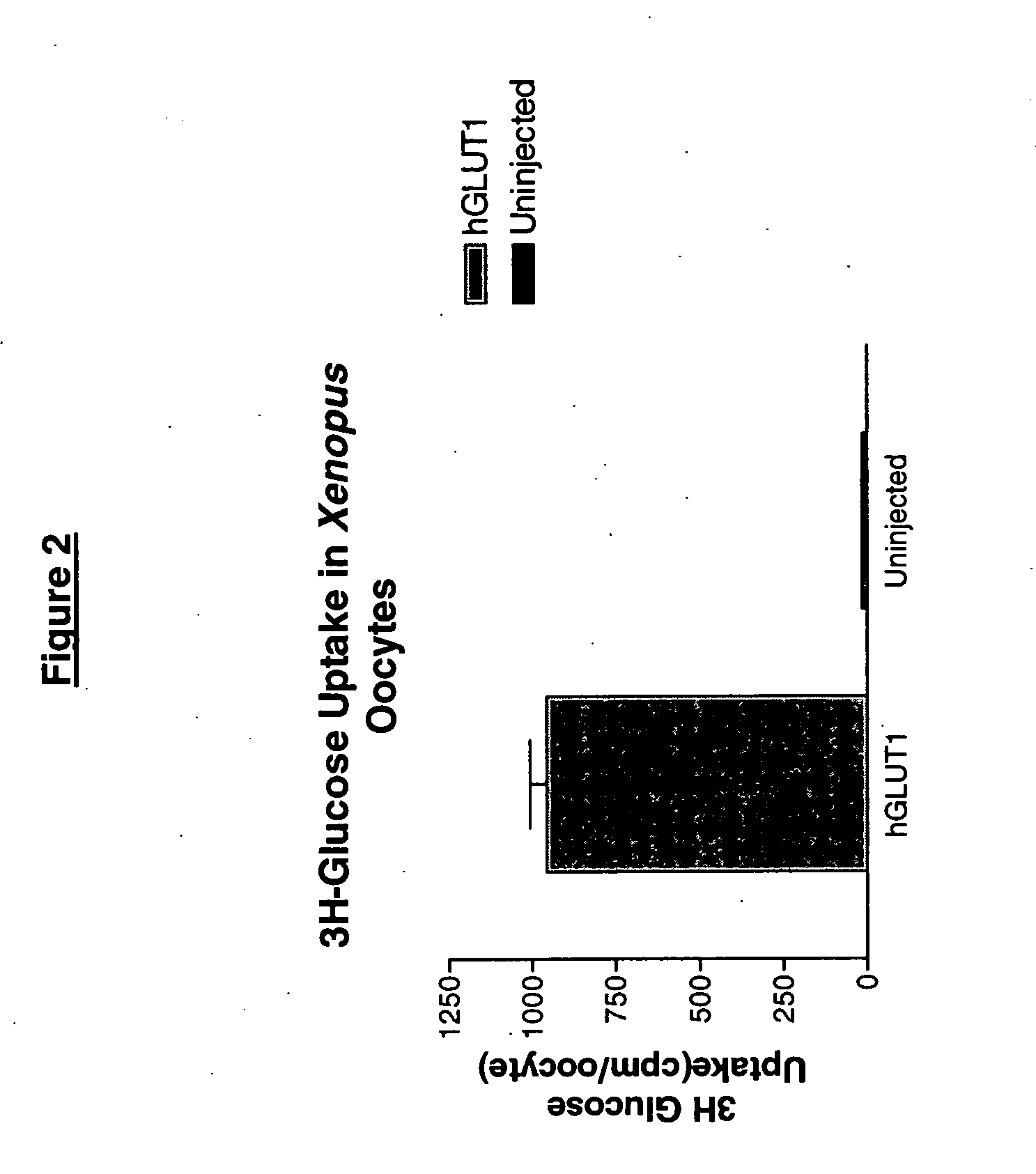
[0087] To assess transport function of a specific transporter protein, it can be desirable to clone the cDNA and express the protein in cells that have low endogenous transport activity. Human GLUT1 was cloned by PCR, fully sequenced, and subcloned into plasmids that can be used for expression in mammalian cells or Xenopus oocytes. Because many cell lines already exhibit high levels of GLUT1 activity, expression in Xenopus oocytes can be advantageous due to the low levels of endogenous sugar transport. For expression in Xenopus oocytes, in vitro GLUT1 cRNA was prepared and injected into defoliculated oocytes.

[0088] Oocytes expressing GLUT1 exhibited higher levels of 3H-glucose uptake than noninjected controls, as shown in FIG. 2. Oocytes expressing GLUT1 or control oocytes not expressing GLUT1 were incubated in an oocyte ringers (ND96) buffer (90 mM NaCl, 10 mM HemiNa HEPES, 2 mM KCl, 1 mM MgCl, 1.8 mM CaCl2) containing 0.5% bovine serum albumin and 3H-...

Example

Example 3

Uptake into Mammalian Cells

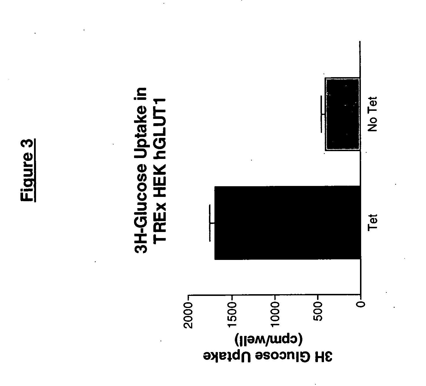
[0089] GLUT1 was subcloned into a plasmid that allows for inducible expression by tetracycline (TREX plasmid, Invitrogen Inc., Carlsbad Calif.). The GLUT1 expression plasmid was transfected into a human embryonic kidney (HEK) cell line and stable clones were isolated by G418 selection and flow activated cell sorting (FACS). An example of glucose uptake in a GLUT1-HEK cell clone is shown in FIG. 3. GLUT1-HEK / TREX cells were plated in 96-well plates at 100,000 cells / well at 37° C. for 24 hours and tetracycline (1 μg / mL) was added to each well for an additional 24 hours to induce GLUT1 transporter expression. Radiolabeled 3H-glucose (˜75,000 cpm / well) was added to each well. Plates were incubated at room temperature for 1 min. Excess 3H-glucose was removed and cells were washed three times with a 96-well plate washer with cold assay buffer. Scintillation fluid was added to each well, and the plates were sealed and counted in a 96-well plate-based ...


PUM

PropertyMeasurementUnit
Levelaaaaaaaaaa
Cytotoxicityaaaaaaaaaa
Toxicityaaaaaaaaaa

Abstract

GLUT1 is consistently expressed at high levels in cancer cells. Disclosed herein are assays for determining whether a test material/molecule is a substrate for, and/or is transported by, the GLUT1 transporter, and therefore a candidate substrate for transport into cancer cells. The assays are useful in screening for cytotoxic agents or imaging components used in the treatment or diagnosis of cancer.

Description

CONTINUITY [0001] This application claims the benefit of U.S. Provisional Application No. 60 / 577,124, filed Jun. 4, 2004, which is incorporated by reference herein in its entirety.TECHNICAL FIELD [0002] The disclosures herein relate to assays and methods of using the same for screening compounds and / or chemical moieties for their ability to be transported into cancer cells. BACKGROUND [0003] Cancer remains the second leading cause of death in the developed world, with solid tumors of the lung, colon, breast, prostate, pancreas, ovary and testis accounting for the majority of cancer deaths. Cancer mortality rates for solid tumors have remained largely unchanged despite the many advances in understanding how solid tumors arise, diagnostic screening, and new cancer drugs. [0004] Small molecule chemotherapeutics typically do not result in a cure for solid tumor cancer, but have clinical value in slowing disease progression, and are an important component of cancer therapy due to their e...

Claims


Application Information

Patent Timeline
IPC IPC(8): C12Q1/68G01N33/574A61K38/00A61K39/00G01N33/50
CPCG01N33/5011G01N2333/62G01N33/6872G01N33/5017
Inventor ZERANGUE, NOAGANGAKHEDKAR, ARCHANA
Owner XENOPORT
Why Eureka
Try Eureka