Novel culture systems for EX vivo development
a culture system and ex vivo technology, applied in the field of cell, tissue, organ culture technology, can solve the problems of poor nutritional support of yolk sac, impractical commercialization of cloned animals, and problems such as problems such as the inability to produce cloned animals,
- Summary
- Abstract
- Description
- Claims
- Application Information
AI Technical Summary
Benefits of technology
Problems solved by technology
Method used
Image
Examples
example 1
[0085] Human Embryo-Derived Cells Differentiated in Juxtaposition to an Embryonated Telolecithal Egg
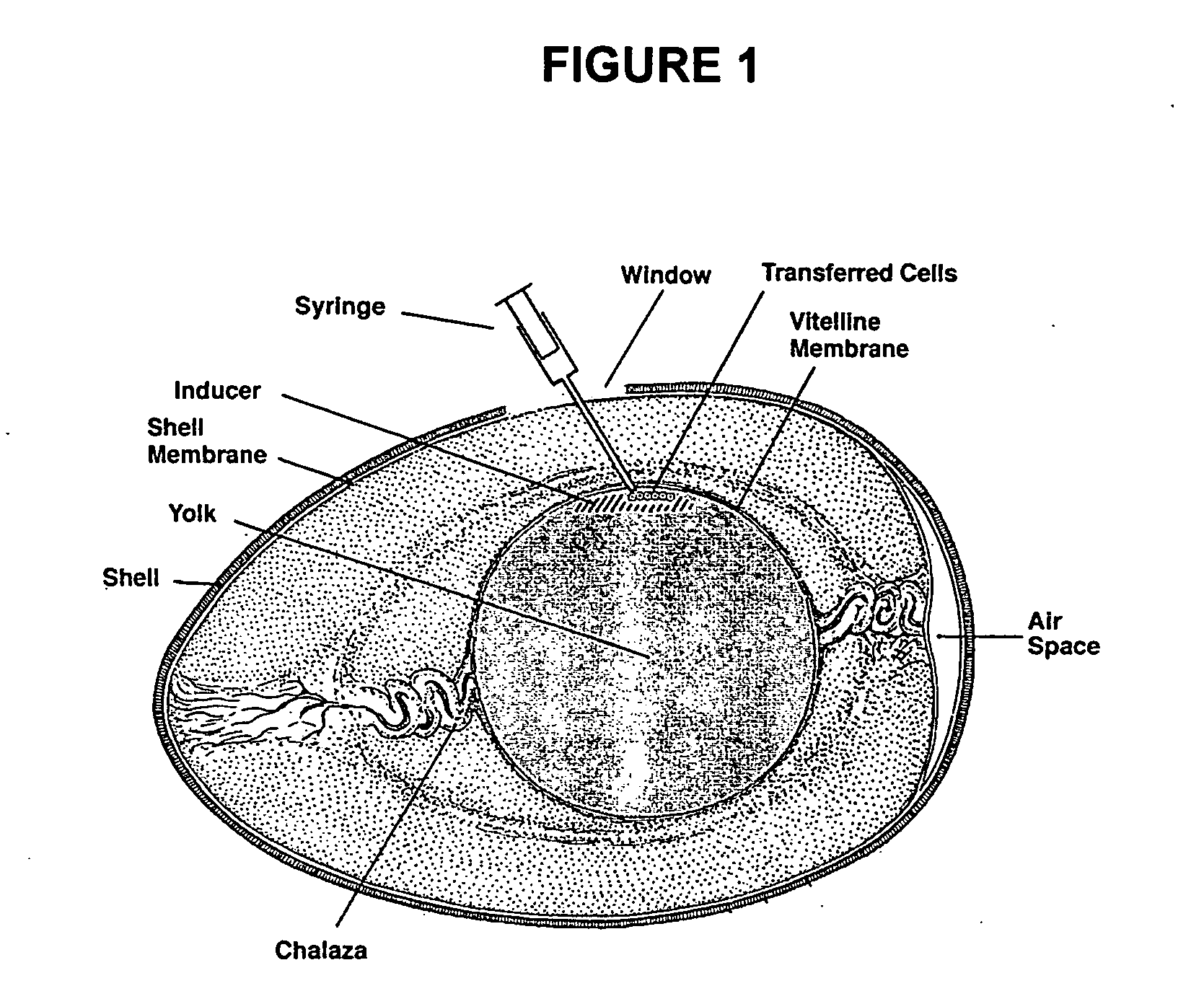
[0086] Approximately 10×106 human ES cells were trypsinized from culture, the trypsin was neutralized with 10% FCS in DMEM and the cells pelleted and resuspended in DMEM. Approximately 1×10⁶ human ES cells were injected within the vitelline membrane of an embryonated SPF egg (Charles River) at two days of incubation at 0.5 cm from the avian embryo. At day 15, the mass of cells were identified beneath the yolk sac membrane, fixed, and Hematoxylin-and-eosin stained. In this example the cells were fixed with formaldehyde, however there are many fixative agents known to those skilled in the art which could be used. As shown in FIG. 5, dense sheets of cells ranging from vacuolated mesenchymal to round cells were visible, consistent with a predisposition to teratoma formation. Yolk sac associated epithelial cells were also observed.
example 2
[0087] Human Embryonic Stem Cell Lines Maintained in the Undifferentiated State Using SPF-Chick Embryonic Feeder Cells
[0088] Preparation of CEF:
[0089] CEF were isolated from 7-8 day old chicken embryos with the heads left on, using the previously described techniques for isolation of mouse embryonic fibroblasts. Briefly, the embryos were eviscerated, the heads left on, digested with trypsin and plated onto gelatin coated plates in DMEM, supplemented with 10% FBS, glutamine and penicillin-streptomycin. The cells were frozen at passage one and used at passage 2 after mitotic inactivation with mitomycin C.
[0090] The hES cell lines, H9, H7 (both NIH-approved) and ACT-4 were consecutively cultured on CEF for 3-6 passages without significant changes in undifferentiated morphology or growth rate. Passages used for the experiment: H-9 & H-7: H-9 started passage 38 through passage 40, H7 started 29 and through passage 35; and ACT 4 derived here from passage 9-11 and 15-19.
[0091] Expressi...
example 3
[0092] Non-Human Embryonic Development Within a SPF Avian Egg and the Use of the Porcine Embryo to Direct the Differentiation of Human Pluripotent Cells
[0093] A cloned or normal porcine blastocyst with or without a transgenic suicide gene is held with an aspiration pipette under low magnification and the trophectoderm is torn opposite the inner cell mass to yield near-planar aggregation of cells. The torn blastocyst is injected with a 200 micron pipette into an unfertilized but fresh SPF windowed avian egg at or near the blastodisc. The resulting reconstructed egg is then resealed with kitchen wrap as is well known in the art and cultured at 37° C. on a racking platform. At the point when cell differentiation of a desired type is occurring in the porcine embryo, hES or hED cells are injected into the porcine embryo. In the case of hematopoietic differentation, the human pluripotent stem cells are injected into the aortic-gonadal-mesonephros region of the porcine embryo to induce di...
PUM
 Abstract
Description
Claims
Application Information
 - R&D
- Intellectual Property
- Life Sciences
- Materials
- Tech Scout
- Unparalleled Data Quality
- Higher Quality Content
- 60% Fewer Hallucinations
Browse by: Latest US Patents, China's latest patents, Technical Efficacy Thesaurus, Application Domain, Technology Topic, Popular Technical Reports.
© 2025 PatSnap. All rights reserved.Legal|Privacy policy|Modern Slavery Act Transparency Statement|Sitemap|About US| Contact US: help@patsnap.com


