
Cleavage of nucleic acids

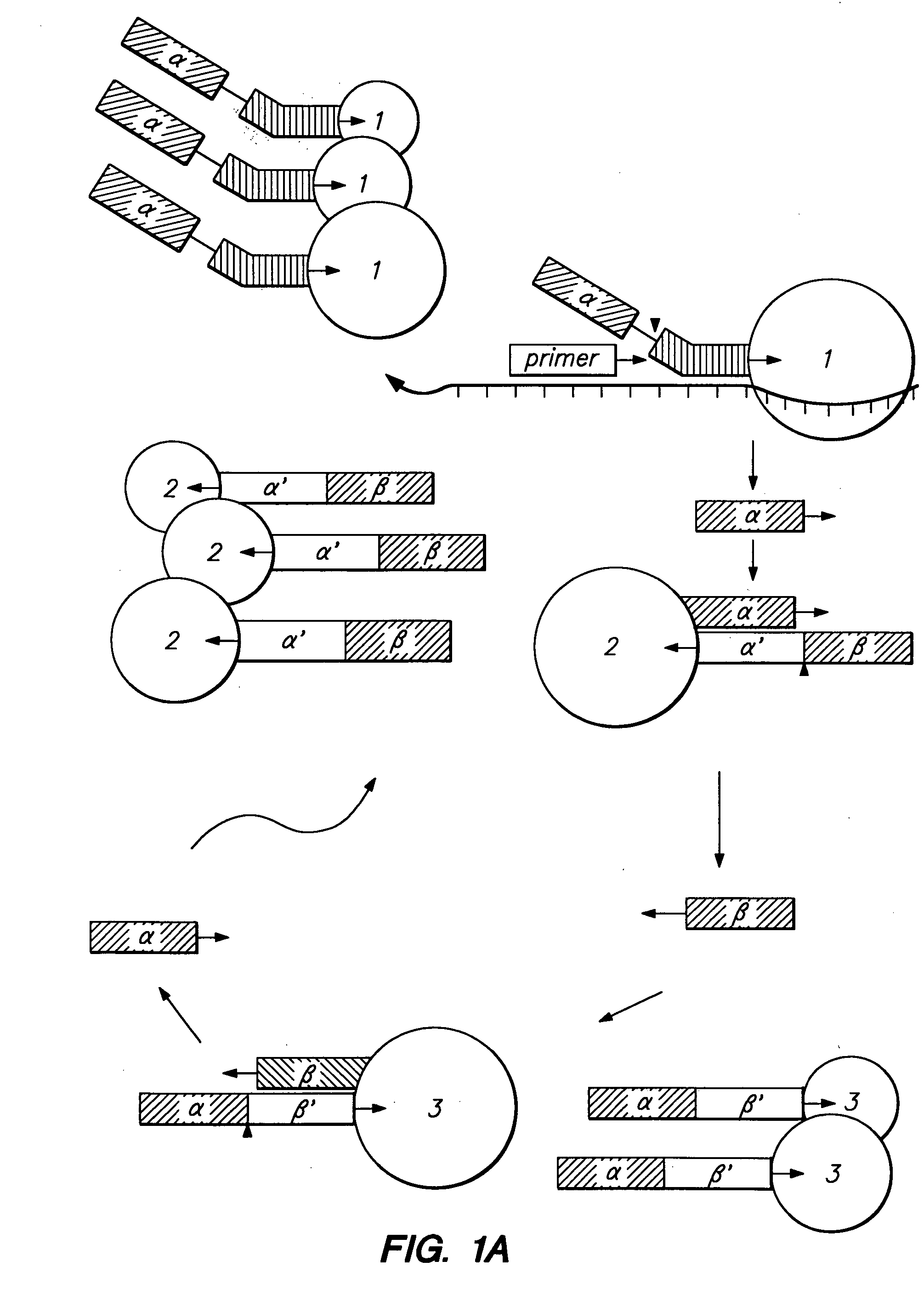
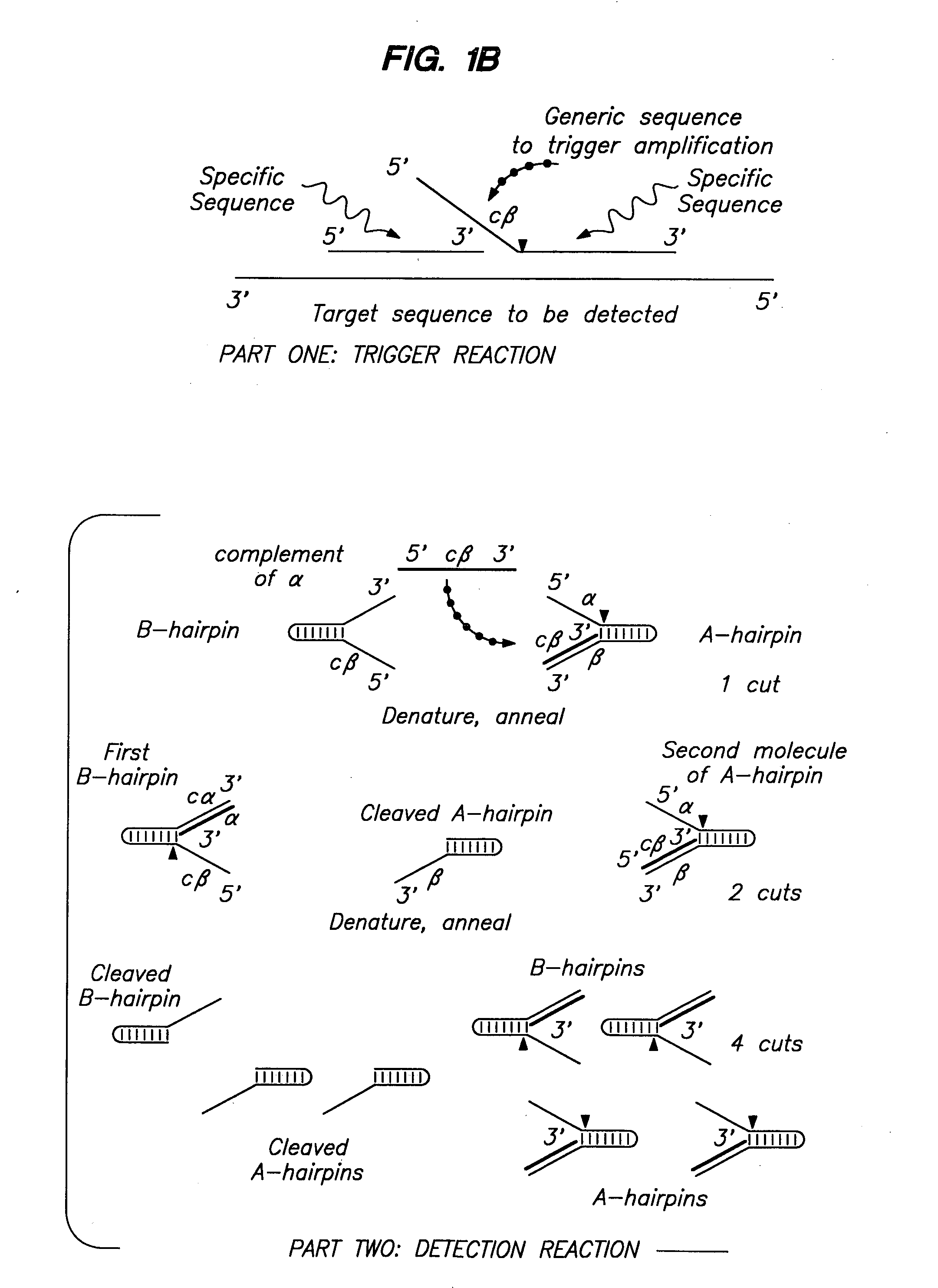
a nucleic acid and cleavage technology, applied in the field of nucleic acid cleavage, detection and characterization of nucleic acid sequences and sequence changes, can solve the problems of ineffective thermostable dna ligases, limited use of lcr for mutant screening, and inability to raise reaction temperatures to prevent, etc., to achieve rapid detection and identification of variants

Inactive Publication Date: 2006-02-16
THIRD WAVE TECH
View PDF17 Cites 22 Cited by
  • Summary
  • Abstract
  • Description
  • Claims
  • Application Information

AI Technical Summary

Benefits of technology

[0061] By the term “genetic fingerprint” it is meant that changes in the sequence of the nucleic acid (e.g., a deletion, insertion or a single point substitution) alter the structures formed, thus changing the banding pattern (i.e., the “fingerprint” or “bar code”) to reflect the difference in the sequence, allowing rapid detection and identification of variants.

Problems solved by technology

The use of LCR for mutant screening is limited to the examination of specific nucleic acid positions.
However, available thermostable DNA ligases are not effective on this RNA substrate, so the ligation must be performed by T4 DNA ligase at low temperatures (37° C.).
Therefore the reaction temperatures cannot be raised to prevent non-specific hybridization of the probes.
In practice, routine polymerase chain reactions rarely achieve the theoretical maximum yield, and PCRs are usually run for more than 20 cycles to compensate for the lower yield.
Reaction conditions must be carefully optimized for each different primer pair and target sequence, and the process can take days, even for an experienced investigator.
The laboriousness of this process, including numerous technical considerations and other factors, presents a significant drawback to using PCR in the clinical setting.
Indeed, PCR has yet to penetrate the clinical market in a significant way.
In addition, both methods require expensive equipment, capable of precise temperature cycling.
This method has a substantial limitation in that the base composition of the mismatch influences the ability to prevent extension across the mismatch, and certain mismatches do not prevent extension or have only a minimal effect (Kwok et al., Nucl.
Any mismatch effectively blocks the action of the thermostable ligase, but LCR still has the drawback of target-independent background ligation products initiating the amplification.
Moreover, the combination of PCR with subsequent LCR to identify the nucleotides at individual positions is also a clearly cumbersome proposition for the clinical laboratory.
Traditional methods of direct detection including Northern and Southern blotting and RNase protection assays usually require the use of radioactivity and are not amenable to automation.
While the repeating process increases the signal, the RNA portion of the oligonucleotide is vulnerable to RNases that may carried through sample preparation.
However, specialized equipment and highly trained personnel are required, and the method is too labor-intense and expensive to be practical and effective in the clinical setting.
However, this method requires the use of osmium tetroxide and piperidine, two highly noxious chemicals which are not suited for use in a clinical laboratory.
RFLP analysis suffers from low sensitivity and requires a large amount of sample.
When RFLP analysis is used for the detection of point mutations, it is, by its nature, limited to the detection of only those single base changes which fall within a restriction sequence of a known restriction endonuclease.
Moreover, the majority of the available enzymes have 4 to 6 base-pair recognition sequences, and cleave too frequently for many large-scale DNA manipulations (Eckstein and Lilley (eds.
Furthermore, the method requires specialized equipment to prepare the gels and maintain the needed high temperatures during electrophoresis.
In addition, long running times are required for DGGE.
This technique is extremely sensitive to variations in gel composition and temperature.
A serious limitation of this method is the relative difficulty encountered in comparing data generated in different laboratories, under apparently similar conditions.
While ddF is an improvement over SSCP in terms of increased sensitivity, ddF requires the use of expensive dideoxynucleotides and this technique is still limited to the analysis of fragments of the size suitable for SSCP (i.e., fragments of 200-300 bases for optimal detection of mutations).
In addition to the above limitations, all of these methods are limited as to the size of the nucleic acid fragment that can be analyzed.
For the direct sequencing approach, sequences of greater than 600 base pairs require cloning, with the consequent delays and expense of either deletion sub-cloning or primer walking, in order to cover the entire fragment.
SSCP and DGGE have even more severe size limitations.
Because of reduced sensitivity to sequence changes, these methods are not considered suitable for larger fragments.
The ddF technique, as a combination of direct sequencing and SSCP, is also limited by the relatively small size of the DNA that can be screened.

Method used


Image

  • Cleavage of nucleic acids
  • Cleavage of nucleic acids
  • Cleavage of nucleic acids

Examples


example 1

Characteristics of Native Thermostable DNA Polymerases

[0391] A. 5′ Nuclease Activity pf DNAPTaq

[0392] During the polymerase chain reaction (PCR) (Saiki et al., Science 239:487 (1988); Mullis and Faloona, Methods in Enzymology 155:335 (1987)), DNAPTaq is able to amplify many, but not all, DNA sequences. One sequence that cannot be amplified using DNAPTaq is shown in FIG. 6 (Hairpin structure is SEQ ID NO:15, PRIMERS are SEQ ID NOS:16-17.) This DNA sequence has the distinguishing characteristic of being able to fold on itself to form a hairpin with two single-stranded arms, which correspond to the primers used in PCR.

[0393] To test whether this failure to amplify is due to the 5′ nuclease activity of the enzyme, we compared the abilities of DNAPTaq and DNAPStf to amplify this DNA sequence during 30 cycles of PCR. Synthetic oligonucleotides were obtained from The Biotechnology Center at the University of Wisconsin-Madison. The DNAPTaq and DNAPStf were from Perkin Elmer (i.e., AMPLIT...

example 2

Generation of 5′ Nucleases from Thermostable DNA Polymerases

[0426] Thermostable DNA polymerases were generated which have reduced synthetic activity, an activity that is an undesirable side-reaction during DNA cleavage in the detection assay of the invention, yet have maintained thermostable nuclease activity. The result is a thermostable polymerase which cleaves nucleic acids DNA with extreme specificity.

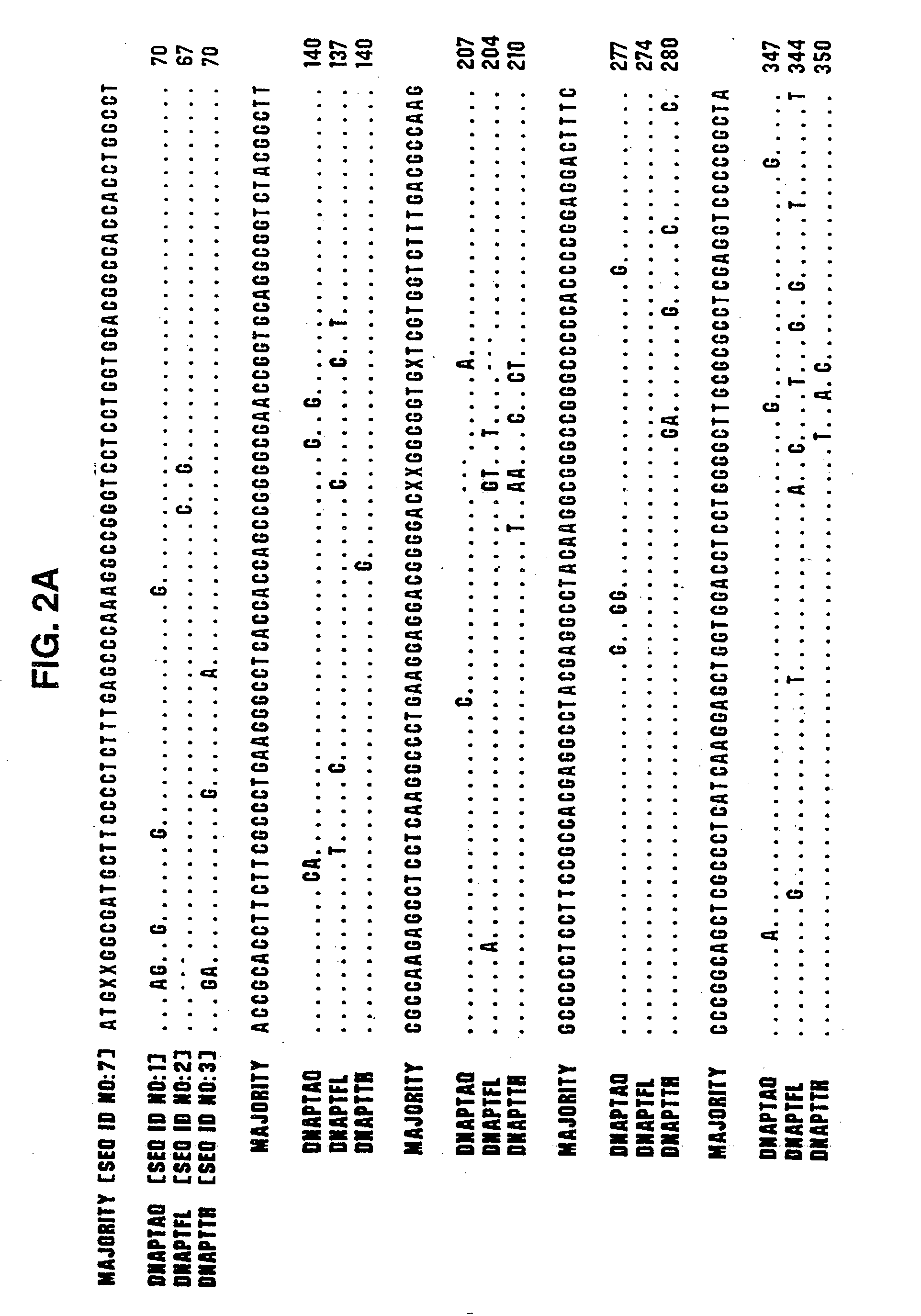
[0427] Type A DNA polymerases from eubacteria of the genus Thermus share extensive protein sequence identity (90% in the polymerization domain, using the Lipman-Pearson method in the DNA analysis software from DNAStar, WI) and behave similarly in both polymerization and nuclease assays. Therefore, we have used the genes for the DNA polymerase of Thermus aquaticus (DNAPTaq) and Thermus flavus (DNAPTfl) as representatives of this class. Polymerase genes from other eubacterial organisms, such as Thermus thermophilus, Thermus sp., Thermotoga maritima, Thermosipho africanus and Bacill...

example 3

5′ Nucleases Derived from Thermostable DNA Polymerases can Cleave Short Hairpin Structures with Specificity

[0489] The ability of the 5′ nucleases to cleave hairpin structures to generate a cleaved hairpin structure suitable as a detection molecule was examined. The structure and sequence of the hairpin test molecule is shown in FIG. 19A (SEQ ID NO:15). The oligonucleotide (labeled “primer” in FIG. 19A, SEQ ID NO:22) is shown annealed to its complementary sequence on the 3′ arm of the hairpin test molecule. The hairpin test molecule was single-end labeled with 32P using a labeled T7 promoter primer in a polymerase chain reaction. The label is present on the 5′ arm of the hairpin test molecule and is represented by the star in FIG. 19A.

[0490] The cleavage reaction was performed by adding 10 fmoles of heat-denatured, end-labeled hairpin test molecule, 0.2 uM of the primer oligonucleotide (complementary to the 3′ arm of the hairpin), 50 μM of each dNTP and 0.5 units of DNAPTaq (Perkin...


PUM

PropertyMeasurementUnit
nucleic acidaaaaaaaaaa
fluorescentaaaaaaaaaa
nucleic acid cleavage structureaaaaaaaaaa

Abstract

The present invention relates to means for cleaving a nucleic acid cleavage structure in a site-specific manner. Enzymes, including 5′ nucleases and 3′ exonucleases, are used to detect and identify nucleic acids derived from microorganisms. Methods are provided which allow for the detection and identification of bacterial and viral pathogens in a sample.

Description

[0001] The present application is a Continuation of co-pending U.S. application Ser. No. 09 / 940,925, filed Aug. 28, 2001, which is a Divisional application of U.S. application Ser. No. 09 / 655,378, filed Sep. 5, 2000, now U.S. Pat. No. 6,673,616, which is a Continuation of U.S. application Ser. No. 08 / 520,946, filed Aug. 30, 1995, now U.S. Pat. No. 6,372,424, which is a Continuation-in-Part application of U.S. appliation Ser. No. 08 / 484,956, filed Jun. 7, 1995, now U.S. Pat. No. 5,843,654, which is a Continuation-in-Part of U.S. application Ser. No. 08 / 402,601, filed Mar. 9, 1995, now abandoned, which is a Continuation-in-Part of U.S. application Serial No. 08 / 337,164, filed Nov. 11, 1994, now abandoned, which is a Continuation-in-Part of U.S. application Serial No. 08 / 254,359, filed Jun. 6, 1994, now U.S. Pat. No. 5,614,402, which is a Continuation-In-Part Application of U.S. application Ser. No. 08 / 073,384, filed Jun. 4, 1993, now U.S. Pat. No. 5,541,311, which is a Continuation-In...

Claims


Application Information

Patent Timeline
Patent Type & Authority Applications(United States)
IPC IPC(8): C12Q1/68C12P19/34C12N9/22
CPCC12N9/1252C12Q1/682C12Q1/6846C12Q2537/149C12Q2525/301C12Q2521/319
Inventor DAHLBERG, JAMESBROW, MARY ANNLYAMICHEV, VICTOR
Owner THIRD WAVE TECH
Learn More