Receptor for lysophosphatidylcholine in vascular endothelial cells and use thereof
a vascular endothelial cell and lysophosphatidylcholine technology, applied in the direction of immunoglobulins, peptides, antibody medical ingredients, etc., can solve the problem of uneven distribution of variable domains and achieve the effect of inhibiting one or more functions
- Summary
- Abstract
- Description
- Claims
- Application Information
AI Technical Summary
Benefits of technology
Problems solved by technology
Method used
Image
Examples
example 1
Antibody Preparation
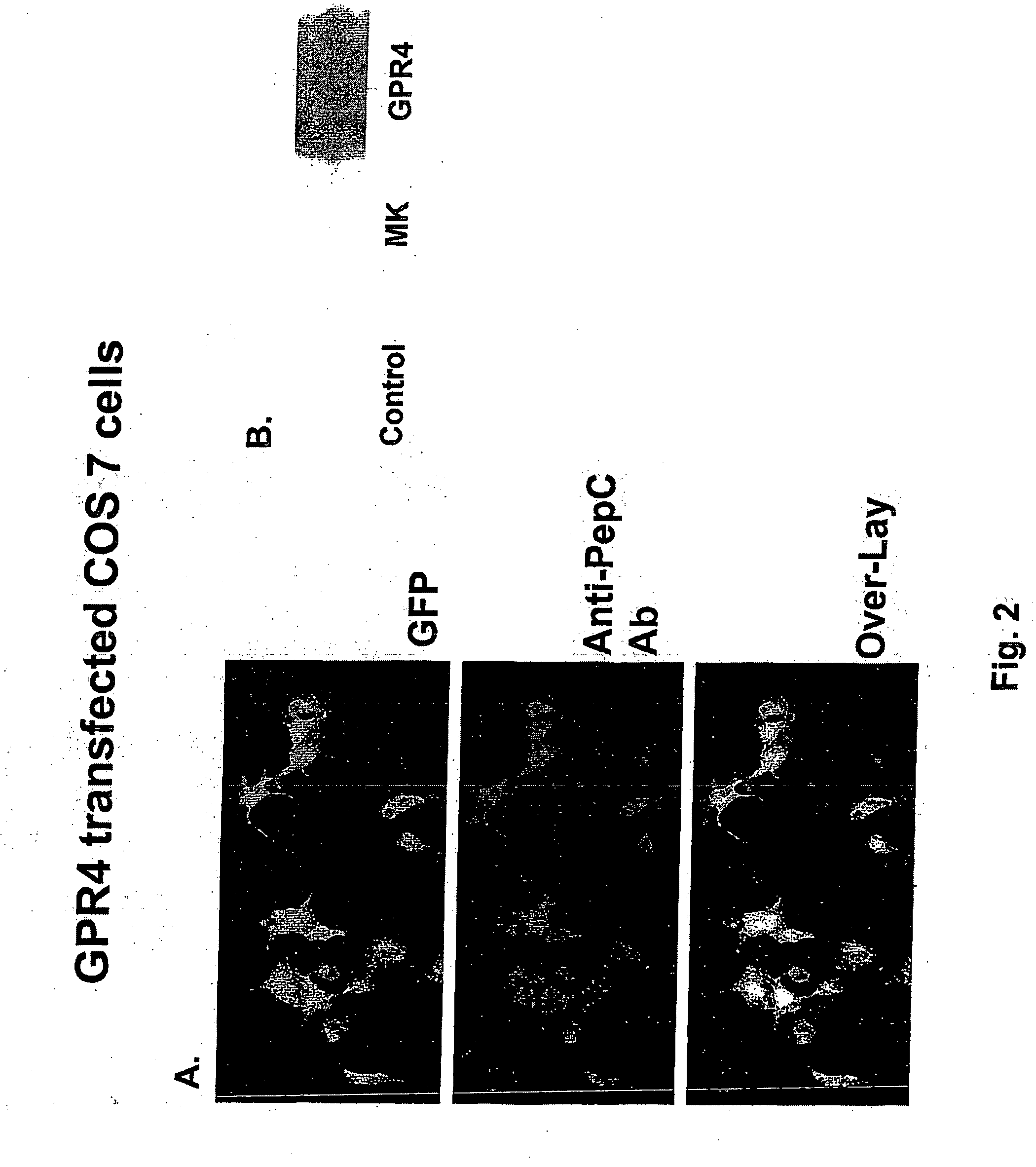
[0099] Polyclonal antibodies to GPR4 were made at the Research Resources Center, University of Illinois at Chicago. Peptides corresponding to either N-terminus (positions 2 through 9) or C-termini (positions 324 through 334) of human GPR4 (GenBank Number U21051) were synthesized on an Applied Biosystems Peptide Synthesizer (Model 433; Foster City, Calif.) using solid phase peptide synthesis with Fmoc (9-fluorenylmethl-oxycarbonyl) chemistry. The peptide was checked and verified by its single peak in the analytical HPLC chromatogram, amino acid composition, and mass spectrum and by NH2-terminal sequencing.
[0100] Each peptide was separately conjugated to keyhole limpet hemocyanin (KLH) using the heterobifunctional coupling reagent m-maleimidobenzoyl-N-hydroxysuccinimide ester (MBS) dissolved (5 mg) in 0.5 ml 0.01 mol / L phosphate buffer (pH 7.0) for immunization in rabbits. Blood was collected before injection to obtain preimmune serum from the rabbits. Booster in...
example 2
GPR4 Expression in HBMEC
[0104] Human microvascular endothelial cells from brain (HBMEC) were cultured to elucidate the induced expression of GPR4 in HBMEC with inflammatory mediators such as TNF-α and oxidants. HBMEC were grown in RPMI 1640 supplemented with 10% FBS, 10% NuSerum (Becton Dickinson; Bedford, Mass.), endothelial cell growth supplement (30 μg / ml), heparin (5 U / ml), 1 mmol / l sodium pyruvate, 1 mmol / l minimal essential media (MEM), nonessential amino acids, 1 mmol / l MEM vitamins, 1% L-glutamine, and 1% penicillin-streptomycin.
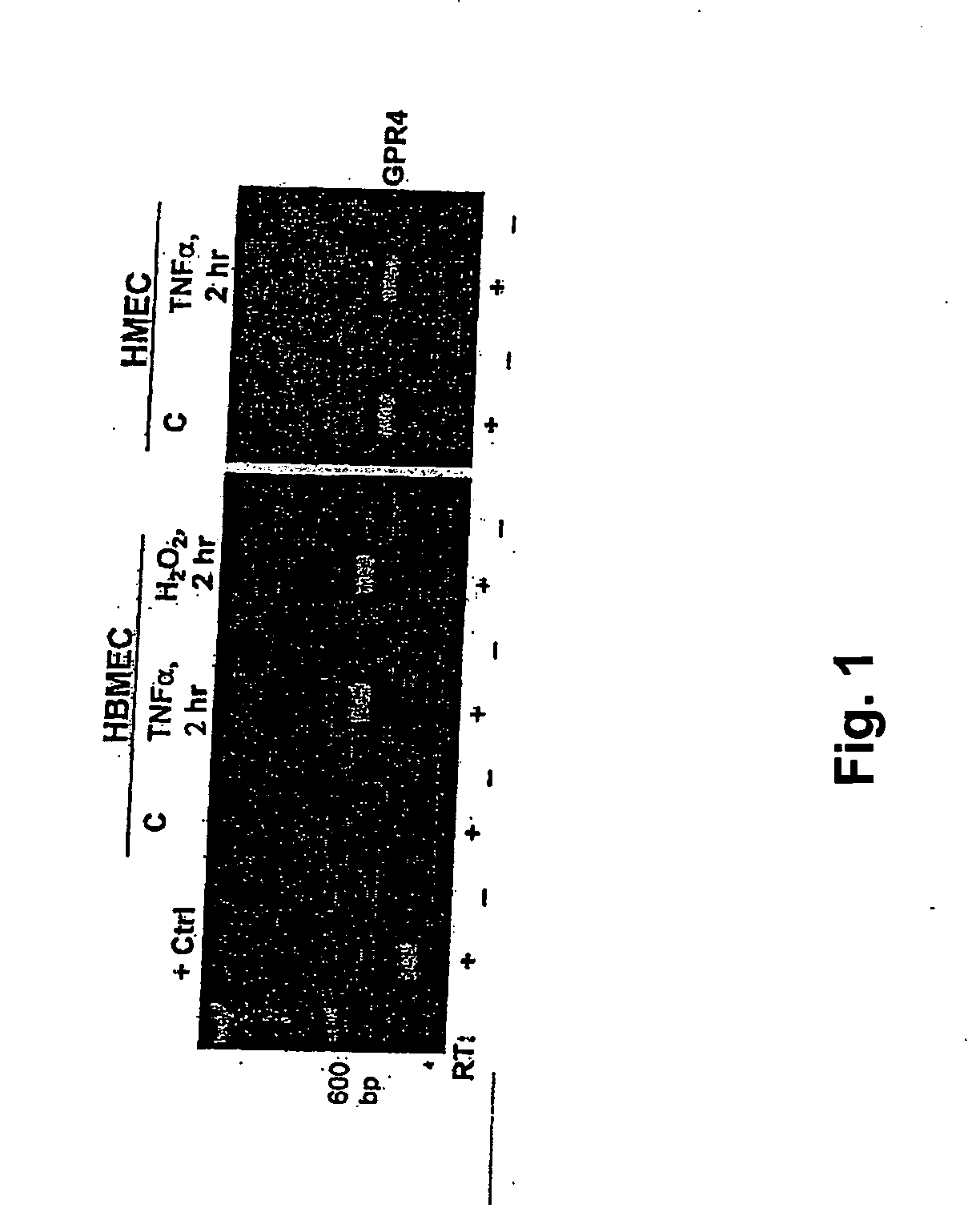
[0105] The expression of GPR4 mRNAs in HBMEC was determined by RT-PCR [Lum H, et al., Am J Physiol Cell Physiol 282:C59-C66 (2002)]. HBMEC were treated with 2 hours or overnight (about 18 hours) with either TNF-α (100 U / ml) or H2O2 (50 pmol / l), and total RNA extracted as well from human lymphocytes as a positive control. Total RNA was reverse transcribed with oligo-dT primers, and PCR was performed with specific primer sets corresponding to GenBank...
example 3
Specific Binding of LPC to Endothelial Cell
Surface by Competition Binding Assay
[0106] Confluent cell monolayers grown in 24-well culture dishes were treated overnight (about 18 hours) with either TNF-α (100 U / ml) or H2O2 (50 μmol / l). The cells were washed and incubated for 60 minutes at 4° C. with HEPES buffer (pH 7.4, 0.1% BSA) containing 0.02 nmol [3]H-LPC plus a 200-fold molar excess of unlabeled LPC. After three washes with cold HEPES buffer, cells were lysed with 0.1 mol / l NaOH, radioactivity was counted, and specific binding from duplicate samples was calculated as (fmol LPC bound / 106 cells). Separate dishes of cells were treated in parallel for cell count determination.
PUM
 Abstract
Description
Claims
Application Information
 - R&D
- Intellectual Property
- Life Sciences
- Materials
- Tech Scout
- Unparalleled Data Quality
- Higher Quality Content
- 60% Fewer Hallucinations
Browse by: Latest US Patents, China's latest patents, Technical Efficacy Thesaurus, Application Domain, Technology Topic, Popular Technical Reports.
© 2025 PatSnap. All rights reserved.Legal|Privacy policy|Modern Slavery Act Transparency Statement|Sitemap|About US| Contact US: help@patsnap.com


