Method for transdifferentiating mesenchymal stem cells into neuronal cells

Active Publication Date: 2006-05-11
AJOU UNIV IND ACADEMIC COOP FOUND
View PDF1 Cites 1 Cited by
  • Summary
  • Abstract
  • Description
  • Claims
  • Application Information

AI Technical Summary

Benefits of technology

[0011] In accordance with one aspect of the present invention, there is provided a method for transdifferentiating mesenchymal stem cells into neuronal cells, which comprises increasing the level of neurogenic basic Helix-Loop-Helix (bHLH) transcription factors in the mesenchymal stem cells.

Problems solved by technology

However, those methods have the problem of highly producing non-neuronal cells together with neuronal cells (Sanchez-Ramos., et al., Exp. Neurol., 164, 247-256(2000); Woodbury., et al., J. Neurosci. Res., 61, 364-370(2000); and Deng., et al., Biochem. Biophys. Res. Comm., 282, 148-152(2001)).
The problem is more pronounced when the cells are transplanted into the brain of experimental animals (Azizi., et al., Proc. Natl. Acad. Sci.

Method used


Image

  • Method for transdifferentiating mesenchymal stem cells into neuronal cells
  • Method for transdifferentiating mesenchymal stem cells into neuronal cells
  • Method for transdifferentiating mesenchymal stem cells into neuronal cells

Examples


example 1

Isolation and Culture of Mesenchymal Stem Cell

(step 1) Extraction of Bone Marrow and Isolation of Mesenchymal Stem Cells

[0044] 4 ml of HISTOPAQUE 1077 (Sigma) and 4 ml of bone marrow obtained from Bone marrow bank (Korean Marrow Donor Program, KMDP) were added to a sterilized 5 ml test-tube. After centrifugation at 400×g for 30 minutes, 0.5 ml of the buffy coat located in the interphase was collected and transferred into a test-tube containing 10 ml of sterilized phosphate buffered saline. The resulting suspension was centrifuged at 250×g for 10 minutes to remove the supernatant and 10 ml of phosphate buffer was added thereto to obtain a suspension, which was centrifuged at 250×g for 10 minutes. The above procedure was repeated twice and DMEM medium (Gibco) containing 10% FBS (Gibco) was added to the resulting precipitate. A portion of the resulting solution corresponding to 1×10⁷ cells was placed in a 100 mm dish and incubated at 37° C. for 4 hours while supplying 5% CO2 and 95%...

example 2

Construction of a Retroviral Vector Expressing Neurogenin 1, a Neurogenic Transcription Factor

(Step 1) Construction of Retroviral Vector Expressing Neurogenin 1 (Ngn1)

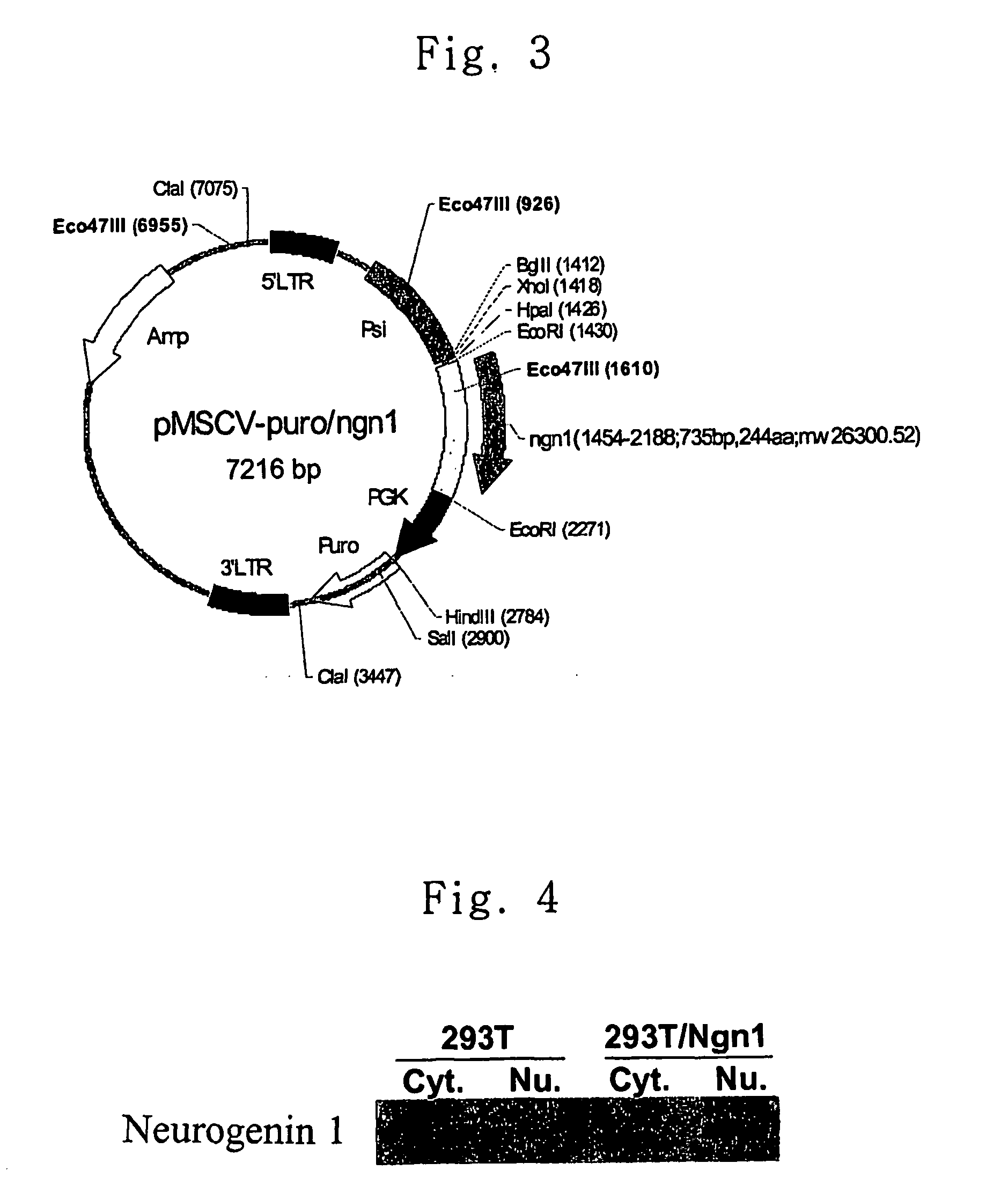
[0053] PCR was carried out using oligonucleotides of SEQ ID NOS.: 1 and 2 as primers and genomic DNA of Balb / c mouse as a template, and the PCR product thus obtained was cloned using TOPO TA cloning kit (Invitrogen). Through sequence analysis of the vector cloned, it was confirmed that the vector contained neurogenin 1 gene. The neurogenin 1 cDNA was isolated with EcoRI and inserted into the EcoRI site of vector pMSCV-puro (Clontech, USA) with T4 DNA ligase (Roche) and transformed into E. coli DH5α to obtain pMSCV-puro / ngn1. The resulting construct of pMSCV-puro / ngn1 is shown in FIG. 3.

(Step 2) Expression of Neurogenin1 Retroviral Vector

[0054] pMSCV-puro / ngn1 vector was transfected into 293T / 17 cell (ATCC CRL-11268) according to the calcium phosphate-coprecipitation method. After 48 hours of transduction, the cel...

example 3

Transduction of Neurogenin1 into MSCs

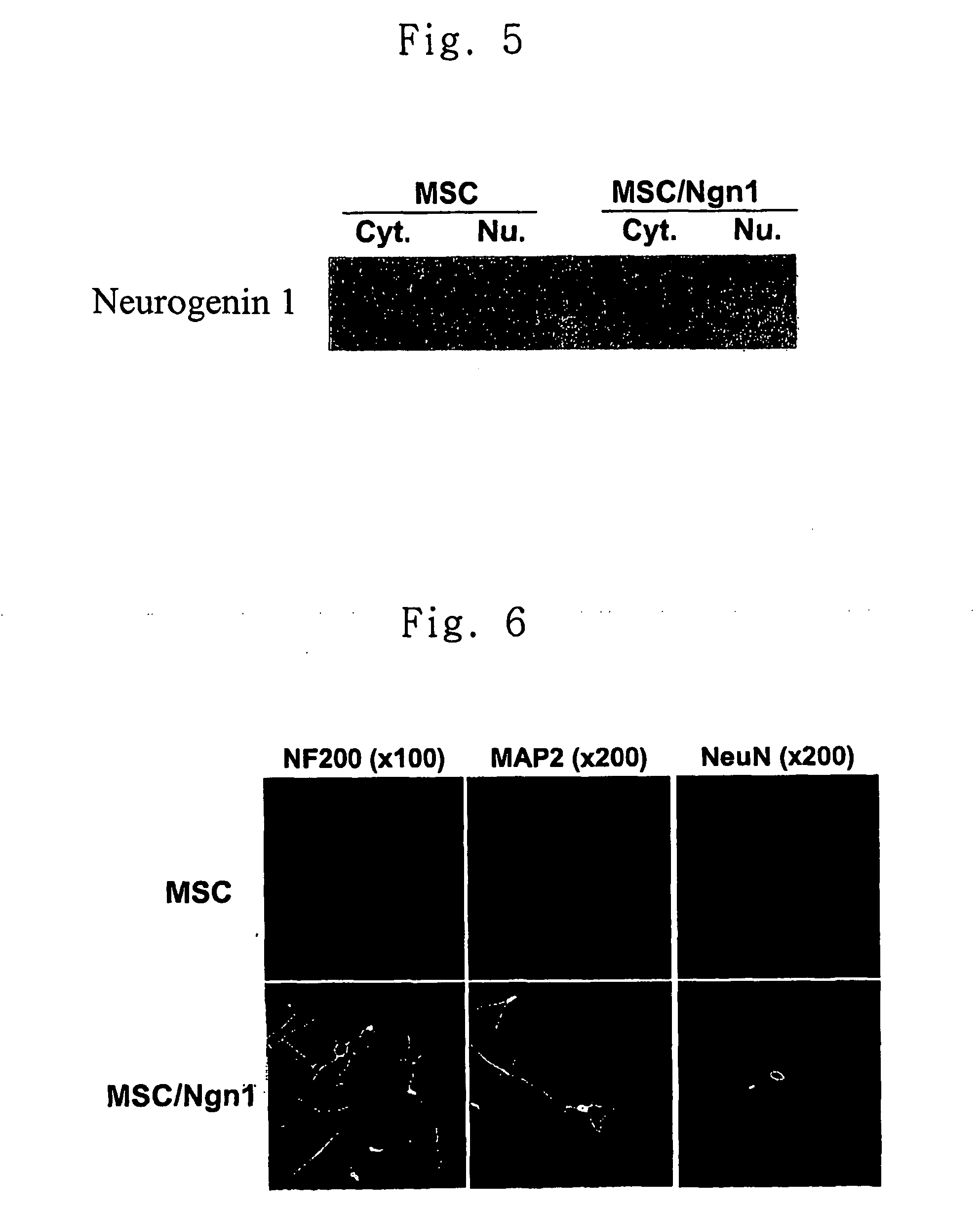
[0056] MSCs were cultured to 70% confluence in 100 mm dishes. Added thereto was 4 ml of the retrovirus solution obtained in Example 2 which was mixed with polybrene (Sigma) to a final concentration of 8 μg / ml and incubated for 8 hours. The retrovirus solution was then removed, the MSCs were cultured in 10 ml of MSC medium for 24 hours. The above procedure was repeated 1-4 times. Then, MSCs were collected using trypsin and diluted 20 fold with the medium. The obtained cells were subcultured in a medium supplemented with 2 μg / ml of puromycin (Sigma) for 2 weeks. The puromycin positive cells were fractionated into cytoplasm and nucleus. In order to confirm the expression of neurogenin1, each fraction was subjected to western blot analysis using anti-neurogenin1 antibody. The result is shown in FIG. 5. As can be seen in FIG. 5, neurogenin1 protein was expressed in the nucleus of MSCs transduced with Ngn1 gene.


PUM

PropertyMeasurementUnit
Timeaaaaaaaaaa
Molar densityaaaaaaaaaa

Abstract

This inventive discloses a method for transdifferentiating mesenchymal stem cells into neuronal cells, which comprises increasing the level of a basic helix-loop-helix (bHLH) transcription factor in the mesenchymal stem cells, said cells being useful in cell therapy or gene therapy for treating brain neurological diseases such as Parkinson's disease, Alzheimer disease, Hungtington's disease, amyotrophic lateral sclerosis, cerebral paralysis and brain ischemia; and spine disfunction caused by a traumatic injury.

Description

FIELD OF THE INVENTION [0001] The present invention relates to a method for transdifferentiating mesenchymal stem cells into neuronal cells and a pharmaceutical composition containing the neuronal cells for cell therapy of neurological diseases. BACKGROUND OF THE INVENTION [0002] Mesenchymal stem cells (MSCs) are multipotent bone marrow stromal cells aiding hematopoiesis and can differentiate into mesodermal lineage cells such as osteocytes, chondrocytes, adipocytes and myocytes. Thus, they can be utilized as sourced for the development of artificial tissues. [0003] It has recently been reported that MSCs have a potential to differentiate into neuroglia in the brain (Azizi., et al., Proc. Natl. Acad. Sci. USA, 94, 4080-4085(1998); and Kopen., et. al., Proc. Natl. Acad. Sci. USA, 96, 10711-10716(1999)), and therefore, it has been proposed the MSCs can be utilized as sources for the treatment of neurological diseases in the central nervous system(Li et al., Neurosci. Lett., 315, 67-70...

Claims


Application Information

Patent Timeline
IPC IPC(8): A61K35/30C12N5/08A61K35/12C12N15/02C12N5/0793
CPCA61K35/12C12N5/0619C12N2501/01C12N2501/06C12N2501/60C12N2510/00C12N2506/1353C12N15/02
Inventor SUH, HAE-YOUNGKIM, SUNG-SOOKIM, JI-WONLEE, YOUNG-DONAHN, SEUNG-CHEOL
Owner AJOU UNIV IND ACADEMIC COOP FOUND
Why Eureka
Try Eureka