Novel culture systems for ex vivo development

a culture system and ex vivo technology, applied in the field of cell, tissue, organ culture technology, can solve the problems of poor nutritional support of yolk sac, impractical commercialization of cloned animals, and problems such as problems such as the inability to produce cloned animals,

Inactive Publication Date: 2006-05-25
WEST MICHAEL D +2
View PDF1 Cites 47 Cited by
  • Summary
  • Abstract
  • Description
  • Claims
  • Application Information

AI Technical Summary

Benefits of technology

[0013] More specifically, this invention provides a novel method of culturing embryos, fetuses, cells, tissues, and organs in ovo in telolecithal or eutelolecithal eggs and for the culture of hES cells, hED cells, and cells differentiated from such cells in co-culture with cells derived from embryonated telolecithal or eutelolecithal eggs for numerous commercial applications that improves yield, efficiency, cost, and risk in each of the above categories.
[0016] In still another aspect of the invention, mammalian embryonic cells can be injected in ovo in juxtaposition to the vitelline membrane and incubated over time to allow the formation of a plurality of mammalian extraembryonic membranes in the avian egg, including the formation of mammalian splanchopleure, somatopleure, chorionic membrane (CAM), allantoic membrane, amniotic membrane, or yolk sac membranes. The generation of such extraembryonic membranes has great utility in supporting the differentiation of hES or hED cells for purposes of research or manufacture, or, in the case of non-human mammalian species, in supporting advanced development of embryos and fetuses for research or production of agricultural animals.

Problems solved by technology

In the case of animal cloning, the high cost of recipient females to gestate the cloned fetuses often makes the commercialization of cloned animals impractical.
In the case of the therapeutic uses of pluripotent stem cells, many pluripotent cells such as human embryonic stem (hES) cells, are problematic to culture using traditional cell culture technology.
However, the embryonic murine fibroblasts are also a potential source of pathogens including uncharacterized retroviruses.
Generally speaking, in most mammals, the yolk sac provides little if any nutritional support.

Method used


Image

  • Novel culture systems for ex vivo development
  • Novel culture systems for ex vivo development
  • Novel culture systems for ex vivo development

Examples


example 1

Human Embryo-Derived Cells Differentiated in Juxtaposition to an Embryonated Telolecithal Egg

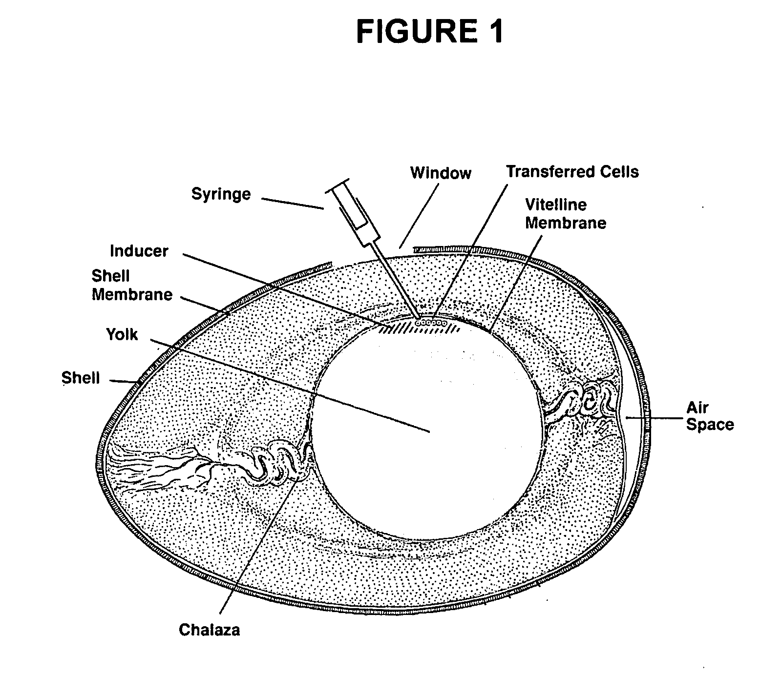
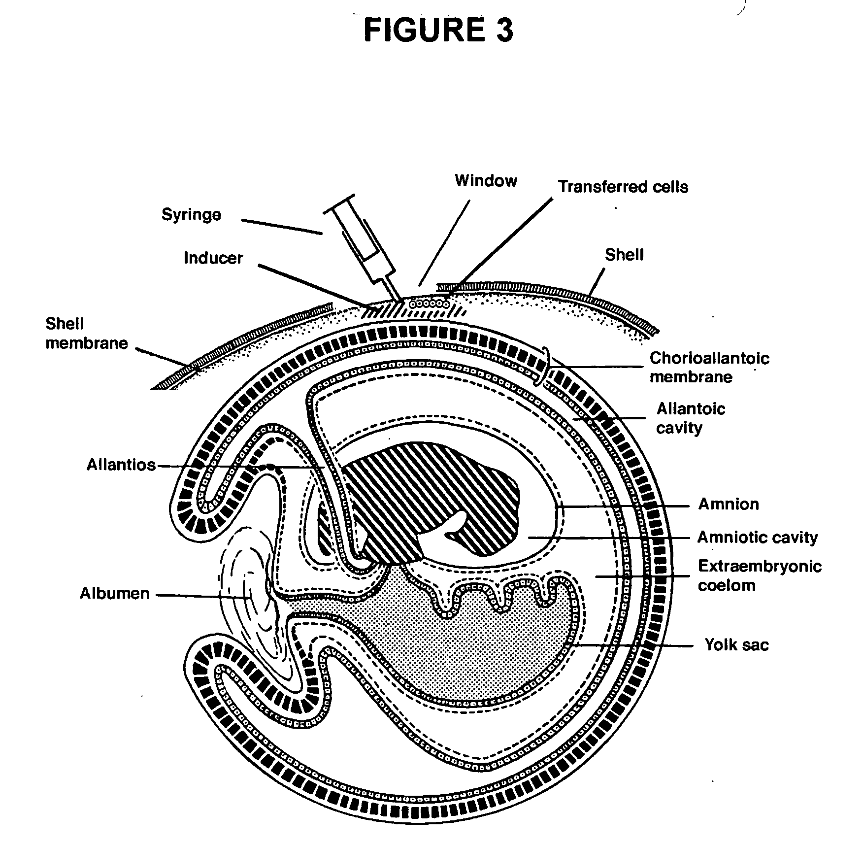
[0074] Approximately 10×106 human ES cells were trypsinized from culture, the trypsin was neutralized with 10% FCS in DMEM and the cells pelleted and resuspended in DMEM. Approximately 1×10⁶ human ES cells were injected within the vitelline membrane of an embryonated SPF egg (Charles River) at two days of incubation at 0.5 cm from the avian embryo. At day 15, the mass of cells were identified beneath the yolk sac membrane, fixed, and Hematoxylin-and-eosin stained. In this example the cells were fixed with formaldehyde, however there are many fixative agents known to those skilled in the art which could be used. As shown in FIG. 5, dense sheets of cells ranging from vacuolated mesenchymal to round cells were visible, consistent with a predisposition to teratoma formation. Yolk sac associated epithelial cells were also observed.

example 2

Human Embryonic Stem Cell Lines Maintained in the Undifferentiated State Using SPF-Chick Embryonic Feeder Cells

[0075] Preparation of CEF:

[0076] CEF were isolated from 7-8 day old chicken embryos with the heads left on, using the previously described techniques for isolation of mouse embryonic fibroblasts. Briefly, the embryos were eviscerated, the heads left on, digested with trypsin and plated onto gelatin coated plates in DMEM, supplemented with 10% FBS, glutamine and penicillin-streptomycin. The cells were frozen at passage one and used at passage 2 after mitotic inactivation with mitomycin C.

[0077] The hES cell lines, H9, H7 (both NIH-approved) and ACT-4 were consecutively cultured on CEF for 3-6 passages without significant changes in undifferentiated morphology or growth rate. Passages used for the experiment: H-9 & H-7: H-9 started passage 38 through passage 40, H7 started 29 and through passage 35; and ACT 4 derived here from passage 9-11 and 15-19.

[0078] Expression of t...

example 3

Non-Human Embryonic Development within a SPF Avian Egg and the Use of the Porcine Embryo to Direct the Differentiation of Human Pluripotent Cells

[0079] A cloned or normal porcine blastocyst with or without a transgenic suicide gene is held with an aspiration pipette under low magnification and the trophectoderm is torn opposite the inner cell mass to yield near-planar aggregation of cells. The torn blastocyst is injected with a 200 micron pipette into an unfertilized but fresh SPF windowed avian egg at or near the blastodisc. The resulting reconstructed egg is then resealed with kitchen wrap as is well known in the art and cultured at 37° C. on a racking platform. At the point when cell differentiation of a desired type is occurring in the porcine embryo, hES or hED cells are injected into the porcine embryo. In the case of hematopoietic differentation, the human pluripotent stem cells are injected into the aortic-gonadal-mesonephros region of the porcine embryo to induce different...


PUM

PropertyMeasurementUnit
thicknessaaaaaaaaaa
diameteraaaaaaaaaa
humidityaaaaaaaaaa

Abstract

The present invention provides methods for the culture of animal pluripotent stem cells and their differentiated progeny cells, tissues, and organs, and nonhuman animal embryos and fetuses.

Description

CROSS REFERENCE TO RELATED APPLICATIONS [0001] This application claims the benefit of priority to U.S. Provisional Application Ser. No. 60 / 534,447, filed Jan. 2, 2004 and U.S. Provisional Application Ser. No. 60 / 539,796, filed Jan. 28, 2004 which are incorporated herein by reference.FIELD OF THE INVENTION [0002] This invention generally relates to cells, tissue, and organ culture technology. More particularly, the invention relates to methods for culturing and differentiating animal pluripotent stem cells and non-human mammalian embryos and fetuses. BACKGROUND OF THE INVENTION [0003] Advances in nuclear transfer and embryonic stem cell technology have facilitated the cloning of non-human animals for diverse applications including agriculture, xenotransplantation, disease models, recombinant protein production, and novel means of manufacturing human cells for use in medical therapies, diagnosis, and discovery research. Each of these practical applications would benefit from new techn...

Claims


Application Information

Patent Timeline
Patent Type & Authority Applications(United States)
IPC IPC(8): A01K67/027C12N5/06C12N5/08C12N5/00C12N5/02C12N5/0735C12N5/10C12N15/00C12N15/873
CPCC12N5/0606C12N2502/04C12N15/873
Inventor WEST, MICHAEL D.CHAPMAN, KAREN B.KLIMANSKAYA, IRINA V.
Owner WEST MICHAEL D
Why Eureka
Try Eureka