
Methods for sterilizing preparations of digestive enzymes

a technology of digestive enzymes and preparations, which is applied in the direction of macromolecular non-active ingredients, lavatory sanitory, pharmaceutical non-active ingredients, etc., can solve the problems of poor sorption, insufficient nutrient-rich substances, and insufficient penetration of ingested meats, so as to reduce the temperature of the preparation, and reduce the residual solvent content of the preparation

Inactive Publication Date: 2006-06-01
CLEARANT
View PDF3 Cites 9 Cited by
  • Summary
  • Abstract
  • Description
  • Claims
  • Application Information

AI Technical Summary

Benefits of technology

"The present invention provides methods for sterilizing preparations of digestive enzymes that are sensitive to radiation without adversely affecting the preparation. The methods involve reducing the level of active biological contaminants or pathogens in the preparation of the enzymes. The invention also provides biological compositions comprising the sterilized preparations of digestive enzymes and stabilizers, which can be used for their intended use after sterilization with radiation. The technical effects of the invention include improved safety and preservation of the preparation of digestive enzymes for use in various applications."

Problems solved by technology

These substances, however, are useless as nutrients without the process of digestion to break down foods.
ingested meats are not well penetrated by these other digestive enzymes and so are poorly absorbed.
Clinical manifestations of GSD-II include progressive muscle weakness due to a build up of glycogen in muscle tissues, eventually resulting in respiratory and / or cardiac failure.
Clinical manifestations include enlargement of the spleen and liver, and frequently results in death, particularly for pediatric patients.
A deficiency in one or more of these enzymes cases a build up of excess amount in the body, causing progressive damage and eventual death.
Preparations of digestive enzymes that are prepared for human, veterinary, diagnostic and / or experimental use may contain unwanted and potentially dangerous biological contaminants or pathogens, such as viruses, bacteria (including inter- and intracellular bacteria, such as mycoplasmas, ureaplasmas, nanobacteria, chlamydia, rickettsias), yeasts, molds, fungi, prions or similar agents responsible, alone or in combination, for TSEs and / or single or multicellular parasites.
Such procedures, however, are not always reliable and are not able to detect the presence of certain viruses, particularly in very low numbers, and in the case of as yet unknown viruses or other contaminants or pathogens that may be in blood.
This reduces the value or certainty of the test in view of the consequences associated with a false negative result.
False negative results can be life threatening in certain cases, for example in the case of Acquired Immune Deficiency Syndrome (AIDS).
Furthermore, in some instances it can take weeks, if not months, to determine whether or not the preparation is contaminated.
This is a result of safety concerns for the workers conducting the tests, and the difficulty and expense associated with the containment facilities and waste disposal.
Heat treatment requires that the product be heated to approximately 60° C. for about 70 hours which can be damaging to sensitive products.
In some instances, heat inactivation can actually destroy 50% or more of the biological activity of the product.
Unfortunately, this method may also remove products that have a high molecular weight.
Further, in certain cases, small viruses and similarly sized contaminants and pathogens, such as prions, may not be removed by the filter.
This procedure requires that unbound sensitizer is washed from products since the sensitizers are toxic, if not mutagenic or carcinogenic, and cannot be administered to a patient.
The published literature in this area, however, teaches that gamma radiation can be damaging to radiation sensitive products, such as blood, blood products, enzymes, protein and protein-containing products.
In particular, it has been shown that high radiation doses are injurious to red cells, platelets and granulocytes (Leitman).
Unfortunately, many sensitive biological materials, such as monoclonal antibodies (Mab), may lose viability and activity if subjected to freezing for irradiation purposes and then thawing prior to administration to a patient.

Method used


Image

  • Methods for sterilizing preparations of digestive enzymes
  • Methods for sterilizing preparations of digestive enzymes
  • Methods for sterilizing preparations of digestive enzymes

Examples


example 1

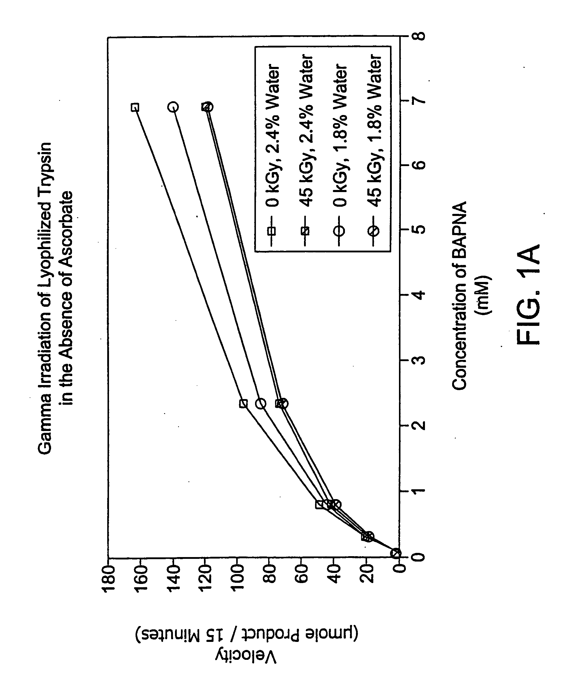
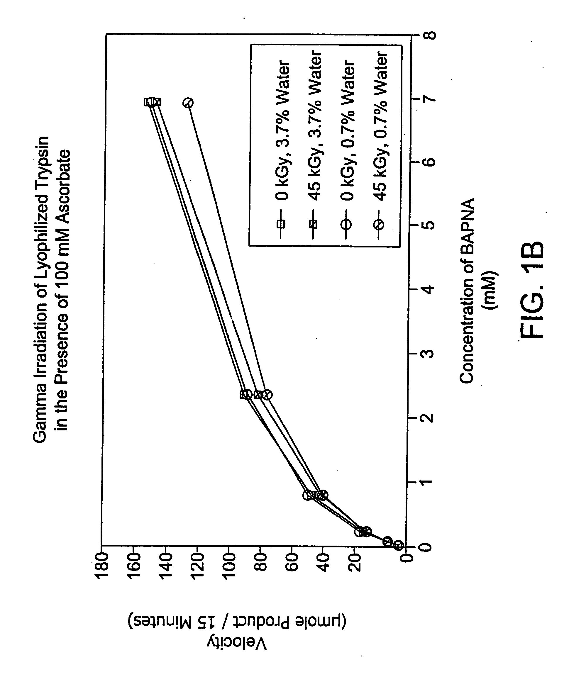
[0107] In this experiment, lyophilized trypsin was irradiated (45 kGy at 1.9 kGy / hr) alone or in the presence of a stabilizer (sodium ascorbate 100 mM) at varying levels of residual solvent content.

Method

[0108] 1 ml aliquots of trypsin alone or with 100 mM sodium ascorbate (10 mg / ml) were placed in 3 ml vials. Samples were prepared in triplicate and subjected to lyophilization, either a primary drying cycle (22 hours, sample temp 0-10° C., shelf temp 35° C., 10 mT) or a combination of a primary drying cycle and a secondary drying cycle (60 hours, sample temp 40° C., shelf temp 40° C., 10 mT).

[0109] All samples were resuspended in 1 ml water, and then diluted 1:10 for assay. Assay conditions: 50 units / ml trypsin per well+BAPNA substrate starting at 3000 μg / ml was serially diluted 3-fold down a 96-well plate. The assay was set up in two 96-well plates and absorption read at both 405 and 620 nm at 5 and 20 minutes. The absorption at 630 nm (background) was subtracted from the value...

example 2

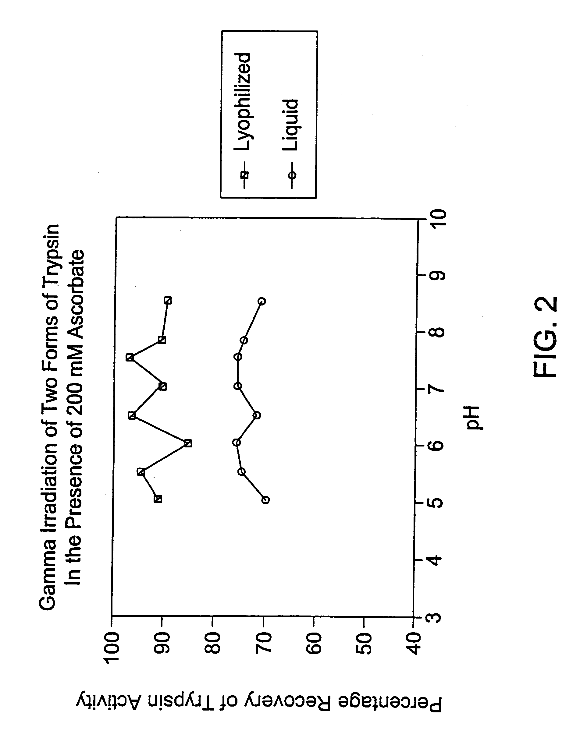
[0113] In this experiment, trypsin was irradiated (45 kGy at 1.6 kGy / hr. and 4° C.) in the presence of a stabilizer (sodium ascorbate 200 mM) as either a liquid or lyophilized preparation at varying pH levels.

Method

[0114] 1 ml of 1 mg / ml (about 3000 IU / ml) trypsin aliquots in the presence of 35 mM phosphate buffer and 200 mM sodium ascorbate were made at varying pH levels between 5 and 8.5, inclusive. 400 μl of each solution was placed in 3 ml vials and then lyophilized and gamma-irradiated. The remaining portion of each solution was gamma-irradiated as a liquid. Lyophilized and liquid samples were assayed at the same time, under the following conditions: Assay conditions: 5 U / well trypsin (50 U / ml)+BATNA substrate (1 mg / ml) was serially diluted 3-fold down a 96-well plate. The assay was set up in two 96-well plates and absorption read at both 405 and 620 nm at 5 and 20 minutes. The absorption at 630 nm (background) was subtracted from the value at 405 nm to obtain a corrected ab...

example 3

[0116] In this experiment, lyophilized trypsin was irradiated (42.7-44.8 kGy at 2.65 kGy / hr at 4° C.) alone or in the presence of a stabilizer (sodium ascorbate 200 mM).

Method

[0117] 1 ml aliquots of trypsin alone or with 200 mM sodium ascorbate (1 mg / ml) were placed in 3 ml vials and frozen overnight at −70° C. Samples were prepared in quadruplicate and subjected to lyophilization, utilizing primary and secondary drying cycles (20 hours total).

[0118] All samples were resuspended in 1 ml water, and then diluted 1:10 for assay. Assay conditions: 50 units / ml trypsin per well+BATNA substrate starting at 3000 μg / ml was serially diluted 3-fold down a 96-well plate. The assay was set up in two 96-well plates and absorption read at both 405 and 620 nm at 5 and 20 minutes. The absorption at 630 nm (background) was subtracted from the value at 405 nm to obtain a corrected absorption value. The change in this value over time between 5 and 15 minutes of reaction time was plotted and Vmax an...


PUM

PropertyMeasurementUnit
temperatureaaaaaaaaaa

Abstract

Methods are disclosed for sterilizing preparations of digestive enzymes to reduce the level of one or more active biological contaminants or pathogens therein, such as viruses, bacteria (including inter- and intracellular bacteria, such as mycoplasmas, ureaplasmas, nanobacteria, chlamydia, rickettsias), yeasts, molds, fungi, prions or similar agents responsible, alone or in combination, for TSEs and / or single or multicellular parasites. These methods involve sterilizing preparations of digestive enzymes, such as trypsin, α-galactosidase and iduronate-2-sulfatase, with irradiation.

Description

FIELD OF THE INVENTION [0001] The present invention relates to methods for sterilizing preparations of digestive enzymes to reduce the level of one or more active biological contaminants or pathogens therein, such as viruses, bacteria (including inter- and intracellular bacteria, such as mycoplasmas, ureaplasmas, nanobacteria, chlamydia, rickettsias), yeasts, molds, fungi, prions or similar agents responsible, alone or in combination, for TSEs and / or single or multicellular parasites. The present invention particularly relates to methods of sterilizing preparations of digestive enzymes, such as trypsin, α-galactosidase and iduronate 2-sulfatase, with irradiation. BACKGROUND OF THE INVENTION [0002] The principal foods upon which an organism, such as a human, survives can be broadly categorized as carbohydrates, fats and proteins. These substances, however, are useless as nutrients without the process of digestion to break down foods. [0003] Digestion of carbohydrates begins in the mo...

Claims


Application Information

Patent Timeline
Patent Type & Authority Applications(United States)
IPC IPC(8): A61L2/08A61K9/08A61K9/19A61K38/46A61K38/48A61K41/00A61K47/12A61K47/14A61K47/20A61K47/22A61K47/36A61K47/42A61L2/00A61L2/10A61L2/12A61P1/06A61P1/12A61P1/14A61P1/16A61P1/18A61P3/00A61P3/08A61P7/00A61P7/06A61P9/00A61P11/00A61P13/12A61P17/00A61P17/16A61P19/00A61P19/02A61P21/00A61P25/04A61P25/28A61P27/02A61P27/16A61P43/00C12N13/00
CPCA61L2/0011A61L2/0029A61L2/0035A61L2/0041A61L2/0047A61L2/0052A61L2/007A61L2/0082C12N13/00A61P1/06A61P1/12A61P1/14A61P1/16A61P1/18A61P3/00A61P3/08A61P7/00A61P7/06A61P9/00A61P11/00A61P13/12A61P17/00A61P17/16A61P19/00A61P19/02A61P21/00A61P25/04A61P25/28A61P27/02A61P27/16A61P43/00
Inventor MANN, DAVID M.BURGESS, WILSONDROHAN, WILLIAM N.GRIKO, YURIMACPHEE, MARTIN J.
Owner CLEARANT
Learn More