Ultrasensitive sensor and rapid detection of analytes

a technology of ultrasonic sensors and analytes, applied in nanoinformatics, instruments, material analysis, etc., can solve the problems of false positives/negatives, complex multiple assay systems with a relatively high cost, and current detection methods that do not meet the requirements of ideal detection systems,

Inactive Publication Date: 2006-06-15
KIM LAB INC
View PDF0 Cites 104 Cited by
  • Summary
  • Abstract
  • Description
  • Claims
  • Application Information

AI Technical Summary

Problems solved by technology

All of these currently available detection methods do not meet all the requirements of an ideal detection system due to their inherent limitations.
If a culturing step is not included, dead cells can be detected, which results in an undesirable outcome.
Also, it is a complex multiple assay system which has a relatively high cost, requires well-trained personnel, and has a longer detection time than other rapid methods.
The presence of various PCR inhibitors in samples or enrichment media may affect the primer binding and amplification and result in false positives/negatives (36, 39).
Thus, the PCR-based tests may not be applicable to food, clinical or environment samples (2, 36).
However, because of the their low sensitivity of some assays (such as dipstick tests and agglutination assays), these methods often generally require relatively long enrichment times (9).
Although the ELISA's sensitivity is relatively high, it still requires a long testing time and involves laborious procedures.
In addition, ELISA assays are expensive since they require expensive instrumentation and high quality purified antigens.
Like other antibody-based assays, IMS also requires an enrichment process and is limited for use on small volume samples (5, 7, 8, 29).
IMS by itself is not a desirable assay system and needs to be modified and incorporated into a much more sensitive and user-friendly system.
ATP detection methods using bioluminescence have limitations in non-selectivity for pathogens, low sensitivity, and indigenous ATP interference (27).
However, FC can encounter problems when applied to detecting bacteria cells in food-based samples.
The vacuum tubes, sheath, and other static parts that come in contact with the bacteria require thorough washing and disinfecting after each sample, thus it is not user-friendly.
In addition, since the calibration of such instrument is a time-consuming and complicated process, this system may not be suitable for untrained personnel not familiar with FC function and analysis.
Furthermore, too many complicated parts can cause difficulty in trouble shooting and frequent breakdowns especially with the vacuum driven system.
This instrument is also quite expensive and the machine itself is large and heavy.
The cost and space requirements would make this instrument suitable only for large and well-established testing labs or organizations.
In spite of the high sensitivity of the FCS technique, there are clear disadvantages associated with the use of the FCS technology as a detection or diagnostic tool for cr

Method used


Image

  • Ultrasensitive sensor and rapid detection of analytes
  • Ultrasensitive sensor and rapid detection of analytes
  • Ultrasensitive sensor and rapid detection of analytes

Examples


example 1

Detection of Fluorescent Microspheres

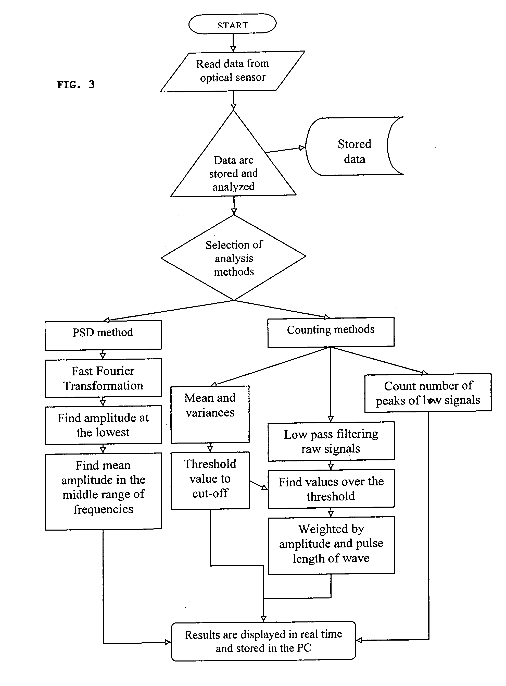
[0112] The number of fluorescent peaks are counted and correlated to the concentration of fluorescent microspheres. The diameter of the microspheres is approximately 2.5 μm and it has a 630 / 640 nm Ex / Em spectrum. A cuvette containing diluted microspheres in a final volume of 4 ml of 0.1M PBS was then subjected to simultaneous vertical and rotational motions. The vertical and rotational speeds of the cuvette movement were 0.8 inch / sec and 300 rpm, respectively. Data was acquired at a sampling rate of 100 kHz for 2 minutes. Raw data was analyzed and the result was displayed as described previously in this application, which enumerated the number of fluorescent signals whose pulse width was between 0.05-0.2 milliseconds and pulse amplitude was greater than that of mean background plus 3 times of standard deviation (mean+3 SD).

[0113] The results are shown in FIG. 9. Five different sets of experiments were carried out in different days. The fluoresc...

example 2

Detection of Salmonella by Using Polyclonal Antibodies Conjugated with Fluorescent Dyes in Phosphate Buffer

[0114]Salmonella typhimurium (ATCC #14028) was grown in 5 ml of LB media overnight at 37° C. The culture was washed and centrifuged in PBS buffer, pH 7.0 twice at 8,000×g for 10 minutes. The cell pellet was reconstituted to its original concentration in PBS. The overnight culture was close to the standard concentration of 5×10⁹ cells / ml. The overnight culture was diluted with 0.1 M PBS buffer to prepare a range of concentrations from 0 cell / ml to 106 cells / ml in 1 ml total volume of PBST (0.1 M PBS, 0.01% Tween 20). A portion of each diluted culture was plated on S. typhimurium selective agar and incubated at 37° C. for overnight to verify actual colony forming unit (CFU).

[0115] In each samples, 100 μl (approximately 105 beads, 0.86 μm in diameter) of magnetic microspheres coated with polyclonal antibodies for Salmonella was added followed by 30 minutes at room temperature wi...

example 3

Detection of Salmonella by Using Polyclonal Antibodies Conjugated with Fluorescent Dyes in Ground Beef

[0117] By using Alexa Fluor conjugated polyclonal antibodies, S. typhimurium was detected in spiked ground beef. 25 g of ground beef was added into 225 ml of PBST in a sterile stomacher bag. After stomaching at 280 rpm for 2 minutes at room temperature, an aliquot of beef homogenate was spiked with S. typhimurium in a total volume of 1 ml of PBST. The samples were incubated with magnetic beads (approximately 105 beads, 0.86 μm in diameter) coated with polyclonal antibodies for Salmonella sp. for 10 minutes at room temperature followed by 2 times of the washing step (2 minutes / each wash). The resulting pellet was resuspended with PBST and incubated with 9 μg of polyclonal antibodies for Salmonella conjugated with AlexaFluor 633 for 30 minutes at room temperature. The sample underwent the same washing step as described above before being transferred into a cuvette.

[0118] The measure...


PUM


Abstract

The present invention relates to systems and methods for real time, rapid detection, identification, and enumeration of a wide variety of analytes, which include but are not limited to, cells (Eukarya, Eubacteria, Archaea), microorganisms, organelles, viruses, proteins (recombinant or natural proteins), nucleic acids, prionss, and any chemical, metabolites, or biological markers. The systems and methods, which include the laser/optic/electronic units, the analytic software, the assay methods and reagents, and the high throughput automation, are particularly adapted to detection, identification, and enumeration of pathogens and non-pathogens in contaminated foods, clinical samples, and environmental samples. Other microorganisms that can be detected with the present invention include clinical pathogens, protozoa and, viruses.

Description

CROSS-REFERENCE TO RELATED APPLICATION [0001] This application claims priority from provisional application Ser. No. 60 / 592,320 filed Jul. 29, 2004, which is incorporated herein by reference and made a part hereof.FEDERALLY SPONSORED RESEARCH OR DEVELOPMENT [0002] Not Applicable. BACKGROUND OF THE INVENTION [0003] 1. Technical Field [0004] The present invention relates to systems and methods for real time, rapid detection, identification, and enumeration of a wide variety of analytes, which include but are not limited to, cells (Eukarya, Eubacteria, Archaea), microorganisms, organelles, viruses, proteins (recombinant or natural proteins), nucleic acids, prions, and any chemicals, metabolic, or biological markers. The systems and methods, which include the laser / optic / electronic units, the analytic software, the assay methods and reagents, and the high throughput automation, are particularly adapted to detection, identification, and enumeration of pathogens and non-pathogens in conta...

Claims


Application Information

Patent Timeline
IPC IPC(8): G06F19/00G06K9/00
CPCB82Y5/00B82Y10/00G01N33/54373
Inventor KIM, MYUNG L.IKRO, JOEPARK, HOSHIN
Owner KIM LAB INC
Why Eureka
Try Eureka