Hemangioblast progenitor cells

Inactive Publication Date: 2006-07-27
NAT UNIV OF SINGAPORE +1
View PDF8 Cites 3 Cited by
  • Summary
  • Abstract
  • Description
  • Claims
  • Application Information

AI Technical Summary

Benefits of technology

[0016] In another aspect there is provided a differentiated committed progenitor cell line that

Problems solved by technology

However, the isolation of hemangioblast from embryos and its prospective isolation have hitherto

Method used


Image

  • Hemangioblast progenitor cells
  • Hemangioblast progenitor cells
  • Hemangioblast progenitor cells

Examples


example 1

Materials and Methods

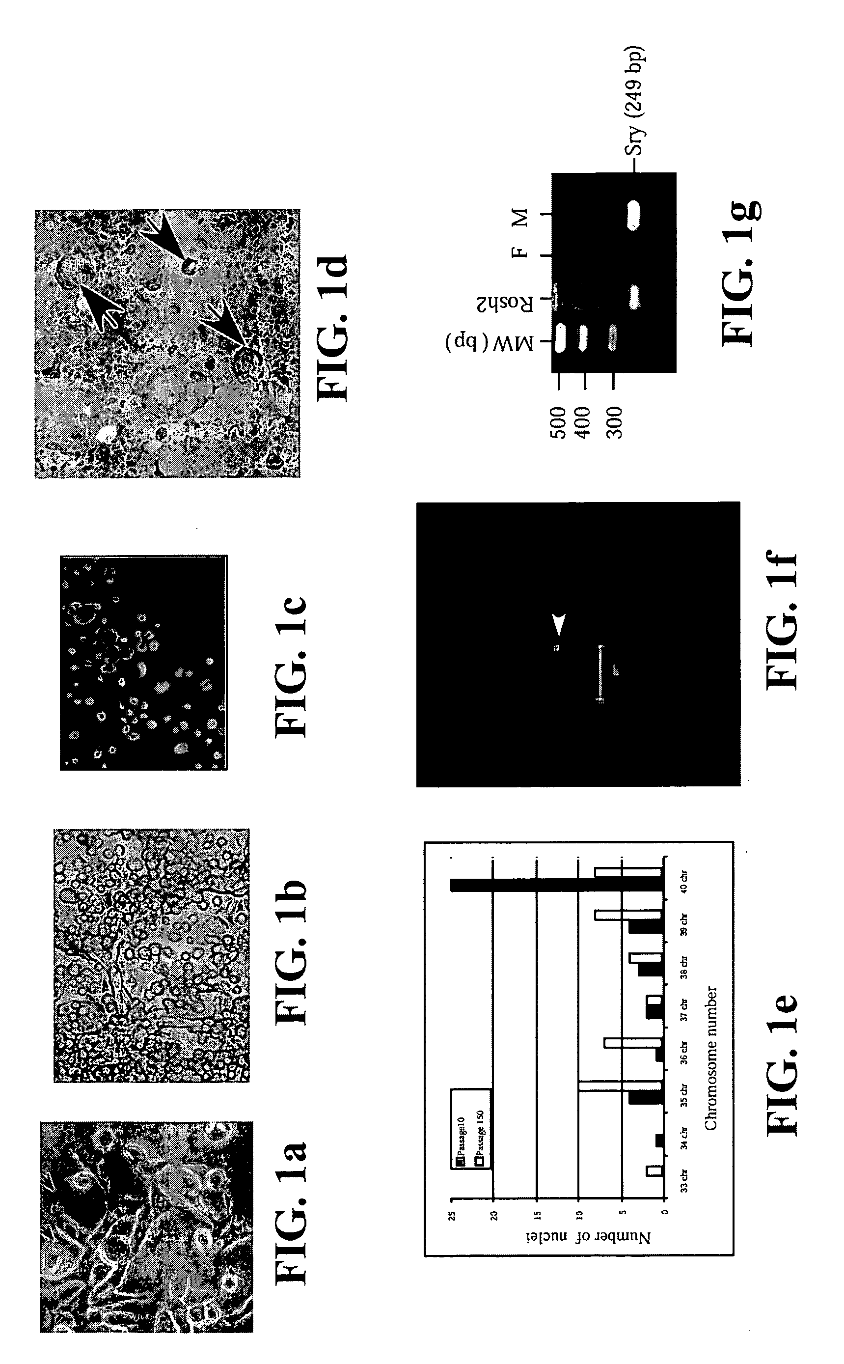
[0088] Derivation of RoSH2 cells: All animal experimentation protocols were approved by National University of Singapore Animal Ethics Research Committee. B6.129S7-GtRosa26 mice were purchased from Jackson Laboratory (Bar Harbor, Me.). 5.5 dpc delayed blastocysts and 6 to 7.5 dpc embryos were prepared as previously described (Robertson, 1987). For 6 to 7.5 dpc embryos, the egg cylinders were dissected out and were placed next to the extra-embryonic tissues for culture. The embryos were cultured on tissue culture dish in ES cell media. For the older embryos, the extra-embryonic tissues were removed after the embryos attached and started proliferation. For 5.5 dpc delayed blastocysts, when the growths reached a size visible to the naked eye, they were disaggregated into cellular clumps with trypsin and then transferred to a 1 cm culture dish with embryonic fibroblast feeder as previously described for isolation of mouse ES cells (Robertson, 1987). The cultures w...

example 2

Preparation of Hemangioblast Cell Lines from Embryonic Stem Cells

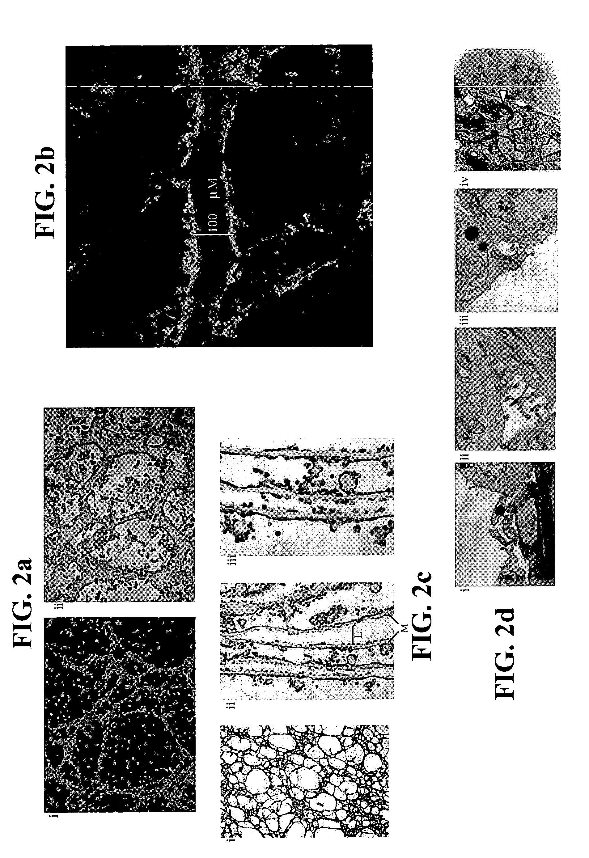
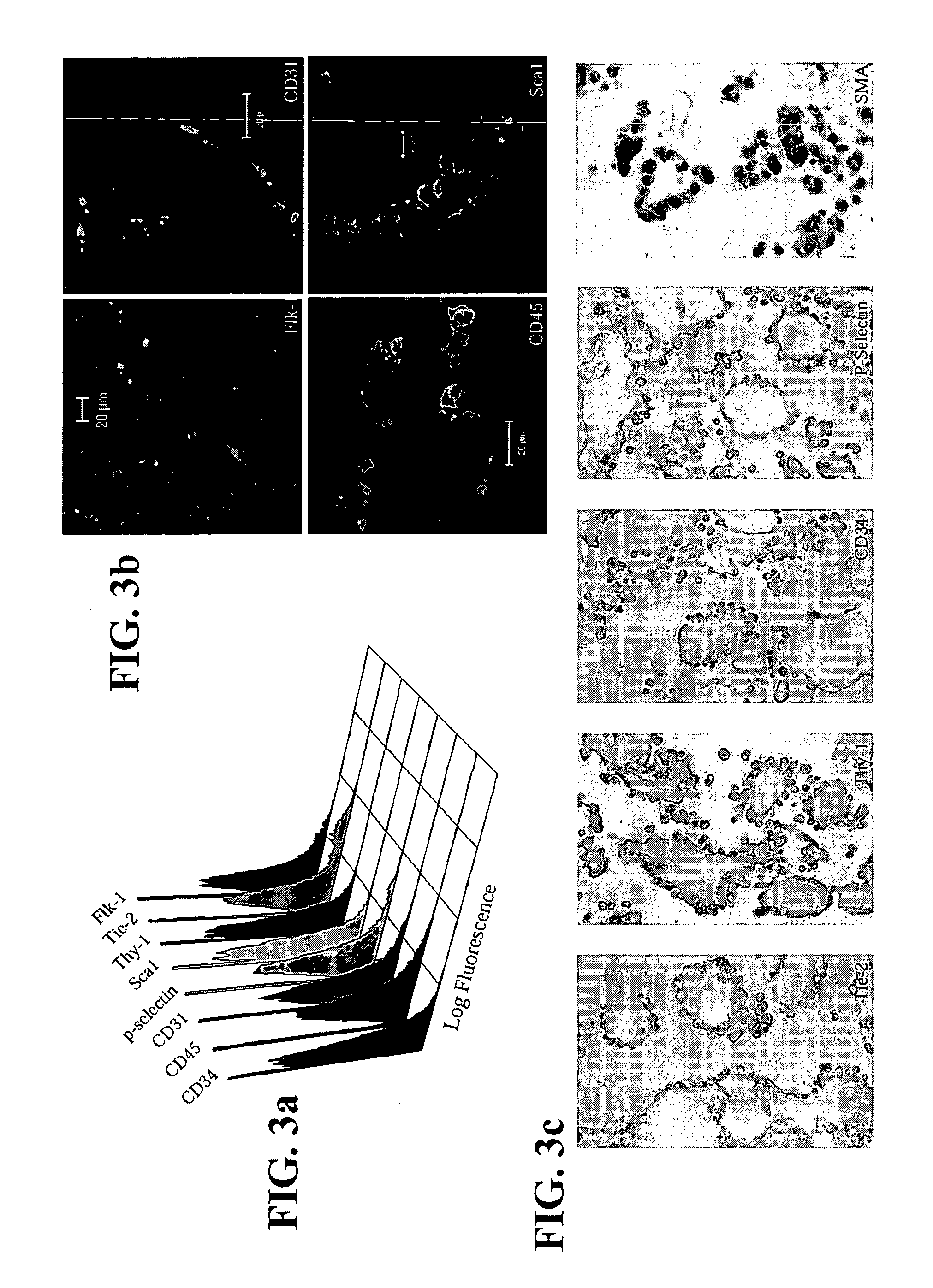
[0110] 2×10⁴ single ES cells in 100 μl ES media ES cells were cultured in 3.9 ml methycellulose media (MethoCult M3134, StemCell Technologies, Inc, Vancouver, Canada), 4.2 ml IMDM (Life Technologies, Rockville, Md.), 1.5 ml Serum, 100 μl monothioglycerol stock solution (37.8 μl in 10 ml PBS) (Sigma, St Louis, Mo.) 100 μl 100× L-glutamine / Penicillin / Streptomycin stock solution (Life Technologies, Rockville, Md.). Six days later, colonies of cells or EBs were clearly visible to the naked eyes. The EBs were then dissociated into cell suspensions by incubating the EBs in 0.15% (w / v) collagenase / PBS supplemented with 20% (v / v) FCS at 37° C. for 30 minutes and then disrupting the cell clumps by passing the solution through a syringe with a 20-gauge needle 3 times. After another 30 minutes of incubation, the disruption was repeated with a 25-gauge needle. These cells were then plated on mitomycin C-treated embryonic fibrobl...

example 3

Preparation of Hemangioblast Cell Lines from Bone Marrow

[0112] Adult bone marrow (BM) was prepared from mice, pigs and humans. For mice, BM was flushed from the femurs of B6.129S7-GtRosa26 with saline using a needle and syringe. In pigs, BM was aspirated from the femur of pigs. Human BM was harvested by scraping from the split sternum of patients undergoing CABG surgery at NUH. The common denominator in all these procedures is the preservation of some BM tissue integrity in tissue clumps of 0.1 to 1 mm3 in volume. Each piece of tissue was cultured individually on 48-well mitomycin C-treated mouse embryonic fibroblast feeder plates in ES cell media. Most of the BM pieces attached to the plates within 24 hours. The cultures were maintained with changes of fresh media every two to four days. Over a period of one week, cells appeared to migrate out of the BM pieces. During the first week, the culture was a complex mix of cell types with much cell proliferation and cell death occurring...


PUM

PropertyMeasurementUnit
Therapeuticaaaaaaaaaa

Abstract

The invention relates to isolated hemangioblast cells. Hematopoietic and endothelial cells are postulated to be derived from a common progenitor, hemangioblast. While hemangioblast has been isolated retrospectively during embryonic stem cell differentiation, it has not been isolated from embryos or from bone marrow. Prospectively stable clonal cell lines have been isolated from mammalian embryos, from embryonic stem cells and from mammalian bone marrow that can differentiate in vitro into tubular structures with both endothelial and hematopoietic markers such as CD34, CD31, Flk-1, TIE2, P-selectin, Sca-1, thy-1, CD45, and smooth muscle actin. Gene expression profiles in the undifferentiated and differentiated cells were consistent with endothelial and hematopoietic differentiation potential. Transplantation studies in isogenic or immunodeficient mice demonstrated that these cells were not tumorigenic. In an appropriate microenvironment, the cells incorporate into the vasculature and participate in hematopoiesis.

Description

FIELD OF THE INVENTION [0001] The present invention relates to the derivation of hemangioblast cell lines which have the potential to differentiate into hematopoietic and endothelial cells in vitro and in vivo. BACKGROUND OF THE DISCLOSURE [0002] Hematopoiesis and vasculogenesis are closely associated events that develop in tandem spatially and temporally during embryogenesis (Murray, 1932; Sabin, 1920). Primitive hematopoiesis and the establishment of the yolk sac vasculature occur simultaneously when mesodermal cells in the presumptive yolk sac proliferate and differentiate to form vascular structures with primitive erythroblasts known collectively as blood islands. Hematopoiesis during mouse development is well characterized (Keller et al., 1999). Blood islands are visible in the yolk sac at 7.5 days post coitus (dpc). By 11.5 dpc, the fetal liver displaces the yolk sac as the major site of hematopoiesis in mouse embryo and also signifies the switchover to definitive hematopoiesi...

Claims


Application Information

Patent Timeline
IPC IPC(8): A61K48/00C12N5/08A61K35/12C12N5/02C12N5/074C12N5/0789
CPCA61K35/12A61K48/00A61K2035/124C12N5/0647C12N5/0692C12N2506/02C12N2502/13
Inventor LIM, SAI
Owner NAT UNIV OF SINGAPORE
Why Eureka
Try Eureka