Label-free methods for performing assays using a colorimetric resonant reflectance optical biosensor

a biosensor and colorimetric resonant technology, applied in the field of label-free methods for performing assays using a colorimetric resonant reflectance optical biosensor, can solve the problem that methods have yet to yield commercially available high-throughput instruments, and achieve the effect of relative effectiveness

Inactive Publication Date: 2006-12-14
X BODY
View PDF99 Cites 38 Cited by
  • Summary
  • Abstract
  • Description
  • Claims
  • Application Information

AI Technical Summary

Benefits of technology

[0010] Another embodiment of the invention provides a method of detecting inhibition activity of one or more molecules against enzymes or binding partners that affect or bind specific binding substances, wherein the specific binding substances are immobilized on a surface of a colorimetric resonant reflectance optical biosensor. The method comprises detecting a colorimetric resonant reflectance optical PWV of a distinct location; applying one or more molecules suspected of having inhibition activity to the distinct location; applying one or more enzymes or binding partners to the distinct location; detecting the colorimetric resonant reflectance optical PWV of the distinct location; and comparing the initial PWV above with the subsequent PWV above. Alternatively, the one or more molecules suspected of having inhibition activity can be mixed with the one or more enzyme or binding partners, which, together, can be applied to the distinct location. The inhibition activity of one or more molecules against enzymes or binding partners which effect or bind one or more specific binding substance is detected. A decrease in the initial colorimetric resonant reflectance optical PWV above in relation to the subsequent colorimetric resonant reflectance optical PWV above is (1) a relative measure of the proportion of specific binding substance that is released from the biosensor surface or binding partners bound to the surface of the biosensor or (2) a measure of relative effectiveness of one or more molecules suspected of having inhibition activity.

Problems solved by technology

With the completion of the sequencing of the human genome, one of the next grand challenges of molecular biology will be to understand how the many protein targets encoded by DNA interact with other proteins, small molecule pharmaceutical candidates, and a large host of enzymes and inhibitors.
However, to date, these methods have yet to yield commercially available high-throughput instruments that can perform high sensitivity assays without any type of label in a format that is readily compatible with the microtiter plate-based or microarray-based infrastructure that is most often used for high-throughput biomolecular interaction analysis.

Method used


Image

  • Label-free methods for performing assays using a colorimetric resonant reflectance optical biosensor
  • Label-free methods for performing assays using a colorimetric resonant reflectance optical biosensor
  • Label-free methods for performing assays using a colorimetric resonant reflectance optical biosensor

Examples


example 1

Immobilized Protein Detection

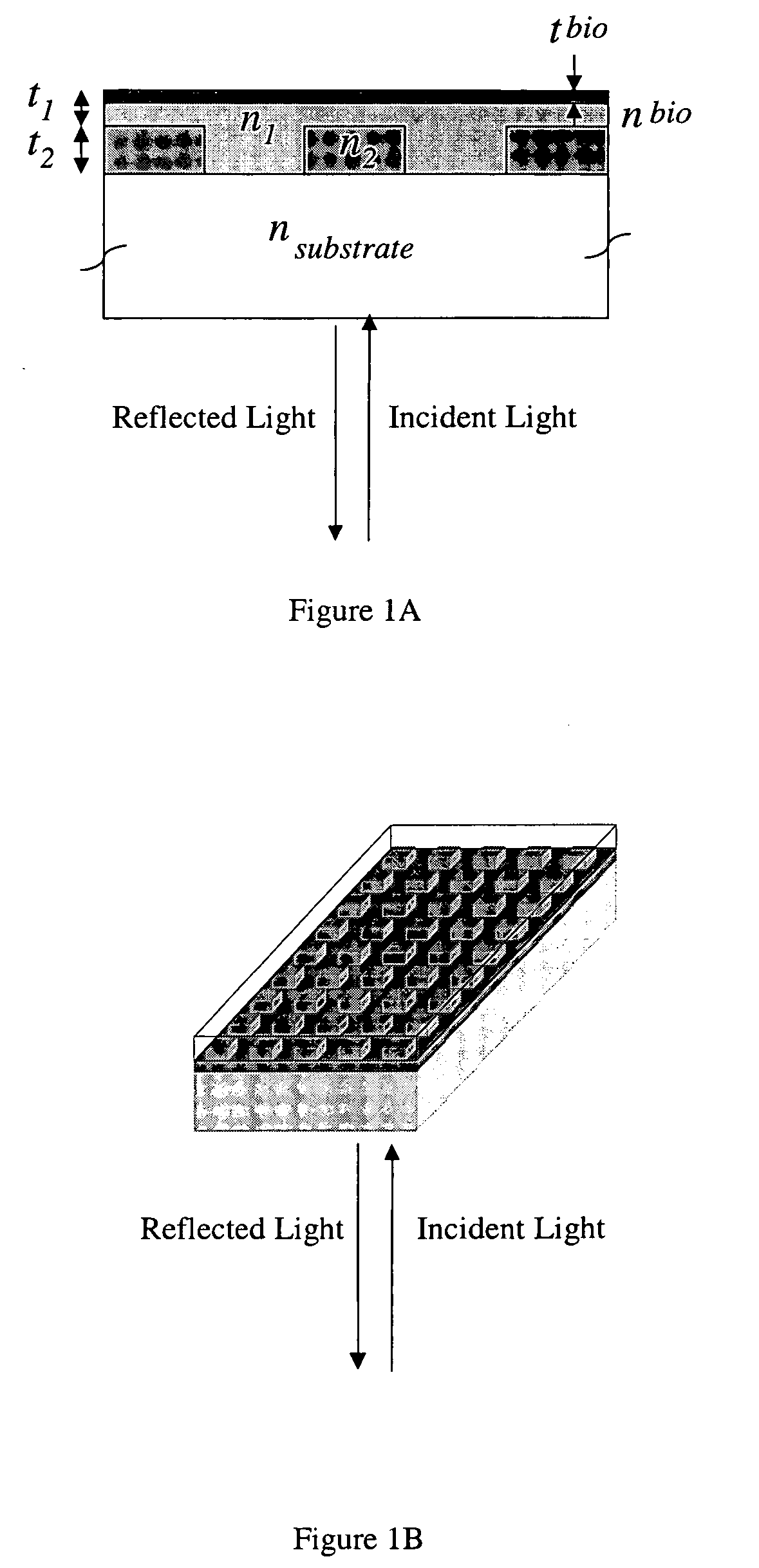
[0168] In order to demonstrate a biosensor's ability to quantify biomolecules on its surface, droplets of BSA dissolved in H2O at various concentrations were applied to a biosensor as shown in FIG. 1. The 3 μl droplets were allowed to dry in air, leaving a small quantity of BSA distributed over a ˜2 mm diameter area. The peak resonant wavelength of each biosensor location was measured before and after droplet deposition, and the peak wavelength shift was recorded. See FIG. 34.

example 2

Immobilization of One or More Specific Binding Substances

[0169] The following protocol was used on a colorimetric resonant reflective biosensor to activate the surface with amine functional groups. Amine groups can be used as a general-purpose surface for subsequent covalent binding of several types of linker molecules.

[0170] A glass substrate biosensor of the invention is cleaned by immersing it into piranha etch (70 / 30% (v / v) concentrated sulfuric acid / 30% hydrogen peroxide) for 12 hours. The biosensor was washed thoroughly with water. The biosensor was dipped in 3% 3-aminopropyltriethoxysilane solution in dry acetone for 1 minute and then rinsed with dry acetone and air-dried. Alternatively, immersion of the biosensor in 10% 3-aminopropyltriethoxysilane (Pierce) solution in ethanol (Aldrich) for 1 min, followed by a brief ethanol rinse. Activated sensors were then dried at 70° C. for 10 minutes. The biosensor was then washed with water.

[0171] A semi-quantitative method is use...

example 3

IgG Assay

[0178] As an initial demonstration for detection of biochemical binding, an assay was performed in which a biosensor was prepared by activation with the amino surface chemistry described in Example 2 followed by attachment of a biotin linker molecule. The biotin linker is used to specifically interact with and effectively tether a streptavidin receptor molecule to the surface by exposure to a 50 μg / ml concentration solution of streptavidin in PBS at room temperature for 2-4 hours. The streptavidin receptor is capable of binding any biotinylated protein to the biosensor surface. For this example, 3 μl droplets of biotinylated anti-human IgG in phosphate buffer solution (PBS) were deposited onto 4 separate locations on the biosensor surface at a concentration of 200 μg / ml. The solution was allowed to incubate on the biosensor for 30 minutes before rinsing thoroughly with PBS. The peak resonant wavelength of the 4 locations were measured after biotin activation, after strept...


PUM

PropertyMeasurementUnit
diameteraaaaaaaaaa
diameteraaaaaaaaaa
molecular weightaaaaaaaaaa

Abstract

Methods are provided for detecting biomolecular interactions. The use of labels is not required and the methods can be performed in a high-throughput manner. The invention also relates to optical devices.

Description

PRIORITY [0001] This application is a divisional of U.S. application Ser. No. 10 / 237,641, filed Sep. 9, 2002, allowed, which is a continuation-in-part of U.S. application Ser. No. 10 / 227,908, filed Aug. 26, 2002, abandoned, and U.S. application Ser. No. 10 / 180,374, filed Jun. 26, 2002, now U.S. Pat. No. 7,023,544, and U.S. application Ser. No. 10 / 180,647, filed Jun. 26, 2002, which are continuations-in-part of U.S. application Ser. No. 10 / 059,060, filed Jan. 28, 2002, now U.S. Pat. No. 7,070,987, and U.S. application Ser. No. 10 / 058,626, filed Jan. 28, 2002, now U.S. Pat. No. 6,951,715, which are continuations-in-part of U.S. application Ser. No. 09 / 930,352, filed Aug. 15, 2001, now U.S. Pat. No. 7,094,595, which claims the benefit of U.S. provisional application 60 / 244,312 filed Oct. 30, 2000; U.S. provisional application 60 / 283,314 filed Apr. 12, 2001; and U.S. provisional application 60 / 303,028 filed Jul. 3, 2001, all of which are incorporated herein by reference in their entiret...

Claims


Application Information

Patent Timeline
Patent Type & Authority Applications(United States)
IPC IPC(8): C12Q1/70C12Q1/68G01N33/53G01N33/554C12M1/34G01N33/569C40B30/06B01L3/00G01N21/25G01N21/47G01N33/543G02B5/18
CPCB01J2219/00315Y10S436/805B01J2219/00605B01J2219/00612B01J2219/00626B01J2219/00637B01J2219/00702B01J2219/0072B01J2219/00722B01J2219/00725B01J2219/00743B01L3/5085B01L2300/0636B01L2300/0829C40B60/12G01N21/253G01N21/4788G01N21/774G01N21/7743G01N33/54373G02B5/008G02B5/1809Y10S436/809Y10S436/81B01J2219/00511
Inventor LIN, BOPEPPER, JANECUNNINGHAM, BRIAN T.GERSTENMAIER, JOHNLI, PETERQIU, JEANPIEN, HOMER
Owner X BODY
Why Eureka
Try Eureka