Comparative genomic hybridization

a genomic hybridization and comparative technology, applied in the field of cytogenetics, can solve the problems of serious mental and physical defects, fetal death, and inability to visualize interphase chromosomes, and achieve the effect of improving the detection sensitivity

Inactive Publication Date: 2006-12-28
RGT UNIV OF CALIFORNIA
View PDF4 Cites 11 Cited by
  • Summary
  • Abstract
  • Description
  • Claims
  • Application Information

AI Technical Summary

Benefits of technology

[0083] Another object of this invention is to provide a method of analyzing cells from a suspected lesion at an early stage of development. An advantage of the methods of this invention is that only a few cells are necessary for the analysis. The early detection of amplifications and / or deletions in cells from a lesion allow for early therapeutic intervention that can be tailored to the extent of, for example, invasiveness known to be associated with such genetic rearrangements. Further, such early detection provides a means to associate the progression of the cells with the genetic rearrangements therein detected by the methods of this invention.
[0085] It is another object of this invention to provide methods of detecting amplifications and / or deletions of nucleic acid sequences wherein certain cell lines termed herein “antenna cell lines”, are used to enhance the sensitivity of the detection.

Problems solved by technology

Abnormalities that involve deletions or additions of chromosomal material alter the gene balance of an organism and generally lead to fetal death or to serious mental and physical defects.
Condensed chromosomes are used because, until recently, it has not been possible to visualize interphase chromosomes due to their dispersed condition and the lack of visible boundaries between them in the cell nucleus.
Unfortunately, such conventional banding analysis requires cell culturing and preparation of high quality metaphase spreads, which is time consuming and labor intensive, and frequently difficult or impossible.
For example, cells from many tumor types are difficult to culture, and it is not clear that the cultured cells are representative of the original tumor cell population.
With the use of such probes, the presence or absence of specific abnormalities can be very efficiently determined; however, it is tedious to test for numerous possible abnormalities or to survey to discover new regions of the genome that are altered in a disease.
A major limitation of Southern analysis and related conventional techniques for analysis of gene amplification is that only specific sites are studied leaving the vast majority of the genome unexamined.
Conventional cytogenetic studies, on the other hand, provide a broad survey of the genome but provide little information about genes that may be involved in amplification events.
Identification of commonly occurring chromosomal changes by analysis of metaphase spreads is often difficult or impossible using conventional banding analysis because of the complexity of the rearrangements and because of the poor quality of the metaphase preparations.
A limitation of such hybridizations is that cytogenetic information is only provided from the regions to which the probes bind.
Such hybridizations are very useful for determining if a particular abnormality is present, for example, the deletion of a specific gene or a duplication among other abnormalities, but it is laborious to search for currently unknown abnormalities on a region by region basis.
Other methods of searching for unknown genetic abnormalities similarly require a lot of work.
Since HSRs in tumors are most often not at the site of the amplified gene(s) in normal cells, standard cytogenetics does not yield any information that could assist with identification of the gene(s).
Discovery of genetic changes involved in the development of solid tumors has proven difficult.
RFLP is also restricted by the availability and informativeness of polymorphic probes.

Method used


Image

  • Comparative genomic hybridization
  • Comparative genomic hybridization
  • Comparative genomic hybridization

Examples


example 1

DNA from Breast Cancer Lines Hybridized to Normal Metaphase Spreads

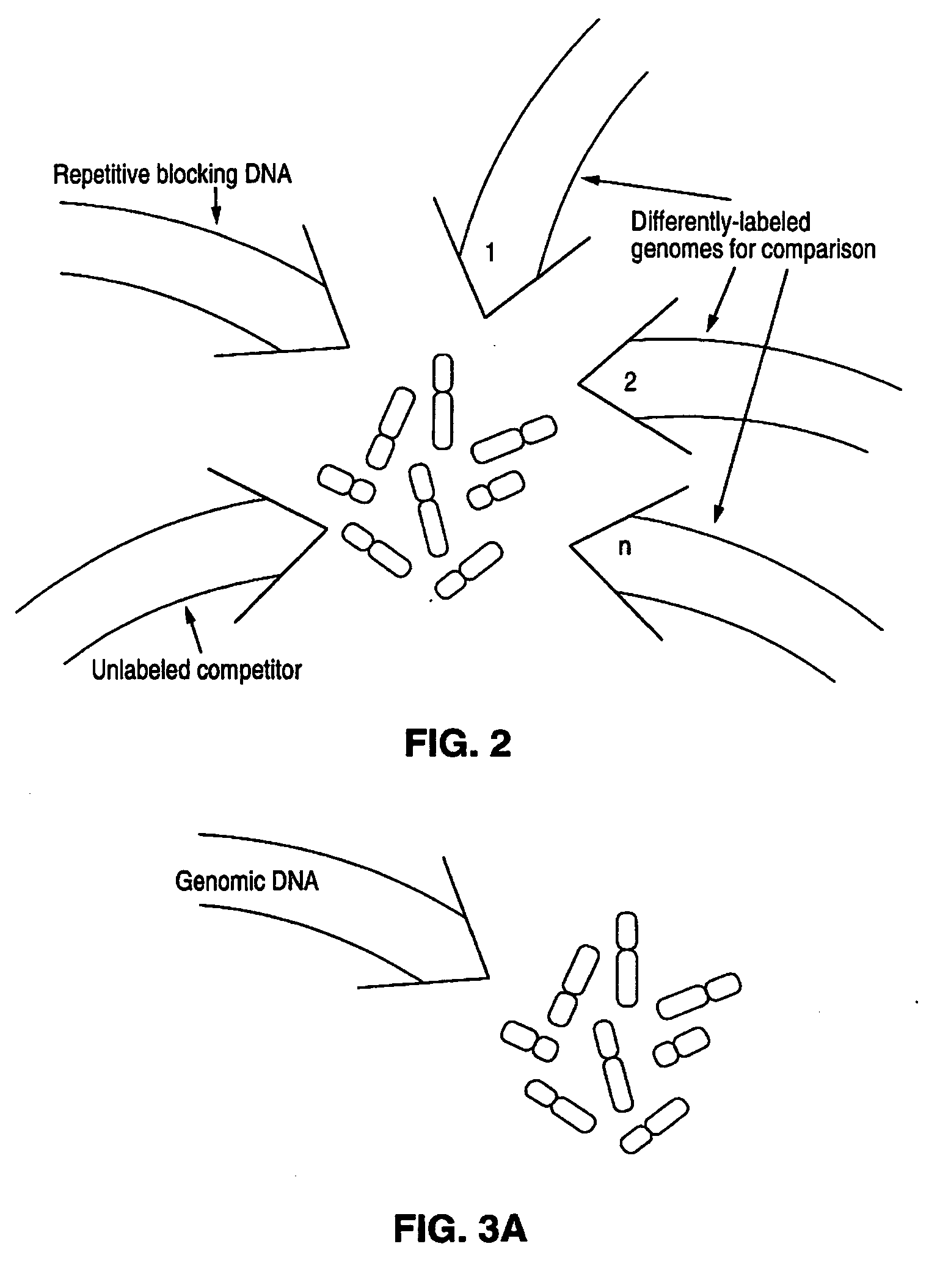
[0249] In this Example, methods of this invention to analyse genomes by Comparative Genomic Hybridization (CGH) are exemplified by hybridizations of breast cancer cell lines to normal metaphase spreads. The target metaphase spreads were pre-hybridized with unlabeled human placental DNA to block the high copy repeat sequences. In this representative example, the hybridization mixture containing the extracted labeled DNA from the cell lines contained unlabeled, repeat-enriched Cot-1 blocking DNA [obtained from Bethesda Research Laboratories (BRL), Gaithersburg, Md. (USA].

[0250] The experiments outlined below include in the hybridization mixture for the subject genomes, that is, the breast cancer cell line DNAs, chromosome-specific repeat sequence probes and chromosome-specific painting probes. Those probes labeled with biotin were included as an adjunct for identifying chromosomes in the metaphase preparations. The e...

example 2

[0277] Hybridizations with two different labeled subject DNAs as schematically outlined in FIGS. 6 and 7 were performed. One of the labeled subject DNAs hybridized was a cell line DNA as described in Example 1 and similarly labeled. The other labeled subject DNA was human genomic DNA labeled with biotin-14-dATP.

[0278] The protocols were essentially the same as in Example 1 except that no chromosome-specific reference probes were used, and the same amount of the labeled human DNA as the labeled cell line DNA, that is, 60 ng, was hybridized. Of course, reference probes could be added to the hybridization mixture, but they need to be differently labeled to be distinguishable.

[0279] The results showed the normal DNA with a red signal and the cell line DNA with a green signal. The green to red ratios were determined along each chromosome. Amplification was indicated by an area where the signal was predominantly green whereas deletions were indicated by more red signals than in other ar...

example 3

Copy Number Karyotypes of Tumor DNA

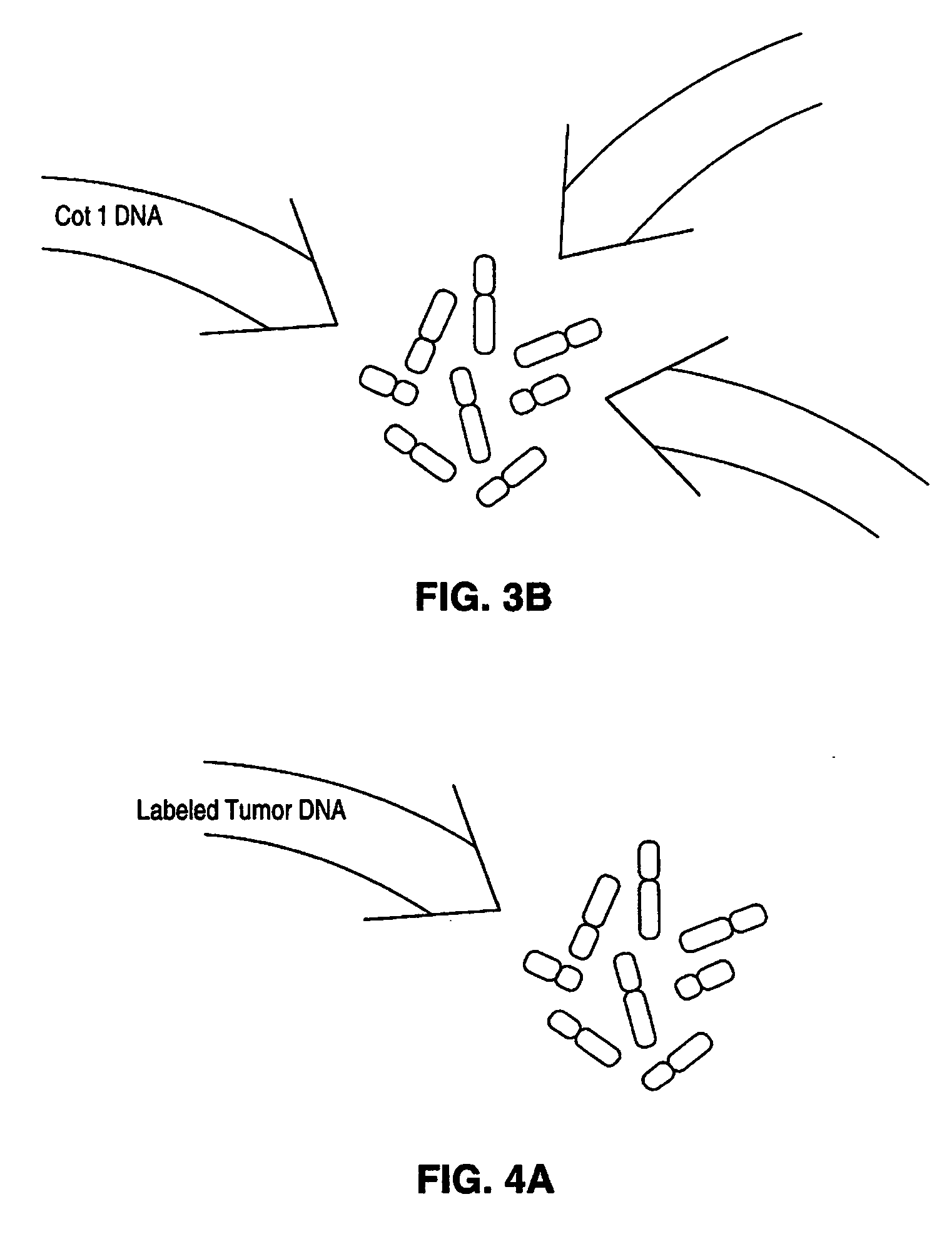
[0284] In the representative experiments of CGH in this example, biotinylated total tumor DNA (cell line and primary tumor DNA) and digoxigenin-labeled normal human genomic DNA are simultaneously hybridized to normal human metaphase spreads in the presence of unlabeled blocking DNA containing high-copy repetitive sequences, specifically unlabeled Cot-1 blocking DNA [BRL, Gaithersburg, Md. (USA)]. The following paragraphs detail the procedures used for the representative CGH experiments of this example.

DNA Labeling:

[0285] DNAs used in this example were labeled essentially as shown above in Example 1. DNAs were labeled with biotin-14-dATP or digoxigenin-11-dUTP by nick translation [Rigby et al., supra; Sambrook et al., supra]. The optimal size for double stranded probe fragments after labeling was 600-1000 bp.

Pretreatment of Metaphase Spreads:

[0286] Lymphocyte metaphase preparations were denatured, dehydrated and air dried, treated with Protei...


PUM

PropertyMeasurementUnit
polarityaaaaaaaaaa
frequencyaaaaaaaaaa
physicalaaaaaaaaaa

Abstract

Disclosed are new methods comprising the use of in situ hybridization to detect abnormal nucleic acid sequence copy numbers in one or more genomes wherein repetitive sequences that bind to multiple loci in a reference chromosome spread are either substantially removed and / or their hybridization signals suppressed. The invention termed Comparative Genomic Hybridization (CGH) provides for methods of determining the relative number of copies of nucleic acid sequences in one or more subject genomes or portions thereof (for example, a tumor cell) as a function of the location of those sequences in a reference genome (for example, a normal human genome). The intensity(ies) of the signals from each labeled subject nucleic acid and / or the differences in the ratios between different signals from the labeled subject nucleic acid sequences are compared to determine the relative copy numbers of the nucleic acid sequences in the one or more subject genomes as a function of position along the reference chromosome spread. Amplifications, duplications and / or deletions in the subject genome(s) can be detected. Also provided is a method of determining the absolute copy numbers of substantially all RNA or DNA sequences in subject cell(s) or cell population(s).

Description

RELATED APPLICATIONS [0001] This application is a continuation-in-part of U.S. Ser. No. 07 / 846,659, filed Mar. 4, 1992, now pending. This application is also a continuation-in-part of U.S. Ser. No. 07 / 670,242, filed Mar. 15, 1991, now pending, which is a continuation-in-part of U.S. Ser. No. 07 / 659,974, filed Feb. 22, 1991, now pending, which is a continuation-in-part of U.S. Ser. No. 07 / 537,305, filed Jun. 12, 1990, now pending, which is a continuation-in-part of U.S. Ser. No. 07 / 497,098, filed Mar. 20, 1990, now pending, which is a continuation-in-part of U.S. Ser. No. 07 / 444,669, filed Dec. 1, 1989, now abandoned, which is a continuation-in-part of U.S. Ser. No. 06 / 937,793, filed Dec. 4, 1986, now abandoned, of which U.S. Ser. No. 07 / 627,707, filed Dec. 14, 1990 is a now pending continuation, and which is a continuation-in-part of U.S. Ser. No. 06 / 819,314 filed Jan. 16, 1986, now abandoned. Priority is claimed in said prior filed applications.[0002] This invention was made with G...

Claims


Application Information

Patent Timeline
Patent Type & Authority Applications(United States)
IPC IPC(8): C12Q1/68C12P19/34C12N15/09C07H21/04
CPCC12Q1/6809C12Q1/6841C12Q1/6853C12Q1/6886C12Q2600/158C12Q2545/114C12Q2545/113C12Q2537/143C12Q2525/151C12Q2563/107
Inventor PINKEL, DANIELGRAY, JOE W.KALLIONIEMI, ANNEKALLIONIEMI, OLLI-PEKKAWALDMAN, FREDERIC
Owner RGT UNIV OF CALIFORNIA
Why Eureka
Try Eureka