Method for cell implantation

a cell implantation and transplantation technology, applied in the direction of skeletal/connective tissue cells, biocide, plant growth regulators, etc., can solve the problems of inferior quality of repair tissue, inability to use as a scaffold, and tisseel® not improving the repair of osteochondral defects

Inactive Publication Date: 2007-04-05
INTERFACE BIOTECH
View PDF1 Cites 19 Cited by
  • Summary
  • Abstract
  • Description
  • Claims
  • Application Information

AI Technical Summary

Benefits of technology

[0033] The invention encompasses the utilization of either implantation of cells simultaneously with a supporting material, into which the cells are mixed during the application,—in the first part of the invention—performed either under a fluid cover, using the general usage of an arthroscope where a fluid is used to keep the joint open for visualization, or as a second part of the invention also in a combination with an arthroscope, which provides a certain pressure of guided sterile air or slight positive pressure to help the surgeon visualize the area under treatment, such as for instance used in other endoscopes—for instance for abdominal exams, etc., where air is supplied under a certain pressure—such as for instance a combination of atmospheric air combined with CO2, in a concentration well tolerated by the mixed cell / coagulating support material. In this second part of the invention, the “cover” used, under which the mixed cell / coagulation support material may be placed under the air pocket, where the pressure may be around 20 -30 mm Hg.
[0045] The method developed by the present inventor is considered novel, due to the new concept of placing a chondrocyte-containing substance at a controlled area in for instance a cartilage defect, such as a chondral defect, an osteochondral defect, or even when applying cells such as osteoblasts / osteocytes in a substance such as for instance hydroxyl-apatite, as a thin bottom layer in a joint suffering from osteoarthritis, and thereafter being able to apply a second layer of chondrocyte / substance as well as when applying cells through an endoscopic procedure to a target organ or target area, such as for instance liver, heart, etc. The substance shall be of a nature that at the same time is not toxic to the cells, and in case of chondrocytes, the substance applied under fluid, shall not be toxic to the chondrocytes or may even be promoting chondrocyte in- growth and / or in a maturation stage in which the cell also produces the matrix structure, that ends up being the new cartilage.

Problems solved by technology

Mats Brittberg et al. have shown that a fibrin adhesive such as Tisseel® with, or without Growth hormone does not have a chondrogenic effect on immature chondrocytes, and it is therefore not suitable as a scaffold to promote repair of chondral or osteochondral defects (Brittberg, M, Sjögren-Jansson, Lindahl, A, Peterson, L, The influence of fibrin sealant on Osteochondral Defect Repair in the Rabbit Knee, from Mats Brittberg's Thesis, Cartilage Repair.
They postulated that fibrin adhesive chemically enhanced cell proliferation, but the repair tissue formed was of inferior quality.
They concluded that Tisseel® did not improve the repair of osteochondral defect, whereas autologous or homologous fibrin clots used as support appeared to show beneficial effect sufficient to merit further in vivo exams.
Methods using scaffold technologies of various forms, where the scaffold (with, or without cells grown in the scaffold) is inserted into the defect, have suffered from difficulties in performing the cell implantation procedure solely guided by arthroscopy.

Method used


Image

  • Method for cell implantation
  • Method for cell implantation

Examples


example 1

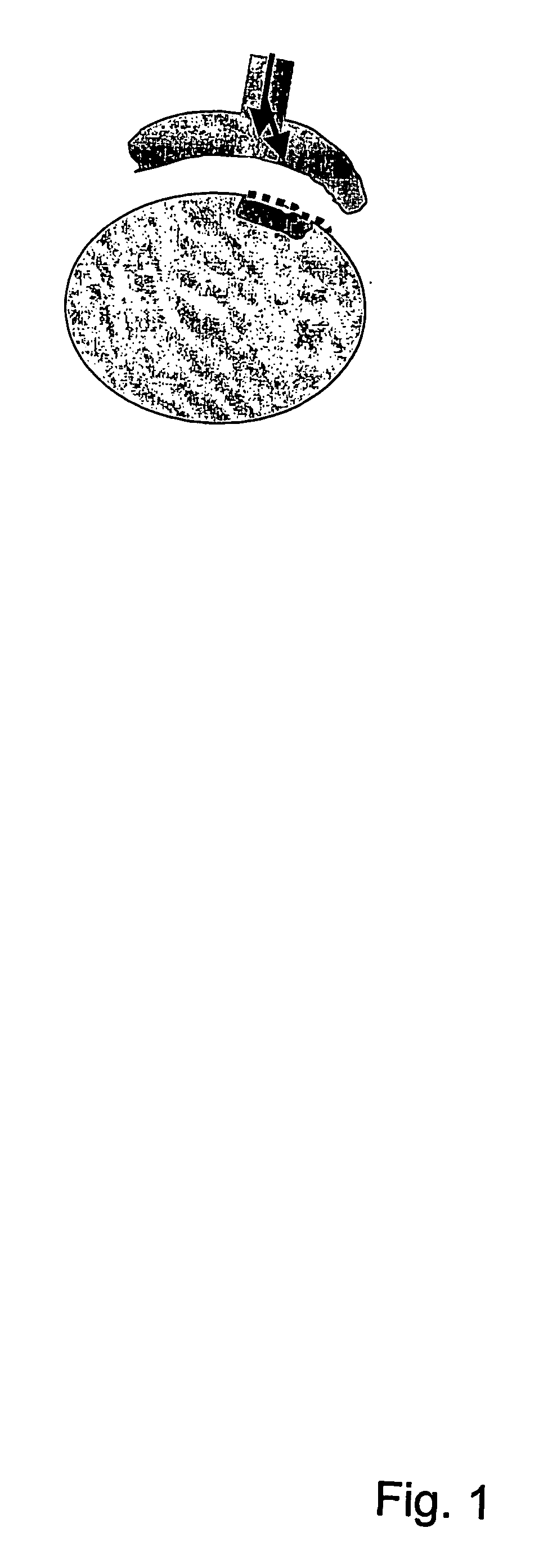
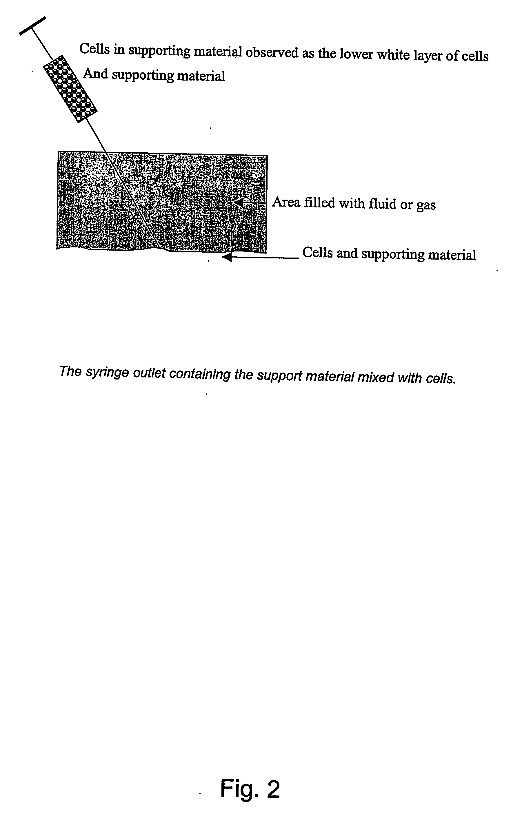
[0101] A joint derived from an animal, such as for instance a knee joint from a pig or a horse or other animals are subjected to arthroscopy and fluid such as 0.9% sterile saline or a gas is filling up the joint to visualize the area targeted for the arthroscopic repair of the area as for instance a cartilage defect in order to enable the application of the supporting material mixed with the cells in such a manner that the cell / supporting material does not mix with the fluid or gas applied via the arthroscope, and with a retained capability to adhere to the target area.

[0102] Alternatively, the cells and or the cells mixed or grown in a hydroxy-apatite suspension (e.g., Bio-Oss), may be delivered through the syringe, and be mixed at the outlet of the syringe into the area to be treated.

example 2

[0103] In this example, the support material may be placed—under pressure from the other liquid, prior to the implantation of the cells. In this case, the supporting material may be placed below the other liquid for instance kept in place by the pressure exerted via the arthroscope until solidification. The cells are then injected at any time after the placement of the liquid substance or after the solidification has occurred, by simply injecting the cells into the supporting material kept in place by the fluid pressure.

example 3

Arthroscopic Autologous Chondrocyte Implantation, Pre-clinical, Combined with Second Fibrin Sealant

First Surgical Arthroscopic Intervention

[0104] Five goats are used for the pre-clinical trial. The goats are subjected to two (2) surgical interventions. The first surgical intervention is an arthroscopic harvest of cartilage biopsy from the knee of the goats. The goats are kept at an approved animal facility at Aalborg Sygehus Syd, Denmark. Following anaesthesia, arthroscopy is performed by either a antero-medial or antero-lateral entrance through a small incision. Two additional incisions are prepared on the medial and the lateral side of the knee. Using an elevator, a 50-200 mg cartilage is removed by a sharp elevator, and aseptically transferred to a Transport tube containing sterile Dulbecco MEM / F12 and fetal calf serum. Using a punching instrument, a 0.6 cm (diameter) circular full thickness cartilage lesion is made, to be used for the implantation of cells 3-5 weeks later. T...


PUM

PropertyMeasurementUnit
pressureaaaaaaaaaa
thicknessaaaaaaaaaa
diameteraaaaaaaaaa

Abstract

An endoscopic method for treating cartilage or bone defects in an animal, said method comprising the steps of: I) identifying the position of the defect, ii) applying cells selected from the group consisting of chondrocytes, chondroblasts, osteocytes and osteoblasts and combinations thereof into the cartilage or bone defect. In particular, the invention relates to a method for arthroscopic or endoscopic implantation of homologous or autologous cells into a defect of an animal body, the method comprising a step of I) arthroscopic or endoscopic application of a fluid to a cavity or surface containing the defect, and the steps of ii) application of the cells to the defect substantially simultaneously with a support material, the application being performed at the defect covered by the fluid, iii) mixing of the cells and the supporting material, iv) solidification of the supporting material so that the defect is covered by a mixture of cells and support material without any significant amount of fluid, and v) optionally, removal of the fluid from the cavity or surface by drainage or suction.

Description

FIELD OF THE INVENTION [0001] The present invention relates to a method for cell implantation or transplantation involving the use of an arthroscope or an endoscope. The method enables arthroscopic and / or endoscopic implantation of cells and is an easy, safe and cheap method. BACKGROUND OF THE INVENTION [0002] Mats Brittberg et al. have shown that a fibrin adhesive such as Tisseel® with, or without Growth hormone does not have a chondrogenic effect on immature chondrocytes, and it is therefore not suitable as a scaffold to promote repair of chondral or osteochondral defects (Brittberg, M, Sjögren-Jansson, Lindahl, A, Peterson, L, The influence of fibrin sealant on Osteochondral Defect Repair in the Rabbit Knee, from Mats Brittberg's Thesis, Cartilage Repair. On cartilaginous tissue engineering with the emphasis on chondrocyte transplantation, Dep. Of Orthopedics, Institute of Surgical Sciences and Dep. Of Clinical Chemistry, Institute of Laboratory Medicine, Göteborg University, Swe...

Claims


Application Information

Patent Timeline
Patent Type & Authority Applications(United States)
IPC IPC(8): A61K35/32A61B1/00A61B17/00A61K35/12A61L27/38A61L27/54C12N5/077
CPCA61B17/00491A61K35/12A61K35/32A61L27/3817A61L27/3821A61L27/3847A61L27/3852A61L27/54A61L2300/414A61L2300/418C12N5/0655C12N2533/54C12N2533/56
Inventor OSTHER, KURT
Owner INTERFACE BIOTECH
Why Eureka
Try Eureka