Optimized proteins that target Ep-CAM

a technology of epithelial cells and proteins, applied in the field of optimized proteins that target epithelial cells, can solve the problems of unsatisfactory potency of curretly available antibodies as anti-cancer agents, limited in vivo accessibility of ep-cam, and other problems, to achieve the effect of increasing activation, increasing activation, and increasing binding to an activating cell

Inactive Publication Date: 2007-05-31
XENCOR
View PDF0 Cites 49 Cited by
  • Summary
  • Abstract
  • Description
  • Claims
  • Application Information

AI Technical Summary

Benefits of technology

[0033] In other aspects, the Fc modification not substantially increase activation of natural killer cells or specifically increases activation by the antibody of dendritic cells.
[0034] In further aspects, the modification increases binding to an activating Fc receptor and does not increase binding to FcγRIIb. In cerain aspects, the modification specifically increases monocyte or macrophage phagocytosis.
[0035] The present invention provides variant Ep-CAM targeting proteins that are optimized for a number of therapeutically relevant properties. A variant Ep-CAM targeting protein comprises one or more amino acid modifications relative to a parent Ep-CAM targeting protein, wherein the amino acid modification(s) provide one or more optimized properties.

Problems solved by technology

It has been postulated that the differential localization of Ep-CAM in normal cells (baso-lateral surface) as compared with cancer cells, accounts for limited in vivo accessibility of Ep-CAM in normal tissues (McLaughlin et al.
Yet another level of complexity is the existence of a number of FcγR polymorphisms in the human proteome.
Despite this arsenal of anti-tumor weapons, the potency of curretly available antibodies as anti-cancer agents is unsatisfactory, particularly given their high cost.

Method used


Image

  • Optimized proteins that target Ep-CAM
  • Optimized proteins that target Ep-CAM
  • Optimized proteins that target Ep-CAM

Examples


example 1

Anti-Ep-CAM Antibodies with Reduced Immunogenicity

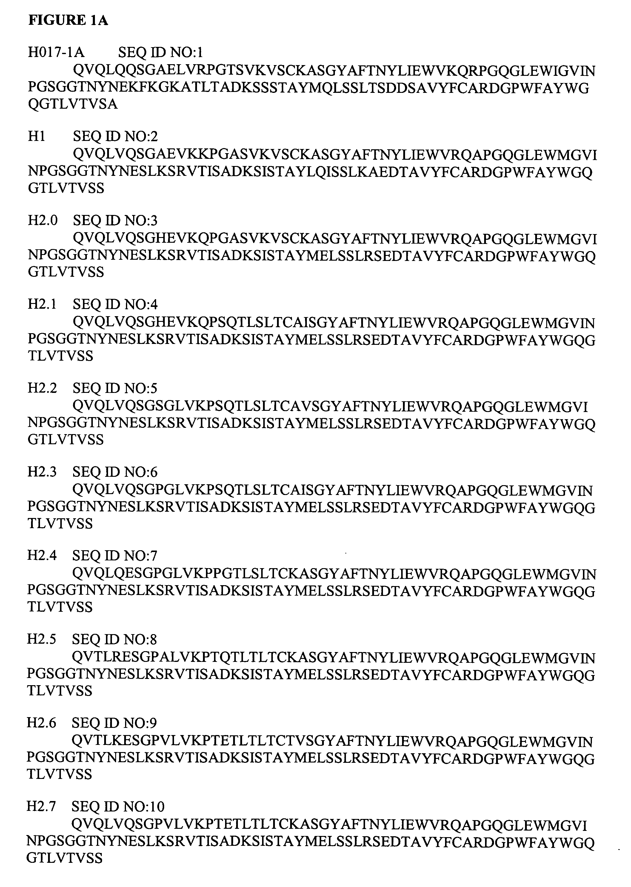
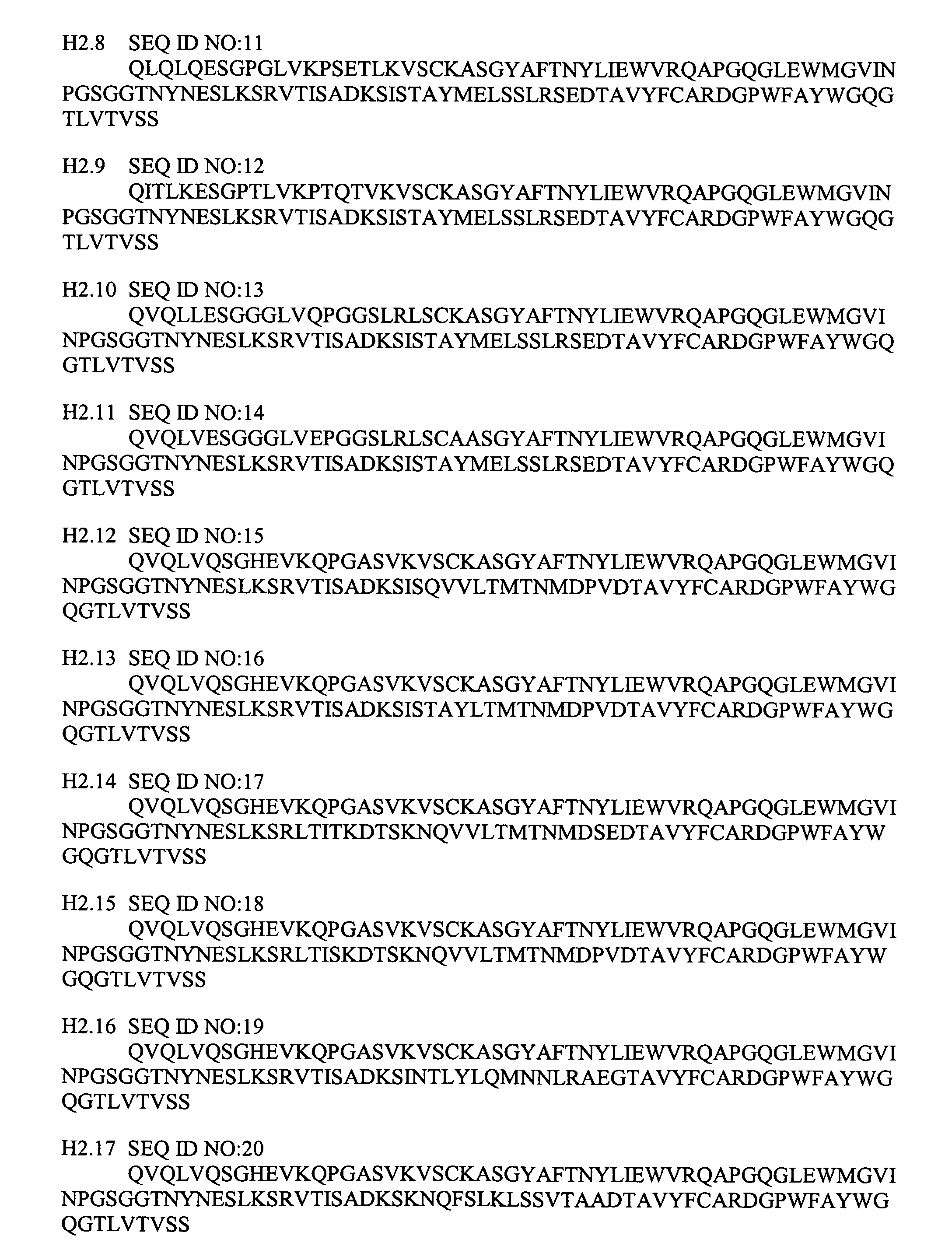
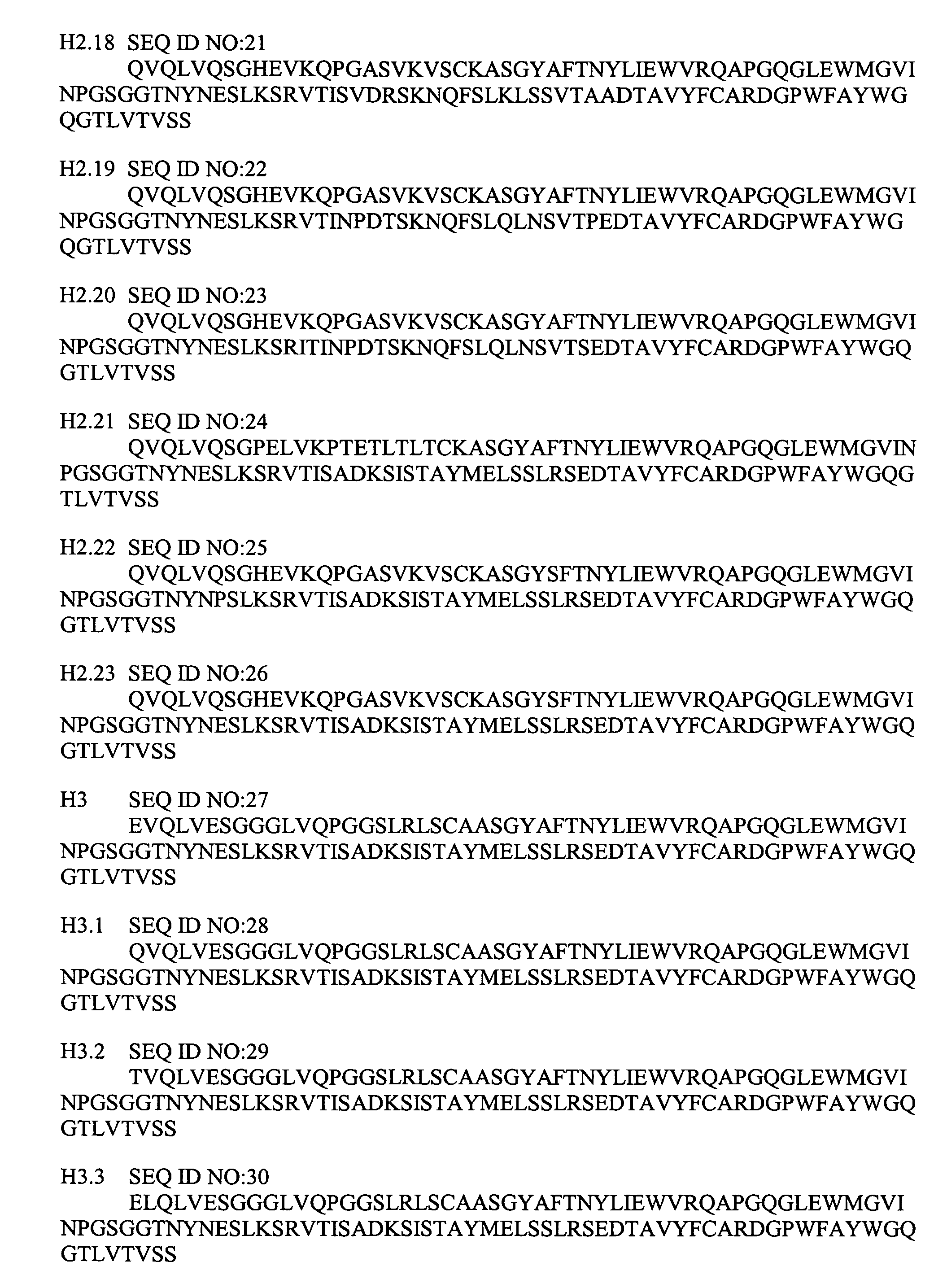
[0354] FIGS. 1 and 2 provide some heavy and light chain variable region sequences of the anti-Ep-CAM antibodies used in the present study. The mouse, parent chimeric heavy and light chains are labeled H0 17-1A and L0 17-1A respectively. Due to the wide use of hybridoma technology, a substantial number of antibodies are derived from nonhuman sources. However, nonhuman proteins are often immunogenic when administered to humans, thereby greatly reducing their therapeutic utility. Immunogenicity is the result of a complex series of responses to a substance that is perceived as foreign, and may include production of neutralizing and non-neutralizing antibodies, formation of immune complexes, complement activation, mast cell activation, inflammation, hypersensitivity responses, and anaphylaxis. Several factors can contribute to protein immunogenicity, including but not limited to protein sequence, route and frequency of administration, a...

example 2

[0356] Combinations of the different heavy and light chains were expressed and the resulting antibodies, with names such as H3L3, H3 / L3 or H3_L3, were purified and examined. Anti-Ep-CAM antibodies were expressed by transient transfection of vectors encoding the heavy and light chains into 293T cells grown in 10% ultra low IgG fetal bovine serum with 1 rmM sodium pyruvate and 1× non-essential amino acids (Gibco®, Invitrogen Hayward Calif.). Five days after transfection, the culture media was removed and ran through a protein A column (Pierce Biotechnology Inc, Rockford Md.). FIG. 4 shows typical yields of some Ep-CAM-binding proteins. FIGS. 5 and 6 contain gels showing the heavy and light chains of some purified antibodies of the present invention. The heavy chains may be made with any type of constant domain including, in humans, IgG1, IgG2 and hybrids comprising IgG1 and IgG2 as well as mouse constant domains such as IgG1 and IgG2a, which may be referred to as mIgG1 and mIgG2a. The...

example 3

Binding Properties of Anti-Ep-CAM Humanized Antibodies

[0357]FIG. 7 shows a schematic representation of the AlphaScreen assay. Binding affinity of anti-Ep-CAM antibodies to the extracellular domain of Ep-CAM was measured using a quantitative and sensitive method, AlphaScreen™ assay. The AlphaScreen™ assay is a bead-based non-radioactive luminescent proximity assay. Laser excitation of a donor bead excites oxygen, which if sufficiently close to the acceptor bead will generate a cascade of chemiluminescent events, ultimately leading to fluorescence emission at 520-620 nm. The AlphaScreen™ assay was applied as a competition assay for screening the antibodies. Wild-type IgG1 Ep-CAM antibody was biotinylated and extracellular domain of Ep-CAM was DIGylated by standard methods for attachment to streptavidin donor beads and anti-DIG acceptor beads. In the absence of competing anti-Ep-CAM antibodies (unlabeled), wild-type antibody (biotinylated) and Ep-CAM (DIGylated) interact and produce ...


PUM

PropertyMeasurementUnit
molecular weightaaaaaaaaaa
pHaaaaaaaaaa

Abstract

Humanized Ep-CAM-targeting antibodies and methods of making and using the same are provided.

Description

[0001] This application claims benefit under 35 U.S.C. §119(e) to U.S. Ser. No. 60 / 697,768 filed Jul. 8, 2005, U.S. Ser. No. 60 / 741,966 filed Dec. 2, 2005, U.S. Ser. No. 60 / 779,961 filed Mar. 6, 2006, and U.S. Ser. No. 60 / 745,078 filed Apr. 18, 2006, each of which is expressly incorporated by reference in its entirety.FIELD OF THE INVENTION [0002] The present invention relates to optimized proteins that target the epithelial cell adhesion molecule (Ep-CAM), and their applications, particularly for therapeutic purposes. BACKGROUND OF THE INVENTION [0003] Epithelial cell adhesion molecule, also known as epithelial glycoprotein 40 [EGP40], epithelial protein 2 [EGP-2], GA733-2, ESA, KSA, 17-1A antigen or other names) is an epithelial transmembrane protein encoded by the GA 733-2 gene (Gottlinger, H. G. et al. 1986, Int. J. Cancer. 15: 47-53; Linnenbach, A. J. et al. 1989, Proc. Natl. Acad. Sci. USA. 86:27-31; Armstrong, A. and Eck, S. 2003. Cancer Biol. Ther. 2: 320-325, Linnenbach, A....

Claims


Application Information

Patent Timeline
Patent Type & Authority Applications(United States)
IPC IPC(8): A61K39/395C07K16/28
CPCC07K16/30C07K16/465C07K2317/24C07K2317/41C07K2317/72C07K2317/732C07K2317/92
Inventor CHAMBERLAIN, AARON K.DESJARLAIS, JOHN R.LAZAR, GREGORY ALAN
Owner XENCOR
Why Eureka
Try Eureka