Multi modal spectroscopy

Inactive Publication Date: 2007-07-19
MASSACHUSETTS INST OF TECH
View PDF17 Cites 65 Cited by
  • Summary
  • Abstract
  • Description
  • Claims
  • Application Information

AI Technical Summary

Benefits of technology

[0011] The combination of TMS and Raman spectroscopy in MMS provides a more complete and complementary biochemical, morphologic and metabolic tissue profiles than either TMS or Raman spectroscopy alone resulting in better diagnostic accuracy. Another key advantage in combining both techniques is the potential for depth sensing. TMS and Raman spectroscopy can use different excitation wavelengths, and therefore sample different tissue volumes because of wavelength-dependent differences in absorption and scattering. A Raman source preferably emits in a range of 750 nm to 1000 nm while the fluorescence source can employ one or more laser sources or a filtered white light source. Reflectance measurements preferably use a broadband source such as xenon flash lamp.
[0012] This difference in sampling volume can be exploited to provide information about the depth (o

Problems solved by technology

On the other hand, vulnerable atherosclerotic plaque is the end result of an inflammatory process that leads to thinning

Method used


Image

  • Multi modal spectroscopy
  • Multi modal spectroscopy
  • Multi modal spectroscopy

Examples


Embodiment Construction

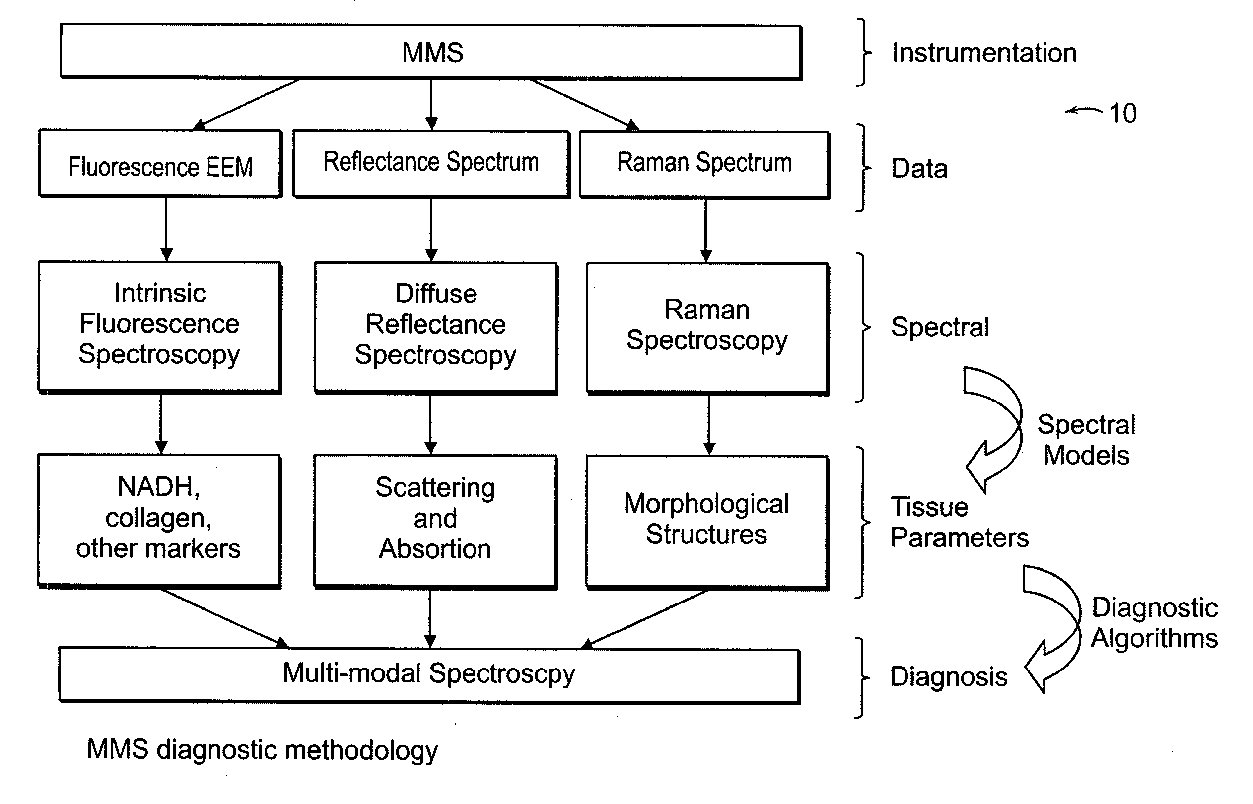
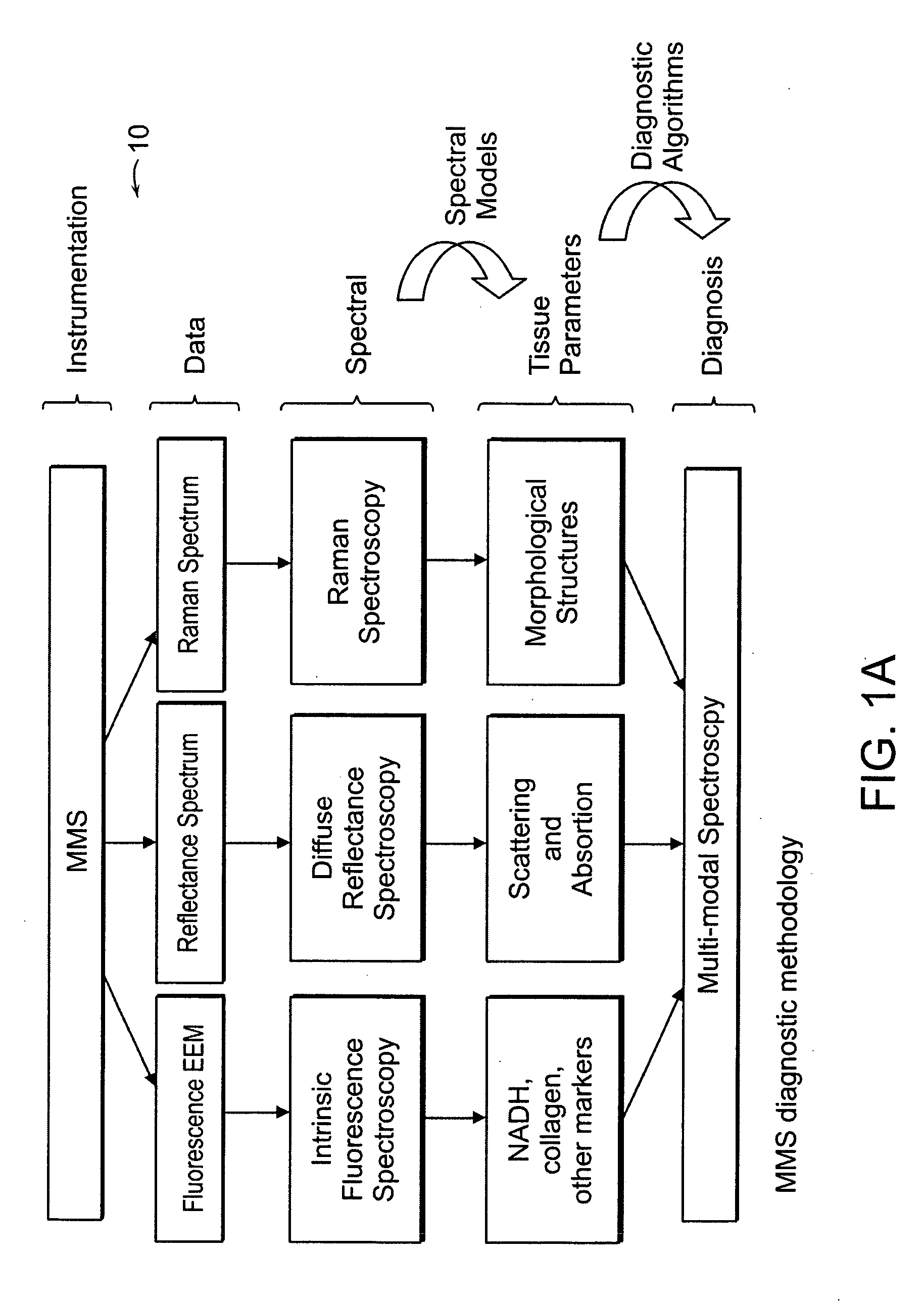
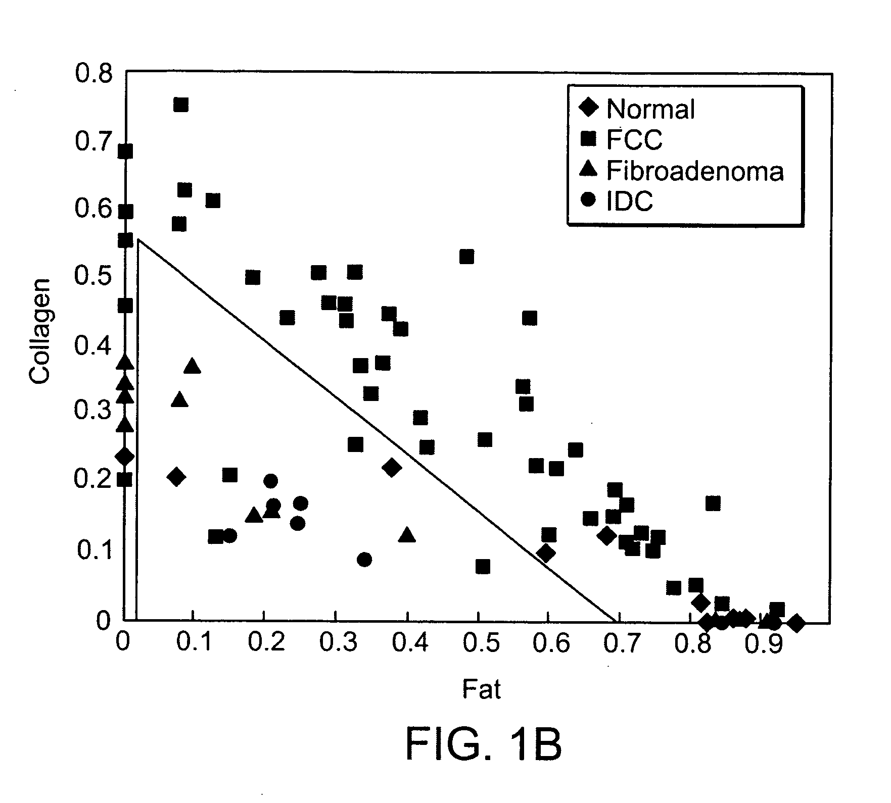
[0038] An MMS system is generally illustrated in FIG. 1A. MMS measurements have been performed on surgical biopsies within 30 minutes of surgical resection. Most of the 30 minute time delay was due to inking and sectioning of the specimen performed as part of the routine pathology consultation performed on these specimens for more information on intra-operative margin assessment in breast cancer patients. IFS, diffuse reflectance and Raman spectra were obtained from a total of 223 spectra from 105 breast tissues from 25 patients. Specimens from patients with pre-operative chemotherapy or who underwent re-excisional biopsy were excluded. DRS and IFS spectra were collected using the FastEEM instrument, followed by collection of Raman spectra with a Raman instrument. Care was taken in placing the Raman probe at the same site on the tissue as the FastEEM probe. Once the spectra were acquired, the exact spot of probe placement was marked with colloidal ink for registration with histopath...


PUM


Abstract

The present invention relates to multimodal spectroscopy (MMS) as a clinical tool for the in vivo diagnosis of disease in humans. The MMS technology combines Raman and fluorescence spectroscopy. A preferred embodiment involves diagnosis cancer of the breast and of vulnerable atherosclerotic plaque, esophageal, colon, cervical and bladder cancer. MMS is used to provide a more comprehensive picture of the metabolic, biochemical and morphological state of a tissue than afforded by either Raman or fluorescence and reflectance spectroscopies alone.

Description

CROSS REFERENCE TO RELATED APPLICATION [0001] This application claims the priority of U.S. Provisional Application No. 60 / 702,248, filed Jul. 25, 2005 entitled, MULTI MODAL SPECTROSCOPY. The entire content of the above application is being incorporated herein by reference.BACKGROUND OF THE INVENTION [0002] Techniques capable of evaluating human disease in a safe, minimally invasive and reproducible way are of importance for clinical disease diagnosis, risk assessment, therapeutic decision-making, and evaluating the effects of therapy, and for investigations of disease pathogenesis and pathophysiology. Among the clinical methods available to diagnose tissue lesions, pathologic examination of cytology preparations, biopsies and surgical specimens is the present day standard. [0003] Pathologists have traditionally based their diagnoses primarily on tissue morphology. However, as the field of diagnostic pathology has evolved, assessment of tissue morphology has become more sophisticated...

Claims


Application Information

Patent Timeline
IPC IPC(8): A61B6/00
CPCA61B5/0068A61B5/0071A61B5/0075A61B5/0084G01N2021/656A61B5/0091A61B5/42A61B5/4312G01N21/64A61B5/0086
Inventor SCEPANOVIC, OBRADGARDECKI, JOSEPHFELD, MICHAEL S.
Owner MASSACHUSETTS INST OF TECH
Why Eureka
Try Eureka