Eureka AIR delivers breakthrough ideas for toughest innovation challenges, trusted by R&D personnel around the world.

Direct differentiation of cardiomyocytes from human embryonic stem cells

a technology of embryonic stem cells and cardiomyocytes, which is applied in the field of stem cell differentiation induction, can solve the problems of chronic heart failure, loss of functional cardiomyocytes, and insufficient replacement of injured or dead cardiomyocytes

Inactive Publication Date: 2007-08-30
ES CELL INT
View PDF6 Cites 17 Cited by
  • Summary
  • Abstract
  • Description
  • Claims
  • Application Information

AI Technical Summary

Benefits of technology

[0019] In another aspect of the present invention there is provided a method of enabling cardiomyocyte differentiation in a stem cell culture said method comprising reducing an effect caused by insulin or an analogue thereof on the stem cell in culture.
[0020] It has now been found that media depleted in insulin is permissive for cardiomyocyte formation from stem cells. Without being limited by theory, the depletion of insulin and possibly other inhibitory factors allows or permits the stem cells to progress toward differentiation to a cardiomyocyte lineage.
[0022] In another aspect the invention also provides for improving yield of cardiomyocytes and cardiac progenitors by adopting the methods described herein.

Problems solved by technology

Although a small percentage of the cells may have proliferative capacity, it is not sufficient to replace injured or dead cardiomyocytes.
Loss of functional cardiomyocytes may lead to chronic heart failure.
The proliferative capacity of the cardiomyocytes is not sufficient to regenerate the heart following myocardial injury.
However, ischemic heart disease is still the most life-threatening disease in western society and alternative therapies will be necessary to improve the clinical outcome for patients with ischemic heart disease further.
However, it is difficult to control and induce the specific differentiation of ES cells solely towards cardiomyocytes (Brand, 2003).
Identifying the END2 inducing factors and other factors involved in the differentiation process has been challenging and illusive since the generation of the visceral-endoderm-like END2 cell line from a mouse P19 embryonic carcinoma cell line (Mummery, 1991).

Method used


Image

  • Direct differentiation of cardiomyocytes from human embryonic stem cells
  • Direct differentiation of cardiomyocytes from human embryonic stem cells
  • Direct differentiation of cardiomyocytes from human embryonic stem cells

Examples


example 1

Characterization of END2 Conditioned Media

I. Materials and Methods

(i) Preparation of END2 conditioned medium

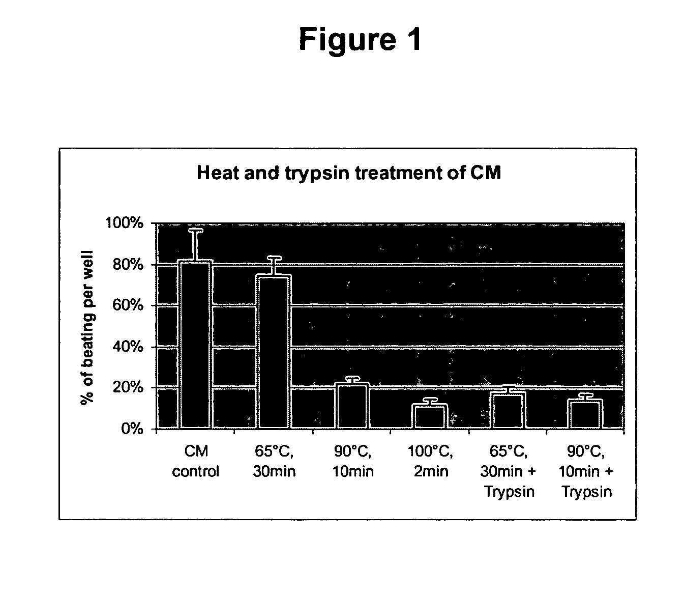
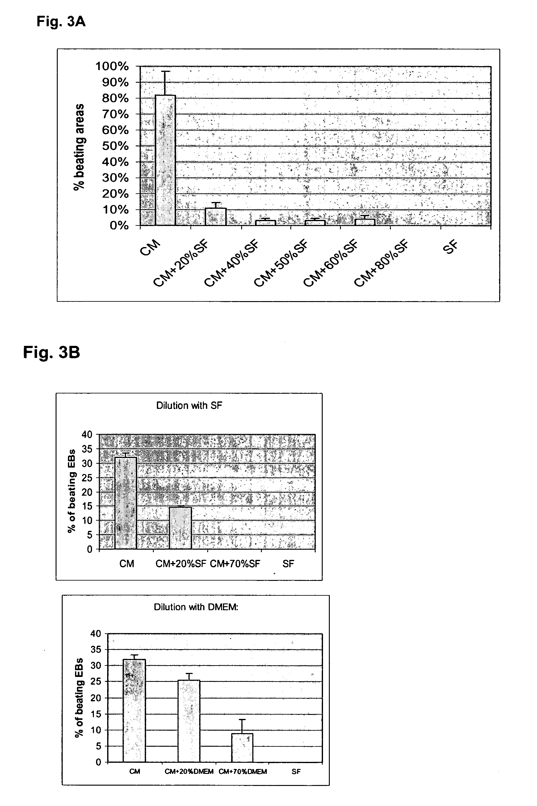
[0123] END2 cells secrete a factor into culture medium that can induce cardiomyocyte differentiation in hES cells. END2 conditioned media (END-CM) is defined as serum-free (SF) media that has been used to culture END-2 cells for four days or more and contains cardiomyocyte-inducing factor / s and / or bioactivity thereafter. hES cells are Human Embryonic Stem Cells. END2 cells are a visceral-endoderm-like cell line derived from P19, a mouse embryonal carcinoma cell line.

[0124] For the production of END2-CM END2 cells were seeded at a density of 1.4×10⁵ cells / cm2 in a T175 flask (Nunc) and were grown for about 3 days in Dulbecco's Modified Eagle's Medium (DMEM) / F12 media (Invitrogen) supplemented with 7.5% fetal calf serum (FCS; Hyclone) until confluency. The confluent END2 cell layer was washed once in PBS+ (Gibco) and serum free (SF) medium was used for conditioning. SF med...

example 2

Microarray Data Analysis to Identify Cardiomyocyte Inducing Factors Produced by END2 Cells

I. Materials and Methods

(i) Microarray Analysis

[0162] The Microarray experiments were conducted using Agilent Mouse 20k Developmental Oligo array or genome 40 k Oligo array technology. MES-1 was used as a reference cell line as it derived from the same parental cell p19 EC cell line as END2 but negative in its induction of cardiac differentiation. Two replica array datasets with dye swap were generated in two independent array experiments. Only features' expression calls were positive and significant above background assigned by the “Agilent G2565AA Feature Extraction Software” were accepted for analysis. The array data files were imported into GeneSpring 7.1 (Silicon Genetics) for data analysis. The expression ratios (Cy5 / Cy3: ratio of the median) were calculated and transformed to log2. Log2 expression ratios were then normalized by intensity-dependent LOWESS normalization. The different...

example 3

Prostaglandin I2 (PGI2) is an Active Component of END2 CM and Able to Increase hES Cell Cardiac Differentiation

I. Materials and Methods

(i) Medium Supplementation with PGI2

[0173] Prostaglandin I2 or PGI2-Na {(5Z, 9α, 11α, 13E, 15S)-6,9-Epoxy-11,15-dihydroxyprosta-5,13-dien-1-oic acid Sodium salt] was obtained from Sigma-Alorich. It was dissolved in 90% Ethanol at 2 mM as stock solution and then diluted with PBS before adding to bSFS at final concentration of 0.2 uM, and 2 uM. Medium change with addition of respective amounts of PGI2 was performed at day 3, 6 and 9 after EB formation.

II. Results

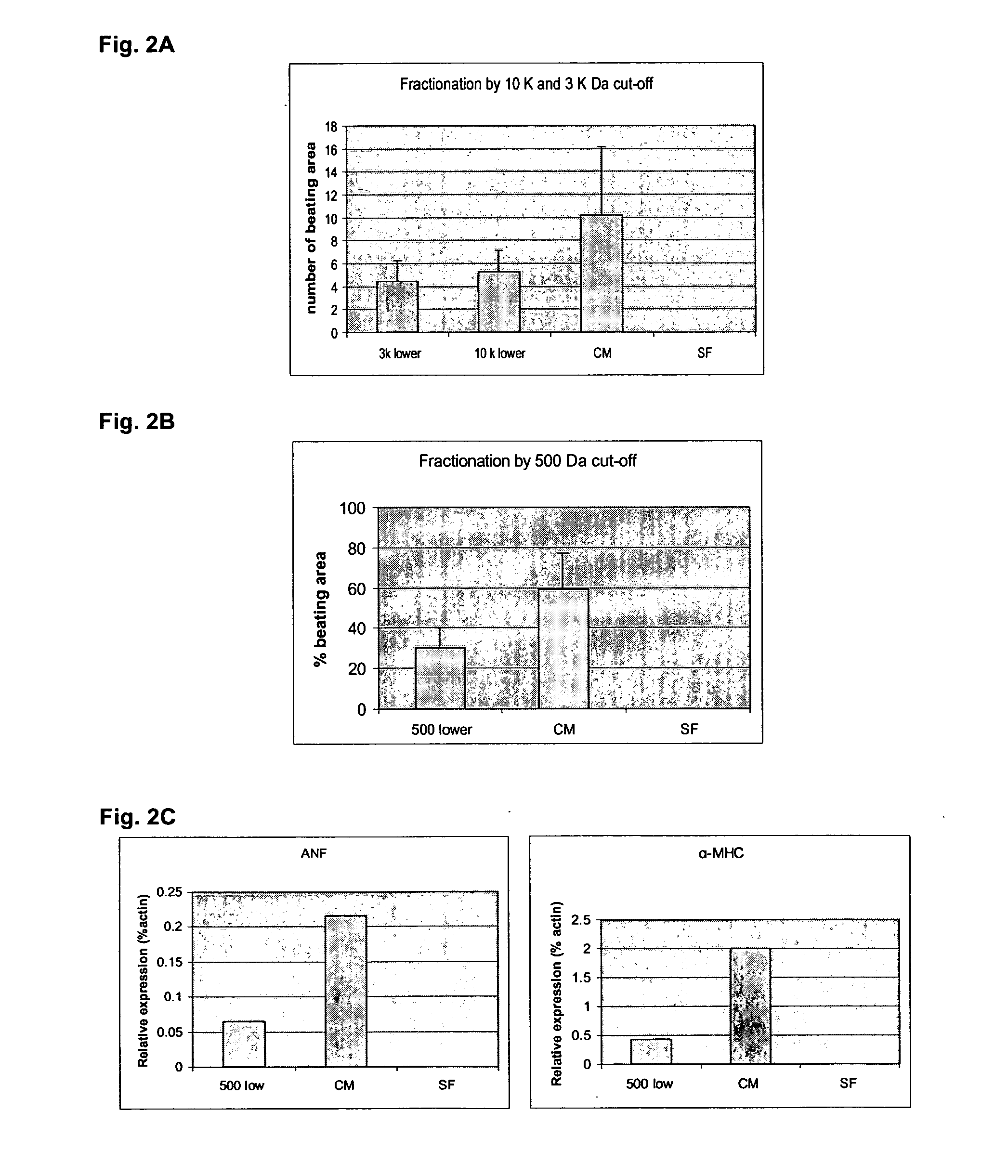
[0174] Factors found in the END2 conditioned media (CM) and involved in the ability of END2 cells to induce hES cell cardiac differentiation were identified. Accumulated data from studies on CM (physiochemical properties and size fractionation) from the previous examples suggested that the inducing factor in END2-CM is potentially a small molecule that is not a protein. Microarray analy...


PUM

PropertyMeasurementUnit
timeaaaaaaaaaa
volumeaaaaaaaaaa
temperatureaaaaaaaaaa

Abstract

The present invention relates to the induction of differentiation in stem cells to cardiomyocytes and factors such as prostaglandin alone or in combination with other factors including essential minerals selected from the group including transferrin and selenium, small molecules selected from the group including a p38 MAPK inhibitor such as SB203580 and protein growth factors of the FGF, IGF and BMP families such as but not limited to IGF1, FGF2, BMP2, BMP4 and BMP6. and insulin that influence the process of differentiation to cardiomyocytes. Media that is appropriate for the induction of differentiation of cardiomyocytes from stem cells is also provided wherein the media contains these factors. The use of cardiomyocytes and cardiac progenitors produced by the directed differentiation in transplantation and screening for cardiac compounds is also provided.

Description

CROSS-REFERENCE TO RELATED APPLICATIONS [0001] This present application is a continuation-in-part of U.S. application Ser. No. 10 / 758,554, filed Jan. 14, 2004. The present application also claims priority from U.S. Provisional Application Ser. No. 60 / 753,434, filed Dec. 22, 2005.FIELD OF THE INVENTION [0002] The present invention relates to the induction of differentiation in stem cells, preferably human embryonic stem (hES) cells to cardiomyocytes and cardiac progenitors and factors that influence the induction of differentiation. Media for the induction of differentiation of cardiomyocytes from stem cells, preferably hES cells is also provided. BACKGROUND [0003] Cardiomyocytes are thought to be terminally differentiated. Although a small percentage of the cells may have proliferative capacity, it is not sufficient to replace injured or dead cardiomyocytes. Death of cardiomyocytes occurs, for example, when a coronary vessel is occluded by a thrombus and the surrounding cardiomyocyt...

Claims


Application Information

Patent Timeline
Patent Type & Authority Applications(United States)
IPC IPC(8): A01K67/027C12N5/08C12N5/06A61K35/12C12N5/077
CPCA61K35/12C12N2502/13C12N2501/02C12N2501/105C12N2501/115C12N2501/125C12N2501/155C12N2501/33C12N2501/999C12N2502/30C12N2506/02C12N2510/00G01N33/502G01N33/5061C12N5/0657A61P9/00C12N5/0606C12N5/0043C12N2500/99C12N2500/25
Inventor DAVIDSON, BRUCE PAULGRAICHEN, RALPH EBERHARDZWEIGERDT, ROBERTXUIQIN, XUMUMMERY, CHRISTINE LINDSAYSUN, WILLIAM
Owner ES CELL INT
Why Eureka
Eureka Blog
Learn More