Apparatus and method for providing information for at least one structure

a technology of at least one structure and an apparatus, applied in the field of apparatus and method for providing information for at least one structure, can solve the problems of limitations, disadvantages and limitations of using conventionally available endoscopes, currently available probes limiting their use to certain procedures and locations,

Inactive Publication Date: 2008-01-17
THE GENERAL HOSPITAL CORP
View PDF83 Cites 155 Cited by
  • Summary
  • Abstract
  • Description
  • Claims
  • Application Information

AI Technical Summary

Benefits of technology

[0008] The present invention discloses a miniature imaging probe or endoscope that is capable of obtaining real-time images with up to or higher than about 100 times the number of resolvable points than a fiber-optic imaging bundle of the same diameter. Another way of describing the present invention is for the same number of resolvable pixels as commercially available imaging bundles, the present invention could have a diameter that is 10 times smaller. In addition, this instrument produces images that do not contain cladding artifacts. Also, this invention allows acquisition of depth (three-dimensional, or 3D) information from the sample. Finally, by acquiring multiple images of the sample, spectroscopic information relating to the chemical composition of the object may be obtained. These properties make this device an enabling technology for performing endoscopic or catheter based imaging in previously inaccessible locations within the body. Important specific applications include fetoscopy, pediatric endoscopy, coronary angioscopy, mini-laparoscopy, mammary ductoscopy, lacrimal ductoscopy, small joint visualization, and other medical and non-medical applications.

Problems solved by technology

Unfortunately, there are still a number of disadvantages and limitations of using conventionally available endoscopes.
The diameter of currently available probes limits their use to certain procedures and locations that can accommodate the large diameter of the endoscope.
Certain procedures cannot currently be done endoscopically because of the diameter of existing probes.
Another problem associated with current probes is the occurrence of adverse reactions, such as in fetoscopy, where the risk to the fetus is high.
Neural imaging carries with it the possibility of brain damage.
Spinal canal and brain ventricular imaging have complications of spinal fluid leakage and headaches which are more frequent and severe with larger diameter probes.
Catheterization of the pancreatic duct is also problematic due to the probe size and resultant complications which include acute pancreatitis.
Also, the size of the incision necessary to insert current probes results in longer healing time and more prominent scarring.
Currently, the clinical use of small diameter endoscopes is limited by poor resolution.
Since optical fibers are of finite diameter, only a limited number of fibers can be incorporated into one imaging bundle, resulting in a limited number of resolvable elements.
In addition, the fill factor is about 85% resulting dead space from the cladding material and causing the image to have a pixelated or “honeycomb” appearance.
These two technical problems have severely limited the clinical use of currently available sub-millimeter diameter imaging probes.
A further disadvantage of fiber bundles is that crosstalk occurs, reducing the signal to noise level.
Also with current fiber optic endoscopes, coupling illumination light into the fiber optic imaging bundle is difficult.
Another disadvantage of optical fiber bundles is that individual fibers may be broken or have defects at their faces, resulting in “dead” pixels.

Method used


Image

  • Apparatus and method for providing information for at least one structure
  • Apparatus and method for providing information for at least one structure
  • Apparatus and method for providing information for at least one structure

Examples


color embodiment

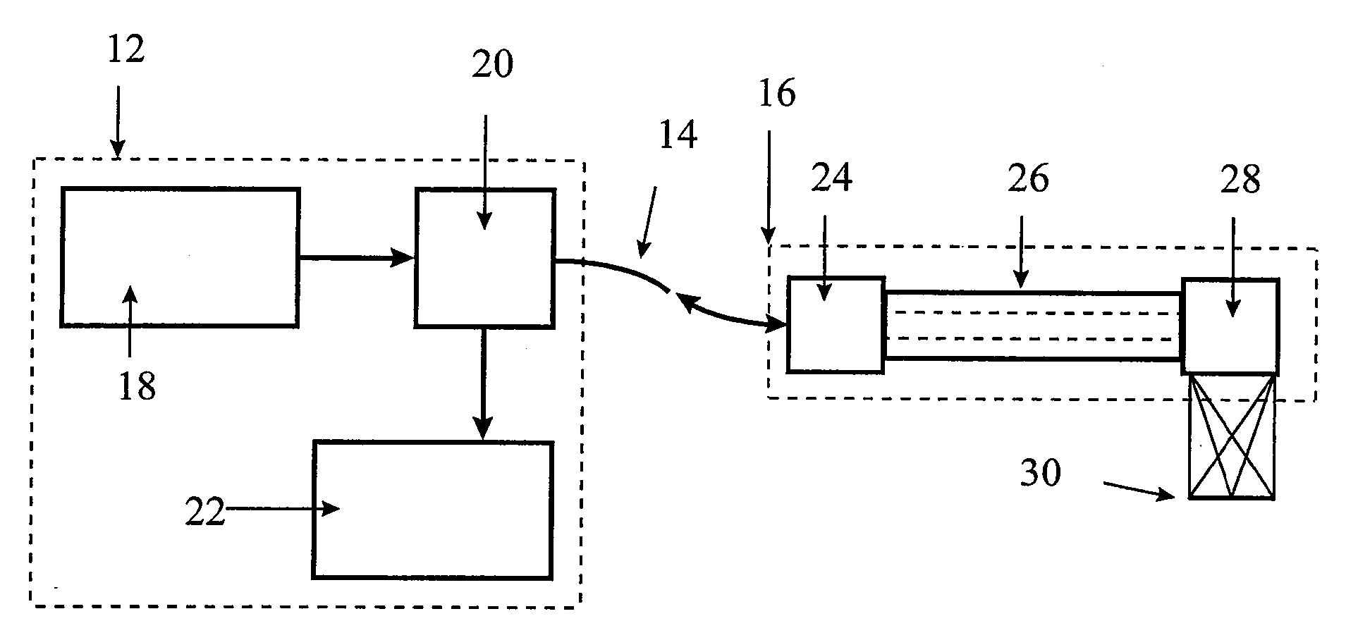
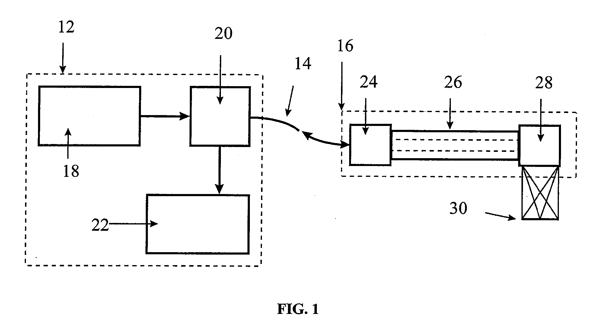
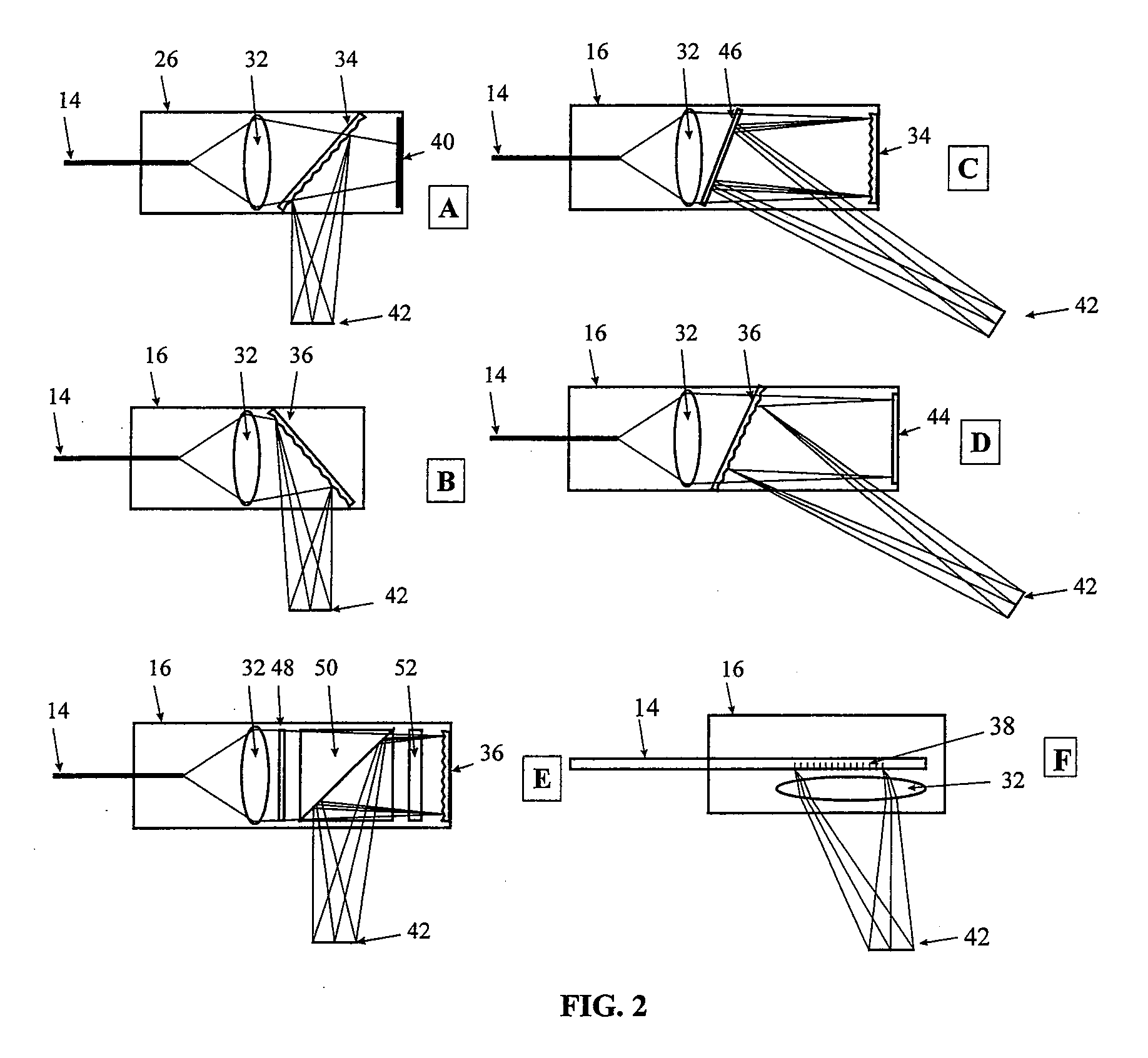
[0057] In an alternative embodiment of the present invention, shown in FIG. 5, a device 100 can be constructed that uses at least two and preferably three or more separate broadband source modules 110, for example, three sources centered at red (630 nm) 102, green (540 nm) 104, and blue (480 nm) 106 to produce color images using this technique. It is to be understood that other colors, wavelengths and number of separate sources can be selected depending on various factors, including, but not limited to, the imaging environment, imaging target, measurements to be obtained, and the like. The three energy components can be separated after reflection from the sample 30 and recombined to form an image. Each of the source / detector modules 102, 104 and 106 for the three spectral bands transmits selected wavelength light to an optical mixer / separator 108, which selectively transmits the light toward the imaging head 109 and to the imaging optics 110 for the different colors. The light refle...


PUM


Abstract

A spectrally encoded endoscopic probe having high resolution and small diameter comprising at least one flexible optical fiber; an energy source; a grating through which said energy is transmitted such that the energy spectrum is dispersed; a lens for focusing the dispersed energy spectrum onto a sample such that the impingement spot for each wavelength is a separate position on the sample, the wavelength spectrum defining a wavelength encoded axis; means for mechanically scanning the sample with focused energy in a direction perpendicular to the wavelength encoded axis; a means for receiving energy reflected from the sample; and, a means for detecting the received reflected energy. The probe grating and lens delivers a beam of multi-spectral light having spectral components extending in one dimension across a target region and which is moved to scan in another direction. The reflected spectrum is measured to provide two dimensional imaging of the region.

Description

BACKGROUND OF THE INVENTION [0001] Clinical use of endoscopic devices and probes has permitted physicians to view and diagnose target bodies, such as tumors, deposits, tears, thrombi, and the like. Unfortunately, there are still a number of disadvantages and limitations of using conventionally available endoscopes. The diameter of currently available probes limits their use to certain procedures and locations that can accommodate the large diameter of the endoscope. Consequently, many procedures currently done surgically could be done endoscopically, if a small enough probe was available with a sufficient number of resolvable points. [0002] Current endoscopic procedures generally require administration of anesthesia and surgical training for insertion. Certain procedures cannot currently be done endoscopically because of the diameter of existing probes. Probe size is proportional to the likelihood of tissue damage occurring during the procedure. Another problem associated with curre...

Claims


Application Information

Patent Timeline
Patent Type & Authority Applications(United States)
IPC IPC(8): H04B10/02A61B1/07A61B5/00
CPCA61B1/00096A61B1/0017A61B1/043A61B1/07G02B21/0064A61B5/0071A61B5/0084G02B21/0036G02B21/0048A61B5/0062A61B5/0059
Inventor TEARNEY, GUILLERMO J.BOUMA, BRETT EUGENESHISHKOV, MILEN STEFANOVROSEN, JONATHAN JAY
Owner THE GENERAL HOSPITAL CORP
Why Eureka
Try Eureka