Endovenous laser treatment generating reduced blood coagulation

a laser treatment and blood coagulation technology, applied in the field of varicose vein laser assisted treatment methods and equipment, can solve the problems of destroying endothelial cells, limiting the efficiency of heat transfer to the vessel wall, and requiring expensive and complicated catheters, so as to reduce the amount of blood present, reduce the formation of blood, and increase the safety and efficacy of endovenous laser treatment

Inactive Publication Date: 2008-01-24
COOLTOUCH
View PDF50 Cites 19 Cited by
  • Summary
  • Abstract
  • Description
  • Claims
  • Application Information

AI Technical Summary

Benefits of technology

[0020] In a first aspect, the present invention includes methods and devices for increasing the safety and efficacy of endovenous laser treatment of varicose veins by providing reduced formation and controlled clearing of the coagulum at the tip of an endovenous fiber. These devices and methods provide a way to substantially prevent the formation of coagulum upon the fiber tip of an endovenous laser treatment device, and to clear the fiber tip of coagulum without causing carbonization and explosive disruption of the vein wall.
[0021] As noted above, residual blood coagulum on the optical fiber tip is a serious impediment to the use of lasers to treat varicose veins. It is not possible to remove all the blood from a vein prior to treatment. Techniques such as elevating the leg, using compression, inducing spasm of the vein and using large amounts of tumescent anesthesia can all reduce the amount of blood present but there will typically always be pockets of blood that the laser must penetrate through to get a good shrinkage and closure of the vein. The use of a non hemoglobin absorbing laser such as one operating at a wavelength of 1320 nm has greatly reduced the coagulum that accumulates on the fiber tip, but there are still some instances where enough blood exists and coagulum could form. This could occur in very large veins or at locations close to the SF Junction where the vein must stay open or is just beginning to need closure.
[0022] Laboratory experiments, including several described herein, have shown that blood coagulates on the fiber tip in direct proportion to the absorption of the laser energy by the hemoglobin, and that there is a threshold where enough energy is delivered to the blood surrounding the fiber tip in a short enough time period such that the blood does not coagulate on the fiber tip and it remains clear and clean.
[0023] The present methods utilize laser pulses that are substantially shorter than those taught in the prior art. For example, the present methods use shorter pulses of 5000 μsec or less, with peak power levels of about 1000 watts. These operational settings provide instantaneous boiling of the thrombus off of the fiber tip rather than the slow cooking taught by the prior art methods, such as the methods taught in the Navarro patent, which lead to baking the thrombus onto the fiber tip. The present exposure times are typically much less than 1% of the exposure times of the prior art methods. The tissue interaction at these very short exposure time intervals is substantially different and preferred in relation to that provided by the prior art methods.
[0024] Laboratory tests have been performed using laser wavelengths of 810 nm, 980 nm, 1064 nm, and 1320 nm of short and long pulse lengths and different energies per pulse and repetition rate. Identical fiber optic delivery devices connected up to these lasers were held in bovine blood preserved with heparin so that the fiber tips were at least 10 mm into the blood. At constant power levels of about 7 watts the formation of coagulum was measured for each laser system. 810 and 980 nm continuous exposure lasers formed coagulum of about 2 cc size at the fiber tip within a few seconds of exposure. This stopped energy from exiting the fiber until the blood charred and then exploded from overheating. The 1320 low energy per pulse laser took about 30 seconds of exposure to form coagulum and it did not explode with increased exposure. The 1320 laser with higher energy per pulse did not develop coagulum over the fiber emitting end no matter how long it was exposed to the blood. This demonstrated the self cleaning nature of the proper wavelength and pulse energy.
[0025] The laser parameters can be adjusted to the levels needed to self clean a fiber tip by adjusting the power supply electronics that control the laser. In particular, the Nd:Yag laser used in the foregoing experiments is a crystalline laser that can be pulsed in a manner such that very high peak powers can be produced to enable this self cleaning action of the fiber tip. Nd:Yag lasers are used in many industrial and medical applications where high energy pulses are needed to drill holes and cut tissue. Lasers described in prior art endovenous treatment methods, such as 810 and 980 Diode lasers, cannot be pulsed in this manner because the facets of the diode bars cannot handle the high peak energy levels.

Problems solved by technology

RF technology has been used to try to heat the vessel wall directly but this technique requires expensive and complicated catheters to deliver electrical energy in direct contact with the vessel wall.
Other lasers at 810 nm and 1.06 um have been used in attempts to penetrate the skin and heat the vessel but they also have the disadvantage of substantial hemoglobin absorption which limits the efficiency of heat transfer to the vessel wall, or in the cases where the vessel is drained of blood prior to treatment of excessive transmission through the wall and damage to surrounding tissue.
All of these prior techniques result in poor efficiency in heating the collagen in the wall and destroying the endothelial cells.
In addition, blood coagulum that accumulates on the tip of the fiber optic energy delivery device is a significant problem associated with the prior art systems.
This is a serious complication referred to as Deep Vein Thrombosis (DVT) that, in the worst cases, is fatal to the patient.
The blood coagulum can also block the energy coming out of the tip of the fiber and thereby reduce the effectiveness of the treatment.
This is a common cause of non-closures or failures of the prior art endovenous treatments.
The blood coagulum is able to absorb so much laser energy that it carbonizes and may explode, causing rupture of the vein wall.
In fact, it has been shown that the carbonized blood actually prevents any direct delivery of laser energy to the vein wall until the carbon explodes, which can cause vein wall perforation.
Research has suggested that these regions of thrombus in a treated vein may not heal in a normal way and result in the vein staying patent or open.
This is considered a treatment failure.
When treating hand veins, these areas of heat induced thrombus left in an otherwise completely closed vein are cosmetically unattractive and need to be surgically punctured and drained post operatively to maintain a good result.
The use of this wavelength region greatly reduces the occurrence of thrombus because of the lower hemoglobin absorption of these wavelengths, but since blood contains a significant amount of water it can still be heated with these water absorbing wavelengths and eventually cause a small thrombus.
These relatively long exposure times at these power levels are needed because the prior art laser wavelengths are not as efficiently coupled to the vessel wall and are instead absorbed in the blood or transmitted through the wall into surrounding tissue.
It will be understood that methods taught in the prior art have been inefficient to such a degree that external cooling is needed on the skin surface to prevent burns.
The high power and long or continuous exposure times associated with systems using high hemoglobin absorbing wavelengths have the result of creating a great deal of coagulum and thrombus.
Most of the energy used by this method passes through the vessel wall and causes damage to surrounding tissue.
This stops the delivery of energy to the blood (or anywhere else) until the carbon explodes, which can cause a vein wall perforation.
Operative complications of this technique include bruising and extensive pain caused by transmitted energy and damage to surrounding tissue.
This prior art technique is also poorly controlled because the amount of residual blood in the vein can vary dramatically.
The blood can boil and explode in the vessel causing occasional perforation of the vein wall and unnecessary damage to healthy tissue.
This is very difficult to achieve and control.
This is required using the described technique because Navarro describes no means for uniformly controlling the rate of energy delivered.
In clinical practice this is very difficult to do and results in excessive perforations and complications.
The RF energy is delivered very slowly in continuous mode only and has caused a significant amount of coagulation or thrombus at the cathode tip.
However, this process actually encourages coagulum formation by heat thrombosis.
One in vitro study model has predicted that thermal gas production by laser heating of blood in a 6 mm tube results in 6 mm of thermal damage.
Patients treated with prior art methods and devices have shown an increase in post-treatment purpura and tenderness.
Since the anesthetic and access techniques for the two procedures are identical, it is believed that non-specific perivascular thermal damage is the probable cause for this increased tenderness.
Slow, uncontrolled pull-back of the catheter is likely one cause for overheating and perforation of the vessel wall, as even the best surgeon may have difficulty retracting the fiber at exactly the correct speed to maintain a vessel wall heating temperature of 85 deg C. This technique prevents damage to surrounding tissue and perforation of the vessel.
Prior art diode lasers are difficult to focus to small spots in high power levels because each diode bar typically needs to be imaged separately into the fiber optic.

Method used


Image

  • Endovenous laser treatment generating reduced blood coagulation
  • Endovenous laser treatment generating reduced blood coagulation
  • Endovenous laser treatment generating reduced blood coagulation

Examples


experiment # 1

Experiment #1: Coagulum Development with 1320 nm vs. 980 nm Laser

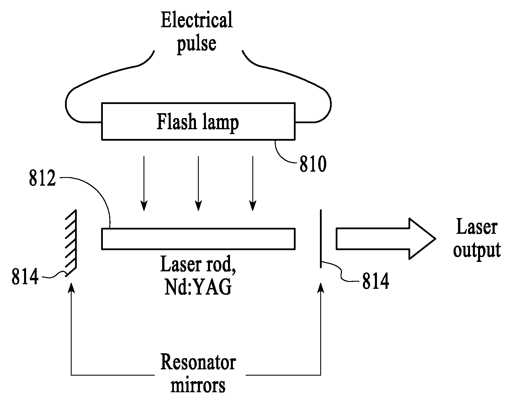
[0081] In a first experiment, coagulation formation on the fiber tip of an Nd:YAG pulsed laser having a wavelength of 1320 nm was compared to coagulation formation on the fiber tip of a Diode laser having a wavelength of 980 nm in continuous mode.

[0082] Method Overview: Approximately 50 cc of porcine blood was stabilized with EDTA and placed in a beaker at room temperature. A standard 600 μm fiber was used on both laser systems. The power levels for both laser systems were measured using a Molectron PowerMax 600 power meter. The porcine blood was stirred between each interval. The fiber was cleaned using 3% hydrogen peroxide and wiped off with a Kim Wipe® between each firing. The fiber was checked using after each test to confirm a circular aiming beam with no tails, and checked by the power meter between each firing. Clot sizes were measured visually using a metric ruler and recorded in a lab notebook and digital pho...

experiment # 2

Experiment #2: Coagulum as a Function of Laser Power and Power Density

[0086] In a second experiment, coagulation formation on a fiber tip was measured as a function of laser peak power and power density for an Nd:YAG laser having a wavelength of 1320 μm.

[0087] Method Overview: Approximately 10 cc of porcine blood was stabilized with EDTA and placed in a graduated cylinder at room temperature. A standard 600 μm fiber and a 365 μm fiber were each used to deliver energy to the blood medium. Clot sizes were measured using a metric ruler.

[0088] First Test: The first test was to compare coagulation accumulations between the 600 μm fiber and the 365 μm fiber.

SystemSettingsTimeFiberPower DensityAiming Beam1320 nm7 W / 50 Hz140 mJ / pulse @ width =5-40 sec600 μm  330,000 W / cm2None thru clot150 μsec1320 nm7 W / 50 Hz140 mJ / pulse @ width =5-40 sec365 μm1,000,000 W / cm2None thru clot150 μsec

[0089] Clot formation was compared at 5-second intervals for both fibers. Coagulum was cleaned off with a Ki...


PUM


Abstract

This invention is an improved method and device for treating varicose veins 200 or the greater saphenous vein 202. The method comprises the use of infrared laser radiation in the region of 1.2 to 2.2 um in a manner from inside the vessel 200 or 202 such that the endothelial cells of the vessel wall 704 are damaged and collagen fibers in the vessel wall 704 are heated to the point where they permanently contract, the vessel 200 or 202 is occluded and ultimately resorbed. The device includes a laser 102 delivering laser energy via a fiber optic catheter 300. A motorized pull back device 104 may be used. The laser is preferably operated under laser energy delivery conditions that substantially prevent the formation of coagulum on the energy emitting tip of the fiber.

Description

RELATED APPLICATION [0001] This Application is related to U.S. patent application Ser. No. 10 / 982,504, filed on Nov. 4, 2004, and titled “ENDOVENOUS CLOSURE OF VARICOSE VEINS WITH MID INFRARED LASER”, Attorney Docket No.15487.4001, which application is a continuation-in-part of and claims the benefit of International Application Number PCT / US2003 / 035178, filed under the Patent Cooperation Treaty on Oct. 30, 2003, Attorney Docket No. NSL-501-PCT, designating the United States of America, and titled “ENDOVENOUS CLOSURE OF VARICOSE VEINS WITH MID INFRARED LASER”, which application claims the benefit of U.S. Provisional Patent Application Ser. No. 60 / 422,566 filed Oct. 31, 2002, entitled “ENDOVENOUS CLOSURE OF VARICOSE VEINS WITH MID INFRARED LASER”, Attorney Docket No. NSL-501-P, each of which applications are fully incorporated herein by reference in their entireties. FIELD OF THE INVENTION [0002] The present invention relates generally laser assisted method and apparatus for treatmen...

Claims


Application Information

Patent Timeline
Patent Type & Authority Applications(United States)
IPC IPC(8): A61N5/067
CPCA61B2018/0097A61B18/24
Inventor HENNINGS, DAVID R.FULLMER, DAVID J.LINDSAY, CRAIG
Owner COOLTOUCH
Why Eureka
Try Eureka