Glossopexy tension system and method

a tension system and tension system technology, applied in the field of glossopexy tension system and method, can solve the problems of excessive daytime sleepiness, increased inspiratory effort, and risk of airway obstruction during sleep, and achieve the effect of preventing undesired hyperextension of the tether

Inactive Publication Date: 2008-03-06
KONINK PHILIPS ELECTRONICS NV
View PDF8 Cites 119 Cited by
  • Summary
  • Abstract
  • Description
  • Claims
  • Application Information

AI Technical Summary

Benefits of technology

[0055] Also disclosed herein is a tongue remodeling system that includes a first body-engaging structure; a second body-engaging structure; an elastic tether configured to be connected to the first body-engaging structure and the second body-engaging structure; and means for preventing overextension of the tether. In some embodiments, the elastic tether includes a spring. In some embodiments, the means for preventing overextension of the tether can include one or more of the following: a controlled deflection spring; a shaft configured to have a portion of the tether wrapped over the shaft; or a tether protection element configured to prevent extension of a portion of the tether adjacent to the tether protection element.
[0056] In some embodiments, disclosed is a tongue remodeling system that includes a first body-engaging structure; a second body-engaging structure; an elastic tether configured to be connected to the first body-engaging structure and the second body-engaging structure; and a tether protection element configured to prevent overextension of the tether. The tether protection element includes a spring, which can be a controlled deflection spring. The tether protection element can be configured to prevent extension of a portion of the tether spaced apart from the tether protection element. In some embodiments, the tether is a spring and the tether protection element is a shaft configured to have a portion of the spring wrapped over the shaft. In some embodiments, the tether protection element is configured to limit tether extension to no more than about 150% of an unstretched length of the tether. In some embodiments, the first body-engaging structure is a tissue anchor.
[0057] Also disclosed is a method of preventing undesired hyperextension of a tether, including the steps of providing a body-engaging structure; providing an elastic tether having a tether-protection element operatively engaged with the tether; implanting the body-engaging structure with the body; extending the tether with the tether connected to the body-engaging structure; and limiting further extension of the tether, wherein limiting further extension is accomplished by the tether protection element. In some embodiments, the tether protection element includes a spring, such as a controlled deflection spring. The tether protection element can prevent extension of a portion of the tether spaced apart from the tether protection element. In some embodiments, the tether can include a spring and the tether protection element includes a shaft configured to have a portion of the spring wrapped over the shaft. In some embodiments, the body-engaging structure is implanted within the tongue, a bony structure, or the mandible.

Problems solved by technology

This muscle inhibition may lead to relaxation of certain muscle groups, including but not limited to muscles that maintain the patency of the upper airways, and create a risk of airway obstruction during sleep.
Because muscle relaxation narrows the lumen of the airway, greater inspiratory effort may be required to overcome airway resistance.
Because these arousal episodes typically occur 10 to 60 times per night, sleep fragmentation occurs which produces excessive daytime sleepiness.
In addition to sleep disruption, OSA may also lead to cardiovascular and pulmonary disease.
Some studies suggest that increased catecholamine release in the body due to the low oxygen saturation causes increases in systemic arterial blood pressure, which in turn causes left ventricular hypertrophy and eventually left heart failure.
OSA is also associated with pulmonary hypertension, which can result in right heart failure.
Other studies, however, have suggested that increased compliance of the lateral walls of the pharynx contributes to airway collapse.
Weight loss may reduce the frequency of apnea in some patients, but weight loss and other behavioral changes are difficult to achieve and maintain.
Progesterone, theophylline and acetozolamide have been used as respiratory stimulants, but each drug is associated with significant side effects and their efficacy in OSA is not well studied.
Protriptyline, a tricyclic antidepressant that reduces the amount of REM sleep, has been shown to decrease the frequency of apnea episodes in severe OSA, but is associated with anti-cholinergic side effects such as impotence, dry mouth, urinary retention and constipation.
Oral appliances aimed at changing the position of the soft palate, jaw or tongue are available, but patient discomfort and low compliance have limited their use.
Although CPAP is effective in treating OSA, patient compliance with these devices is low for several reasons.
Sleeping with a sealed nasal mask is uncomfortable for patients.
Smaller sealed nasal masks may be more comfortable to patients but are ineffective in patients who sleep with their mouths open, as the air pressure will enter the nasopharynx and then exit the oropharynx.
CPAP also causes dry nasal passages and congestion.
UPPP also exposes patients to the risks of general anesthesia and postoperative swelling of the airway that will require a tracheostomy.
Excessive tissue removal may also cause velo-pharyngeal insufficiency where food and liquids enter into the nasopharynx during swallowing.
For patients who fail UPPP or LAUP, other surgical treatments are available but these surgeries entail significantly higher risks of morbidity and mortality.
This involves significant morbidity and has shown only limited effectiveness.
These patients are predisposed to OSA because the posterior mandible position produces posterior tongue displacement that causes airway obstruction.

Method used


Image

  • Glossopexy tension system and method
  • Glossopexy tension system and method
  • Glossopexy tension system and method

Examples


Embodiment Construction

A. Anatomy of the Pharynx

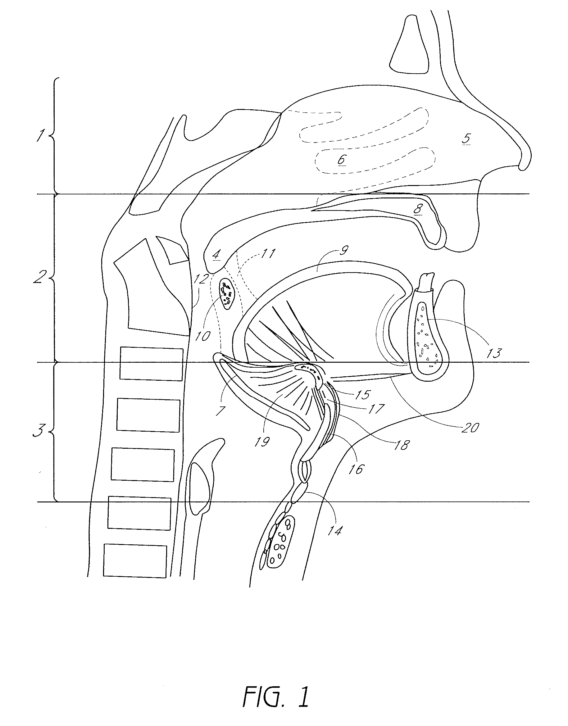
[0144]FIG. 1 is a sagittal view of the structures that comprise the pharyngeal airway and may be involved in obstructive sleep apnea. The pharynx is divided, from superior to inferior, into the nasopharynx 1, the oropharynx 2 and the hypopharynx 3. The nasopharynx 1 is a less common source of obstruction in OSA. The nasopharynx is the portion of the pharynx above the soft palate 4. In the nasopharynx, a deviated nasal septum 5 or enlarged nasal turbinates 6 may occasionally contribute to upper airway resistance or blockage. Only rarely, a nasal mass, such as a polyp, cyst or tumor may be a source of obstruction.

[0145] The oropharynx 2 comprises structures from the soft palate 4 to the upper border of the epiglottis 7 and includes the hard palate 8, tongue 9, tonsils 10, palatoglossal arch 11, the posterior pharyngeal wall 12 and the mandible 13. The mandible typically has a bone thickness of about 5 mm to about 10 mm anteriorly with similar thicknesses late...


PUM


Abstract

Methods and devices are disclosed for manipulating the tongue. An implant is positioned within at least a portion of the tongue and may be secured to other surrounding structures such as the mandible and / or hyoid bone. In general, the implant is manipulated to displace at least a portion of the posterior tongue in an anterior or lateral direction, or to alter the tissue tension or compliance of the tongue. Methods and devices for tensioning a glossopexy system are also disclosed. A tether can include a tether protection element configured to prevent overextension of the tether.

Description

CROSS-REFERENCE TO RELATED APPLICATIONS [0001] The present application claims priority under 35 U.S.C. § 119(e) to U.S. Provisional Application Nos. 60 / 813,230, 60 / 813,285 and 60 / 813,058, all filed on Jun. 13, 2006. All of the priority applications are hereby incorporated by reference in their entirety. [0002] The present application also incorporates by reference in its entirety U.S. patent application Ser. No. 11 / 349,067, filed Feb. 7, 2006.BACKGROUND OF THE INVENTION [0003] 1. Field of the Invention [0004] This invention relates generally to a system and method for treating upper airway obstruction, sleep disordered breathing, upper airway resistance syndrome and snoring by manipulating the structures of the oropharynx, including the tongue. [0005] 2. Description of the Related Art [0006] Respiratory disorders during sleep are recognized as a common disorder with significant clinical consequences. During the various stages of sleep, the human body exhibits different patterns of b...

Claims


Application Information

Patent Timeline
Patent Type & Authority Applications(United States)
IPC IPC(8): A61F5/56
CPCA61B17/0401A61F5/566A61B2017/00814A61B2017/0412A61B2017/042A61B2017/0422A61B2017/0427A61B2017/0433A61B2017/0435A61B2017/0437A61B2017/0438A61B2017/044A61B2017/0443A61B2017/045A61B2017/0451A61B2017/0453A61B2017/0458A61B2017/0461A61B2017/0462A61B2017/0464A61B2017/0496A61B2017/06057A61B2017/0618A61B2017/248A61B2018/00327A61B17/24
Inventor HIROTSUKA, MARKJACKSON, JASPERFRAZIER, ANDREWROUE, CHADVAN DER BURG, ERIKDINEEN, MICHAEL
Owner KONINK PHILIPS ELECTRONICS NV
Why Eureka
Try Eureka