Raman Spectral Analysis Of Sub-Surface Tissues And Fluids

a subsurface tissue and spectral analysis technology, applied in the direction of optical radiation measurement, diagnostics using spectroscopy, instruments, etc., can solve the problems of few non-invasive or minimally invasive methods for examining bone details, the risk of bone fracture in the normal population of people with osteoporosis is significantly increased, and the sacrificial procedure is clearly not an option in examining human patients

Active Publication Date: 2008-03-27
UK RES & INNOVATION LTD
View PDF12 Cites 71 Cited by
  • Summary
  • Abstract
  • Description
  • Claims
  • Application Information

AI Technical Summary

Benefits of technology

[0011] It is an object of this invention, therefore, to provide an analysis technique based on Raman spectroscopy, that is capable of extracting sub-surface chemical compositional information of in-vivo and living tissues such as bone tissue. It is also an object of the invention to enable such analysis without requiring any incision, puncture or other surgical intervention to expose the target sub-surface tissue.

Problems solved by technology

For instance, a person with osteoporosis has a significantly increased risk of bone fracture in comparison to the normal population.
To date however there are few non-invasive or minimally-invasive methods for examining details of bone or composition of other tissues.
Sacrificial procedures are clearly not an option in examining human patients.
This technique however is only able to measure the inorganic phase of bone (hydroxyapatite) and the organic phase (primarily collagen I) is largely invisible.
Both of these can often cause discomfort or pain to the patient.
Unfortunately analysis by these methods has been limited to surface studies i.e. on exposed bone.
The near-infrared range (700-850 nm) penetrates further, but multiple scattering causes loss of detailed spatial information.
Confocal microscopy, a standard technique used to probe sample depths, is precluded on living tissue as a tight focal spot causes local heating and tissue damage.
Moreover this technique is substantially less effective in diffusely scattering media, such as biological tissue, in which it is only practicable to depths of around ten times the mean free path of photons in the medium.
The elastic scattering technique of Das et al. is however not directly extendable to Raman spectroscopy.
This results in a much weaker signal.
Furthermore the Raman signal is far more susceptible to interference from luminescence or sample damage.

Method used


Image

  • Raman Spectral Analysis Of Sub-Surface Tissues And Fluids
  • Raman Spectral Analysis Of Sub-Surface Tissues And Fluids
  • Raman Spectral Analysis Of Sub-Surface Tissues And Fluids

Examples


Embodiment Construction

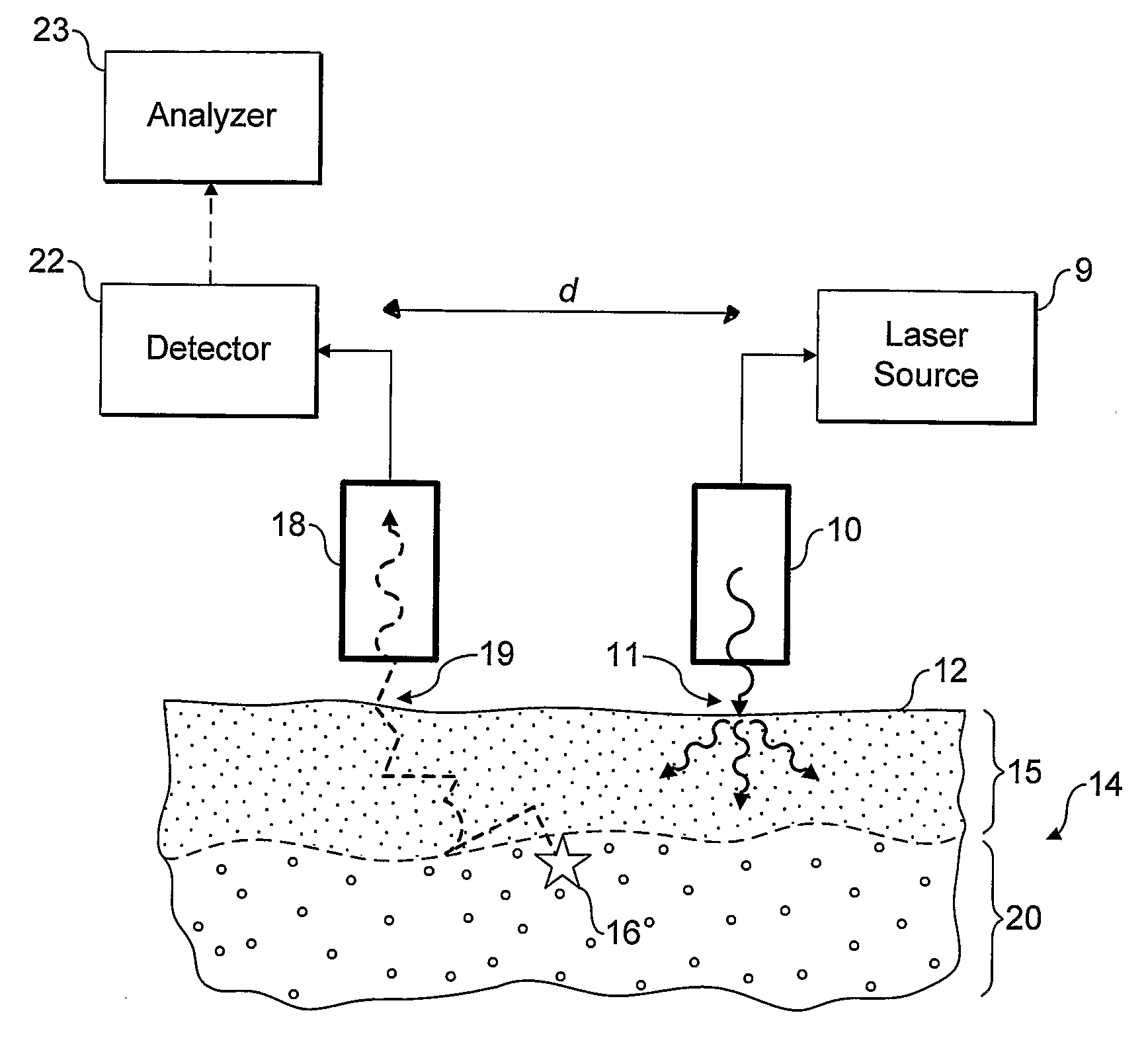
[0058] Referring now to FIG. 1 an embodiment of the invention is shown in operation, in schematic cross section. A light source 10, incorporating or supplied by laser 9, is used to irradiate localised entry region of a surface 12 of in-vivo tissue 14, which in the present example is made up of a skin layer 15 and underlying bone tissue 20. The incident radiation from the light source is scattered diffusely through the sample, especially the upper skin layer. Some of the radiation may be absorbed by the tissue, some may give rise to optical emissions for example by fluorescence, and some re-emerges unchanged through the tissue surface 12.

[0059] A small proportion of the photons of the incident radiation are inelastically scattered giving rise to Raman photons, for example as illustrated by Raman event 16. The Raman photons in turn are diffusively scattered through the tissue. Some may be absorbed, for example giving rise to fluorescence, but some emerge unchanged through the surface...


PUM

PropertyMeasurementUnit
wavelengthaaaaaaaaaa

Abstract

Apparatus and methods for determining, in-vivo, characteristics of sub-surface tissues or fluids in the human or animal body are disclosed. Incident radiation is supplied at one or more entry regions on a surface, and light is collected from one or more collection regions spaced from the entry regions. Raman features are detected in the collected light and depth related information derived therefrom.

Description

FIELD OF THE INVENTION [0001] The present invention relates to methods and apparatus for determining characteristics of in-vivo sub-surface tissues by detecting Raman features of diffusely scattered light. For example, the described methods and apparatus are suitable for detecting Raman spectral features of in-vivo bone through skin, nail and other surface tissues, without requiring exposure of the bone tissue by incision or puncture. DISCUSSION OF THE PRIOR ART [0002] The field of investigative studies of bone and other biological and living tissues encompasses a myriad of analytical techniques. Such techniques have been developed in response to the many situations in which it has been found to be important to assess the bone quality or tissue composition of a particular patient, human or animal. For instance, a person with osteoporosis has a significantly increased risk of bone fracture in comparison to the normal population. Diagnosis of degenerative skeletal diseases, for exampl...

Claims


Application Information

Patent Timeline
Patent Type & Authority Applications(United States)
IPC IPC(8): A61B5/00A61B5/103G01N21/65
CPCA61B5/0059A61B5/0071A61B5/0075G01N2021/656A61B5/4504G01N21/65A61B5/441
Inventor MATOUSEK, PAVELPARKER, ANTHONY WILLIAM
Owner UK RES & INNOVATION LTD
Why Eureka
Try Eureka