Method for Obtaining Modified Proteins and Viruses with Intact Native Binding Site
a technology of native binding site and protein, which is applied in the field of obtaining modified proteins and viruses with intact native binding site, can solve the problems of low yield, slow protease digestion of antibodies, and inability to extend serum half-life to useful levels, and achieve the effect of reducing antigenicity
- Summary
- Abstract
- Description
- Claims
- Application Information
AI Technical Summary
Benefits of technology
Problems solved by technology
Method used
Image
Examples
example 1
Preparation of Antibody Coated by Mannose
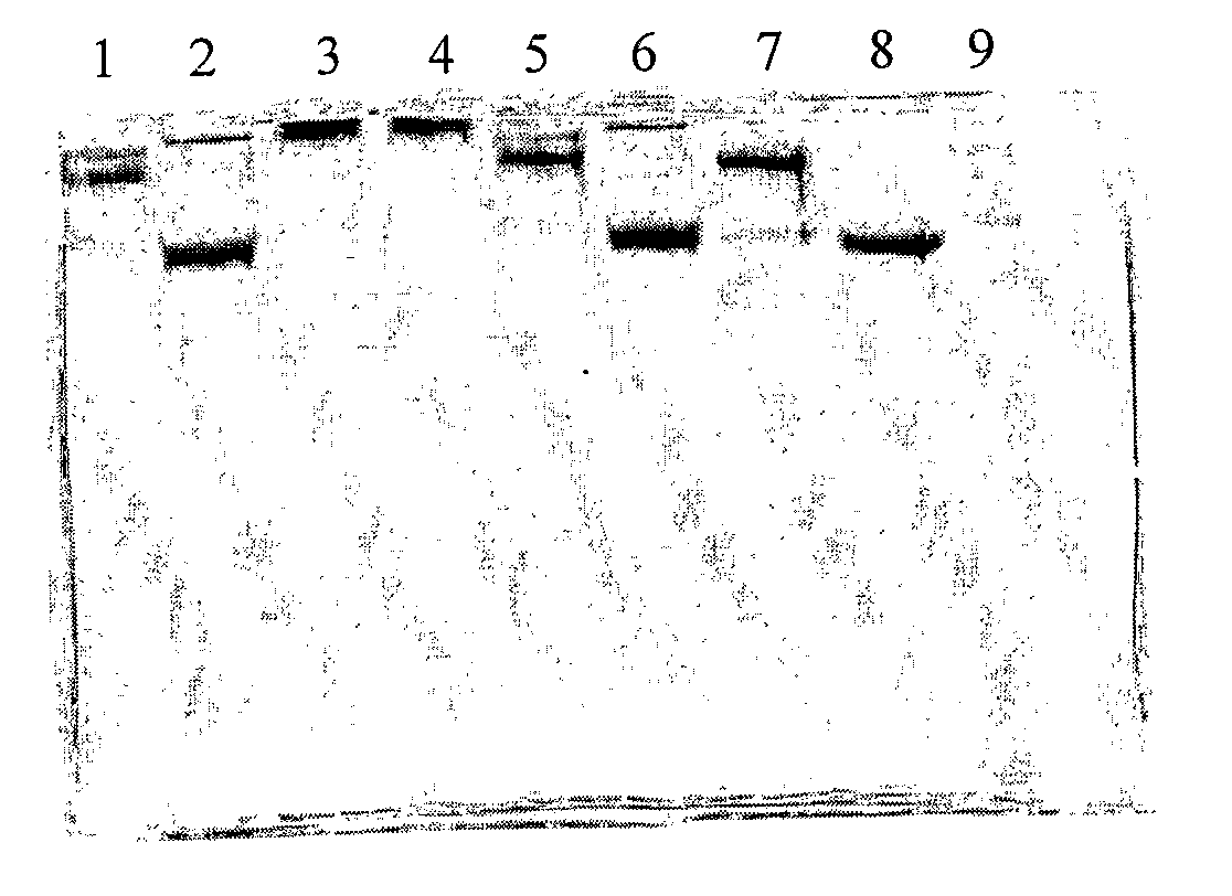
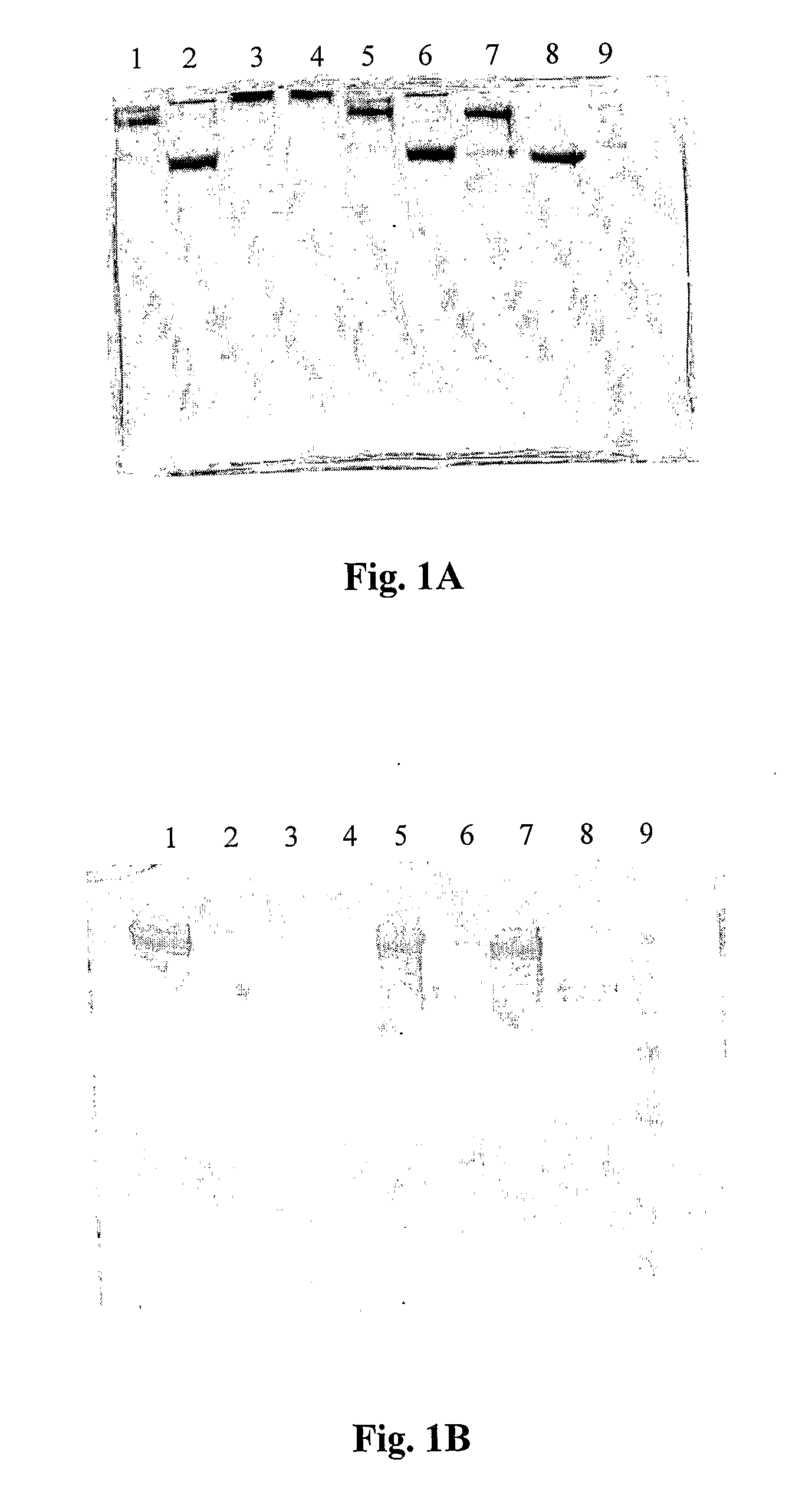
[0074]In this preliminary example, cow IgG or chicken IgY was coated with mannose by three different coating procedures: (i) mannose+EDC; (ii) mannose+EDC+pTSA; (iii) mannose+EDC+NaBH3CN.
[0075](i) Coating with mannose+EDC
[0076]D(+)-mannose (10 mg) was added to a 0.5 ml solution of cow IgG or chicken IgY (225 μg / ml solution in PBS 50 mM, pH=7.0), the solution was shaken for 12 hours at room temperature, then 9 mg of EDC was added, and the mixing was continued for additional 12 hours.
[0077](ii) Coating with mannose+EDC+pTSA
[0078]D(+)-mannose (10 mg) was added to a 0.5 ml solution of cow IgG or chicken IgY (225 μg / ml solution in PBS 50 mM, pH=7.0), the solution was shaken for 12 hours at room temperature, then 3 mg of p-TSA and 9 mg of EDC were added, and the mixing was continued for additional 12 hours.
[0079](iii) Coating with mannose+EDC+NaBH3CN
[0080]D(+)-mannose (10 mg) was added to a 0.5 ml solution of chicken IgY (1 mg / ml solution in PBS 50...
example 2
Preparation of Antibody Coated by Mannose and Oleic Acid
[0082]In this preliminary example, cow IgG was coated with mannose and oleic acid. D(+)-mannose (10 mg) was added to a 0.5 ml solution of cow IgG (225 μg / ml solution in PBS 50 mM, pH=7.0), the solution was shaken for 12 hours at room temperature, then 3 mg of p-TSA and 9 mg of EDC were added, and the mixing was continued for additional 12 hours. The reaction mixture was dialyzed against 1 liter of PBS solution (50 mM, pH=7.0) three times. To the remaining mixture, 5 μl of oleic acid, 3 mg of p-TSA and 9 mg EDC were added, and the solution was mixed for additional 12 hours.
example 3
Masking of Antibody Surface in an Antibody-Antigen Complex
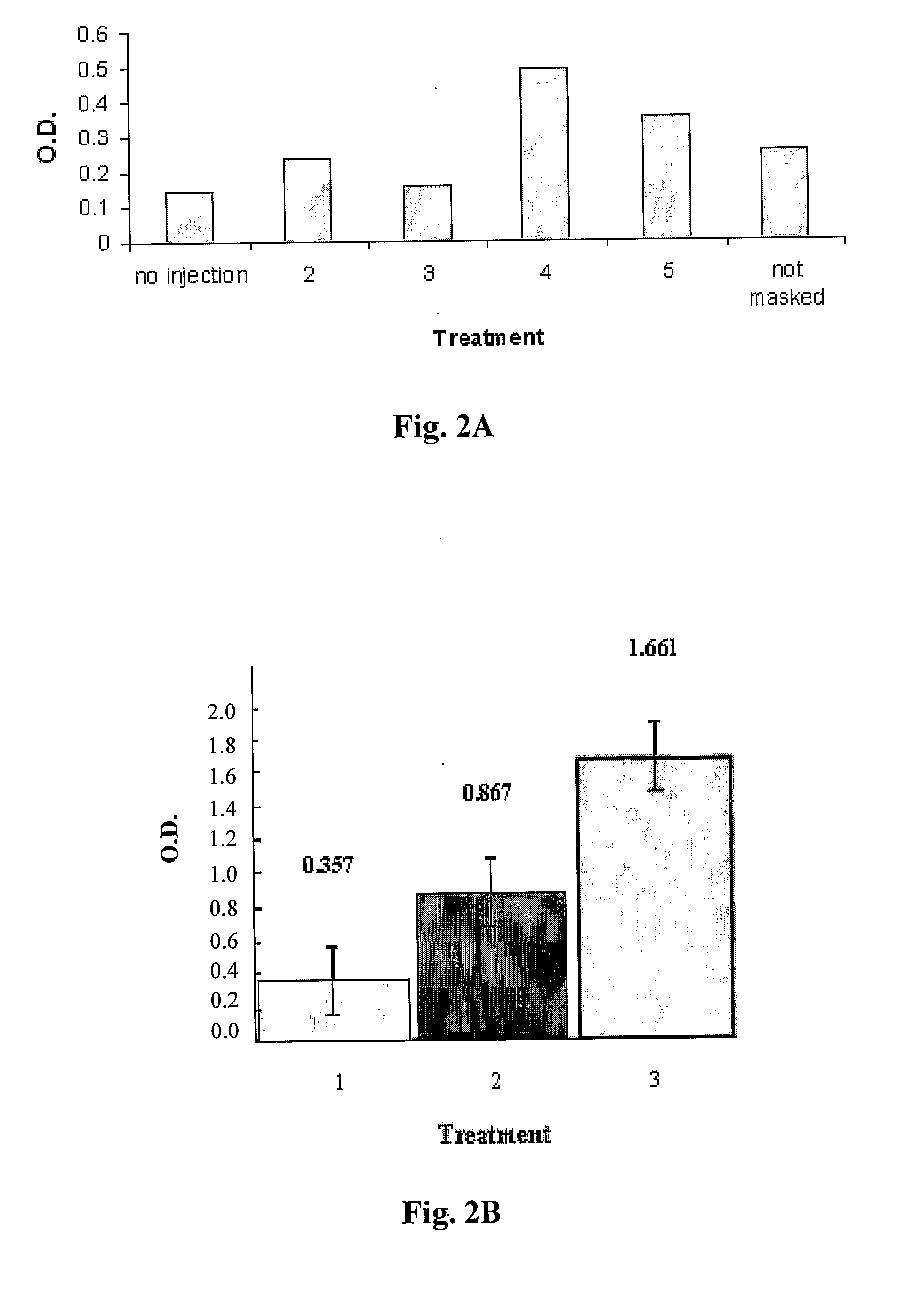
[0083]After the preparation of antibody molecules coated by small molecules as described in Examples 1 and 2 above, it was of interest to mask the antibody surface in an antibody-antigen complex according to the method of the present invention. For this purpose, IgY obtained from chicken injected with E. coli was complexed with the whole bacteria as described below.
[0084]Heat killed virulent E. coli O78:K80 was injected to chicken (Leghom layers, n=4), twice in two-weeks interval and antibodies were isolated from egg yolk. The purified chicken IgY fraction obtained from the egg yolk was incubated with the virulent E. coli O78:K80 (108) for 2 hours at 37° C. The complex was centrifuged and washed twice in PBS. The pellet containing the complex was suspended in PBS buffer and coated with D(+)-mannose as described in Example 1 (ii). The mannose-coated complex was washed twice following centrifugation, to separate free unattached...
PUM
| Property | Measurement | Unit |
|---|---|---|
| pH | aaaaa | aaaaa |
| size | aaaaa | aaaaa |
| pH | aaaaa | aaaaa |
Abstract
Description
Claims
Application Information
 - R&D
- Intellectual Property
- Life Sciences
- Materials
- Tech Scout
- Unparalleled Data Quality
- Higher Quality Content
- 60% Fewer Hallucinations
Browse by: Latest US Patents, China's latest patents, Technical Efficacy Thesaurus, Application Domain, Technology Topic, Popular Technical Reports.
© 2025 PatSnap. All rights reserved.Legal|Privacy policy|Modern Slavery Act Transparency Statement|Sitemap|About US| Contact US: help@patsnap.com


