Methods and systems of combining magnetic resonance and nuclear imaging

a magnetic resonance and nuclear imaging technology, applied in the field of methods, can solve the problems of increasing signal noise and distortion, theoretic benefits of trying to combine spect and mri within a single system, and the inability to combine a gamma photon detector with a single system

Active Publication Date: 2008-05-15
GAMMA MEDICA +1
View PDF5 Cites 3 Cited by
  • Summary
  • Abstract
  • Description
  • Claims
  • Application Information

AI Technical Summary

Benefits of technology

[0010] An aspect of the present invention provides a dual-modality, fused image dataset from MRI and single-photon nuclear medicine imaging modalities in a single imaging session. The single imaging session allows a body (e.g., a human or animal body) or other object being scanned to remain motionless for sequential scanning while using the same body position on the same bed, thereby minimizing mis-registration artifacts from changes in body orientation between imaging studies. The single session also allows the simultaneous operation of the two modalities, providing exact co-registration in both position as well as in time. The ability to perform fused dual-modality imaging is helpful in both clinical imaging as well as pre-clinical research studies involving humans or laboratory animals for the development of drugs and therapies or the general study of biological processes.
[0030] In one embodiment, the method further includes the steps of generating at least one direct detection signal in response to detecting the gamma photons by the at least one semiconductor detector; and receiving the detection signal by a signal processor having a plurality of electronics adapted to amplify, address, and process the detection signal; and removing an interference effect of the magnetic filed suitable for magnetic resonance imaging on the single photon nuclear imaging by positioning the signal processor away from the magnetic field suitable for magnetic resonance imaging.

Problems solved by technology

Although there may be benefits to combine SPECT and MRI, any theoretical benefits of trying to combine SPECT and MRI within a single system have been mostly dismissed because the functions of the PMTs in a typical SPECT system are severely compromised by the high magnetic fields needed for MRI and because magnetic field uniformity needed for MRI is distorted by the PMTs (i.e., the ferro-magnets in the PMTs).
However, combining a CZT detector for detecting gamma photons is still not a trivial task because the electrons and holes of the CZT detector need to travel non-negligible distances to generate their signals (e.g., travel distances of 2-5 mm and even larger).
However, the electronics located away from the magnetic field need to be connected via relatively long cables that result in an increased signal noise and distortion.

Method used


Image

  • Methods and systems of combining magnetic resonance and nuclear imaging
  • Methods and systems of combining magnetic resonance and nuclear imaging
  • Methods and systems of combining magnetic resonance and nuclear imaging

Examples


Embodiment Construction

[0050] In the following detailed description, only certain exemplary embodiments of the present invention are shown and described, by way of illustration. As those skilled in the art would recognize, the described exemplary embodiments may be modified in various ways, all without departing from the spirit or scope of the present invention. Accordingly, the drawings and description are to be regarded as illustrative in nature, and not restrictive.

[0051] An embodiment of the present invention is designed to enhance the MRI imaging by incorporating an additional modality in the same gantry as operated by an MRI machine. The added modality is SPECT, limited-angle SPECT, or planar imaging based on the single photon emission principle.

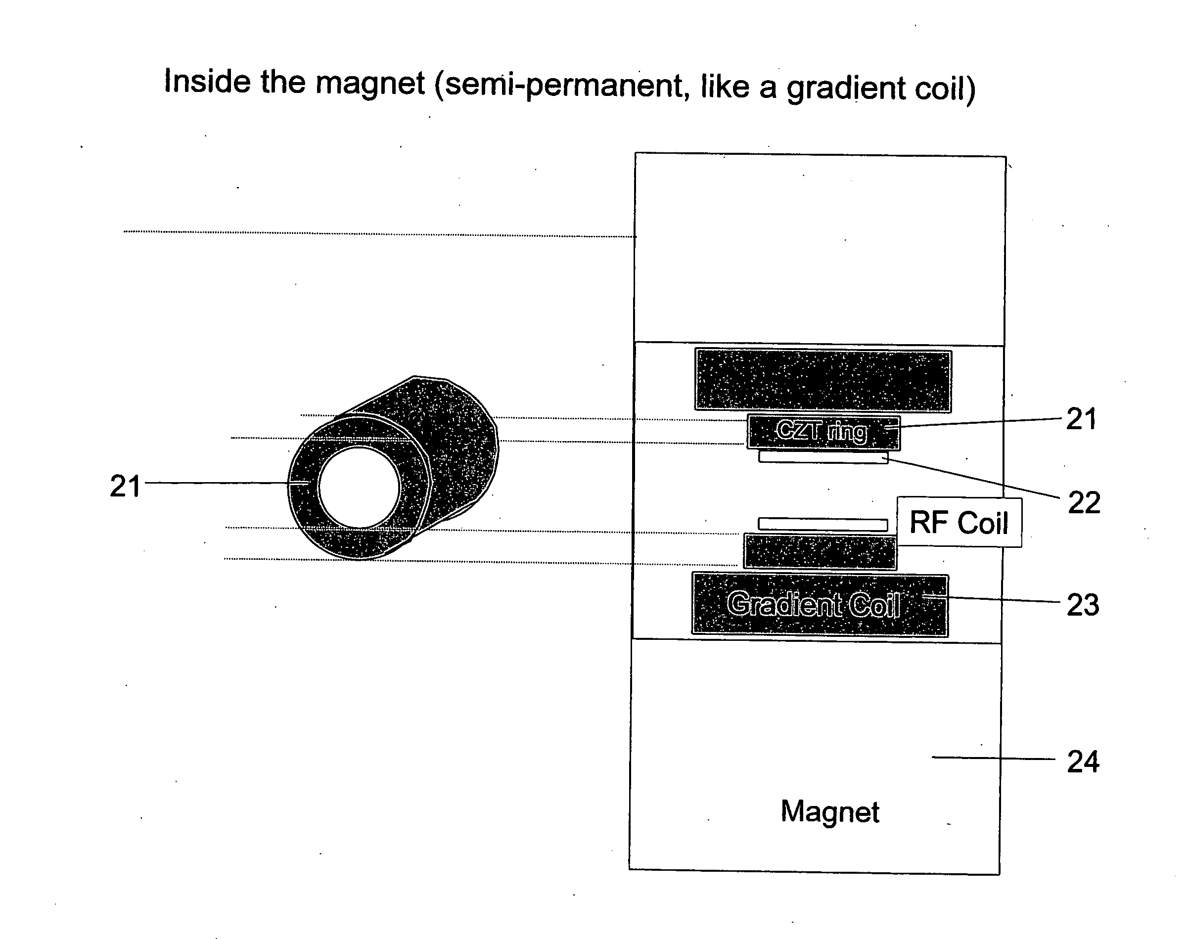
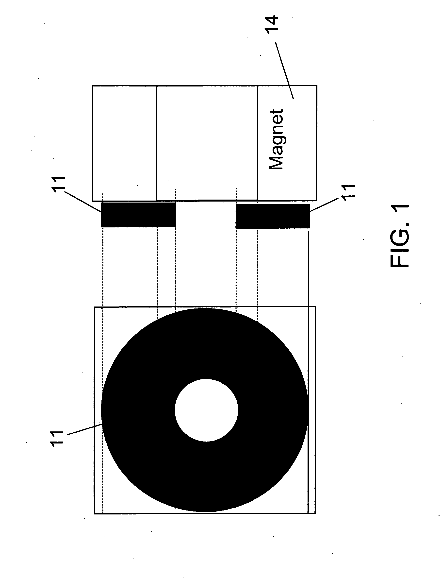
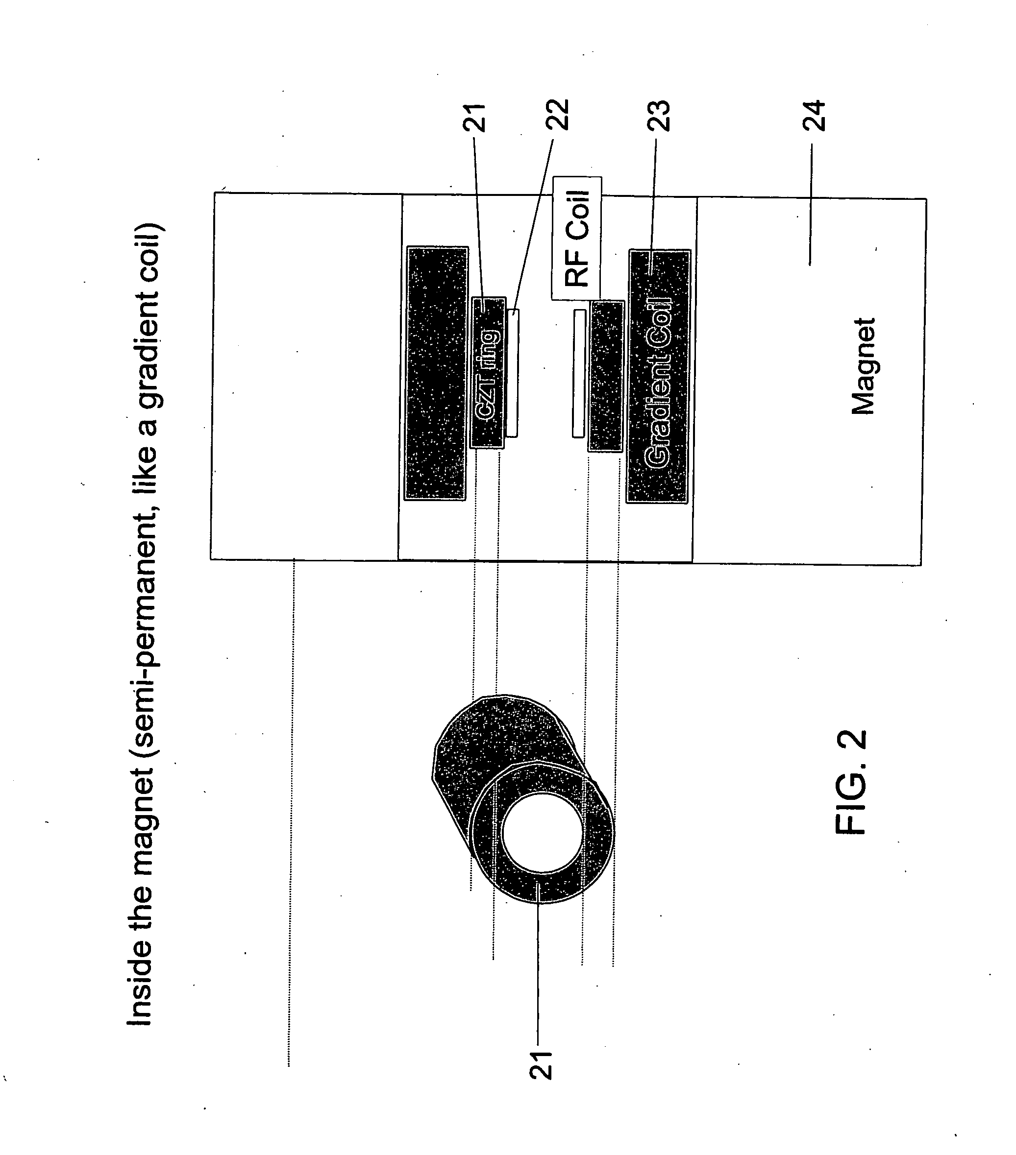
[0052] In one embodiment of the present invention, the single photon emission imagers are based on a semiconductor direct conversion detector, such as a cadmium zinc telluride (CZT) detector. The embodiment of the present invention reduces the possibility ...


PUM


Abstract

A multi-modality imaging system for imaging of an object under study, e.g., a whole body or parts of the body of animals such as humans, other primates, swine, dogs, rodents (especially mice and rats), that includes a magnetic resonance imaging apparatus and a cadmium zinc telluride (CZT)-family semiconductor, single-photon imaging apparatus in close proximity such that sequential or simultaneous imaging can be done with the two modalities using the same support bed of the object under study in the same, uninterrupted imaging session.

Description

CROSS-REFERENCE TO RELATED APPLICATION [0001] This application claims priority to and the benefit of U.S. Provisional Application No. 60 / 858,773 filed Nov. 10, 2006, the entire content of which is incorporated herein by reference.FIELD OF THE INVENTION [0002] The invention relates generally to multi-modality medical imaging. More particularly, the invention relates to methods and systems for combining magnetic resonance imaging (MRI) with single photon nuclear imaging, such as single photon emission computed tomography (SPECT). BACKGROUND OF THE INVENTION [0003] Magnetic resonance imaging is an imaging technique used to visualize the inside of an object (or subject) under study (e.g., a human or animal body or a body part or an entire laboratory animal or specimen from the animal or a plastic test phantom). MRI relies on the relaxation properties of excited hydrogen nuclei in water and fat. When the object to be imaged is placed in a powerful, uniform magnetic field, the spins of th...

Claims


Application Information

Patent Timeline
Patent Type & Authority Applications(United States)
IPC IPC(8): G01T1/24
CPCG01R33/481G01T1/249G01T1/1603
Inventor WAGENAAR, DOUGLAS J.PATT, BRADLEY E.
Owner GAMMA MEDICA
Why Eureka
Try Eureka