Mechanical tissue device and method

a tissue device and mechanical technology, applied in the field of mechanical tissue devices and methods, can solve the problems of high resistance to blood flow, high hemodynamic problems that can be fatal, and very rare patent ductus venosus after birth, and achieve the effect of increasing the length of the device, reducing the width, and ensuring the patient's comfor

Inactive Publication Date: 2008-05-22
SEPTRX
View PDF2 Cites 249 Cited by
  • Summary
  • Abstract
  • Description
  • Claims
  • Application Information

AI Technical Summary

Benefits of technology

[0020]The mesh is removably is secured to at least one or more bases of the frame, and positioned between the arms thereof. The bases of the frame and the fasteners which secure the tubular mesh can be collars, for example, having central lumens. The aforementioned third-fastener is insertable into the lumen of at least one of the bases of the frame in order to secure the mesh to the frame. The lumens of the fasteners and bases are aligned along a common axis in order that a the embolic filtering device can be loaded onto a guide wire.
[0021]The frame can include at least one base and at least two arms which extend therefrom, between which the mesh is at least partially disposed. The frame can be made of metal, fabric and / or a polymer. The arms are positioned opposite one another and, in their resting state, are spaced apart from one another. When the device is composed of a shape memory metal, such as nitinol, the device can be collapsed into a catheter tube by compressing the arms of the frame toward one another, causing the length of the device to increase, and the width to decrease. As the device is released from the catheter tube, it reverts to its functional, relaxed state. The embolic filtering device may also be composed of non-shape memory metals, such as Elgiloy, cobalt chromium, and stainless steel, for example. Each arm includes at least one anchor positioned on the arms of the frames. The anchors can either be arcuate or linear in formation, depending on the shape of the patent foramen ovale to be treated, and are of sufficient rigidity to secure the device within the lumen of a septal defect.
[0022]To allow for non-invasive visualization of the device within a subject at least a portion of the frame or mesh is composed of or coated with a radiopaque material, such as tantalum. The device may also be treated with thrombin, collagen, hyluron, or a host growth factor to encourage and facilitate growth of tissue onto the device so as to further secure the device within the septal defect. The device can also be coated with an anticoagulant to deter formation of blood clots on the surface of the device.
[0023]In an exemplary embodiment, the mesh is composed of at least 96 strands of 0.002″ diameter wire braided such that the wires are situated at an angle of 35.degree. relative to the longitudinal axis of the device. The interstices created by the braided wires are small enough such as to effectively filter emboli, thereby preventing emboli from passing through the patent foramen ovale, or other septal defect.

Problems solved by technology

Unfortunately, they sometimes fail to close and create hemodynamic problems that can be fatal if left untreated.
However, the fetal lungs are collapsed which causes a high resistance to blood flow.
A patent ductus venosus after birth is very rare and almost always fatal.
Unfortunately, each of these devices have distinct disadvantages and limitations ranging from the size of the device delivery sheath, ease of implantation, feasibility, safety and effectiveness.
The major disadvantage with this device is the lack of a centering mechanism.
Consequently, closure of defects may become difficult because the required size may be too large for the atrial septum to accommodate, or the device may impinge critical structures.
There are also reports that the retrieval of the Sideris button device after incorrect deployment is difficult.
The major disadvantage of using this device is the attendant risk of aortic perforation cause by its sharp eyelet corners.
In fact, the Angel Wings device was withdrawn from further clinical trials because of this problem.
The device is also ill-suited for treating fenestrated defects.
While the device is retrievable in the event of malpositioning before release of the device, it requires a complex procedure to implant, and the components are known to have a high incidences of thrombosis.
It is also reported that frame fractures have been detected in 20% of the patients treated with this device.
The primary disadvantage with this device is that it is ill-suited for closing fenestrated defects.
Moreover, the device is a thick, bulky profile which dramatically increases the chances that the device will interfere with the heart's operation.
Another disadvantage is its known capacity for incomplete endothelialisation with thrombus formation.
While the CardioSEAL is deemed to be relative easy to use, it is reported that, of all the devices, the CardioSEAL device has the highest incidence of arm fractures, which has raised serious issues concerning its safety.
Moreover, the CardioSEAL device, like the Amplatzer device is relatively large, and requiring at least a 10 F or 11 F delivery systems, and an undue amount of hardware within the heart.
These characteristics increase the chance that the device will interfere with the heart's operation, lend to residual shunting and / or embolization.
The size of the CardioSEAL device also renders it less suitable for small children.
While this added feature may reduce the instances of residual shunting, the aforementioned defects and disadvantages of the CardioSEAL are still a concern.
Surgery, however, is obviously associated with the usually risks of general anesthesia, open heart procedures, infections, etc.

Method used


Image

  • Mechanical tissue device and method
  • Mechanical tissue device and method
  • Mechanical tissue device and method

Examples


Embodiment Construction

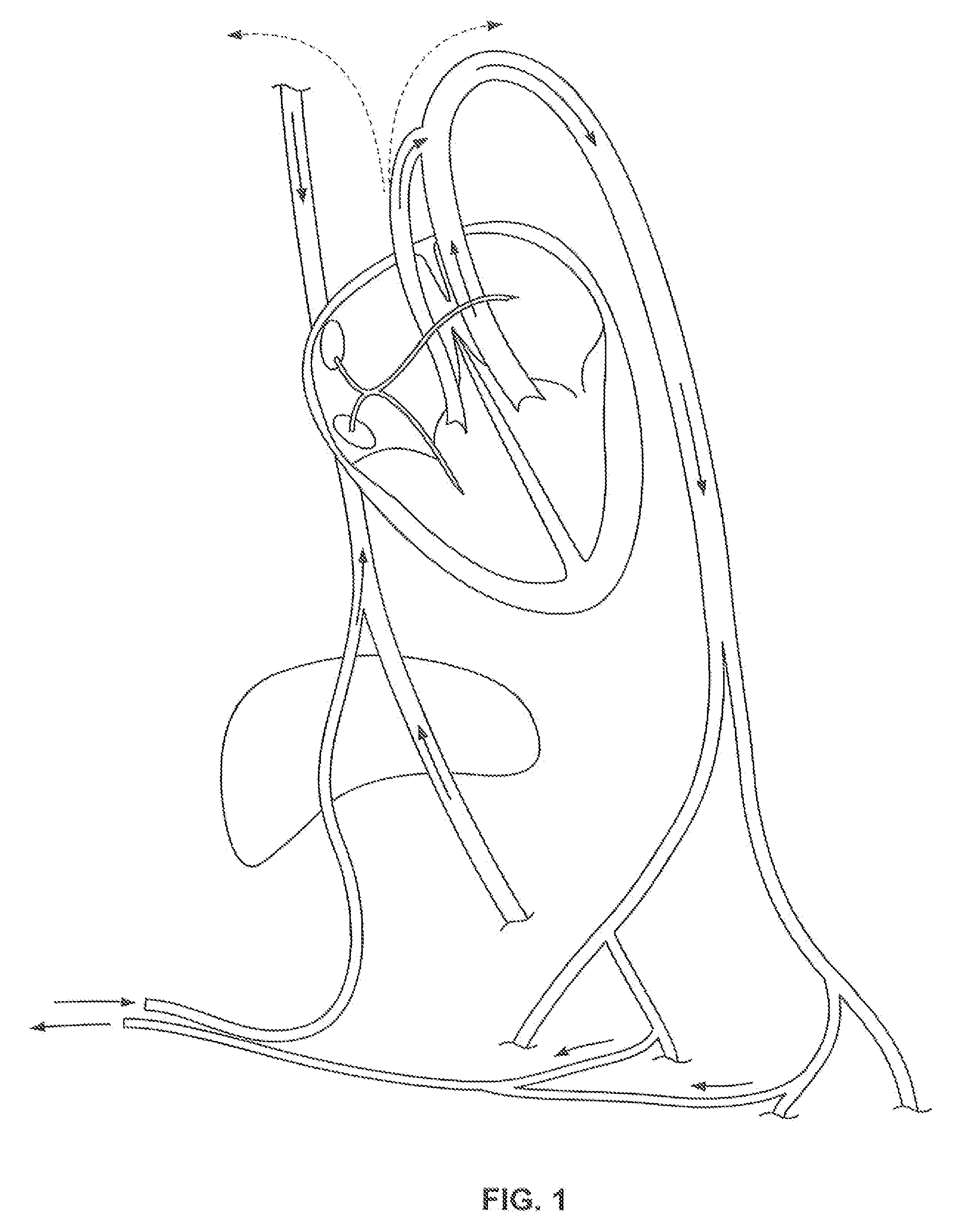
[0039]Disclosed are methods and apparatuses for preventing the passage of emboli between a venous blood pool and an arterial blood pools using devices for creating a barrier to the conducting of emboli at a passage between a venous blood pool and an arterial blood pool. The device can treat cardiac defects, such as patent foramen ovale or other atrium septal defects. Although referred to as a filtering device, the device can work by any mechanism including or not including filtering. For example, the embolic filtering device can act as a scaffold for tissue to grow.

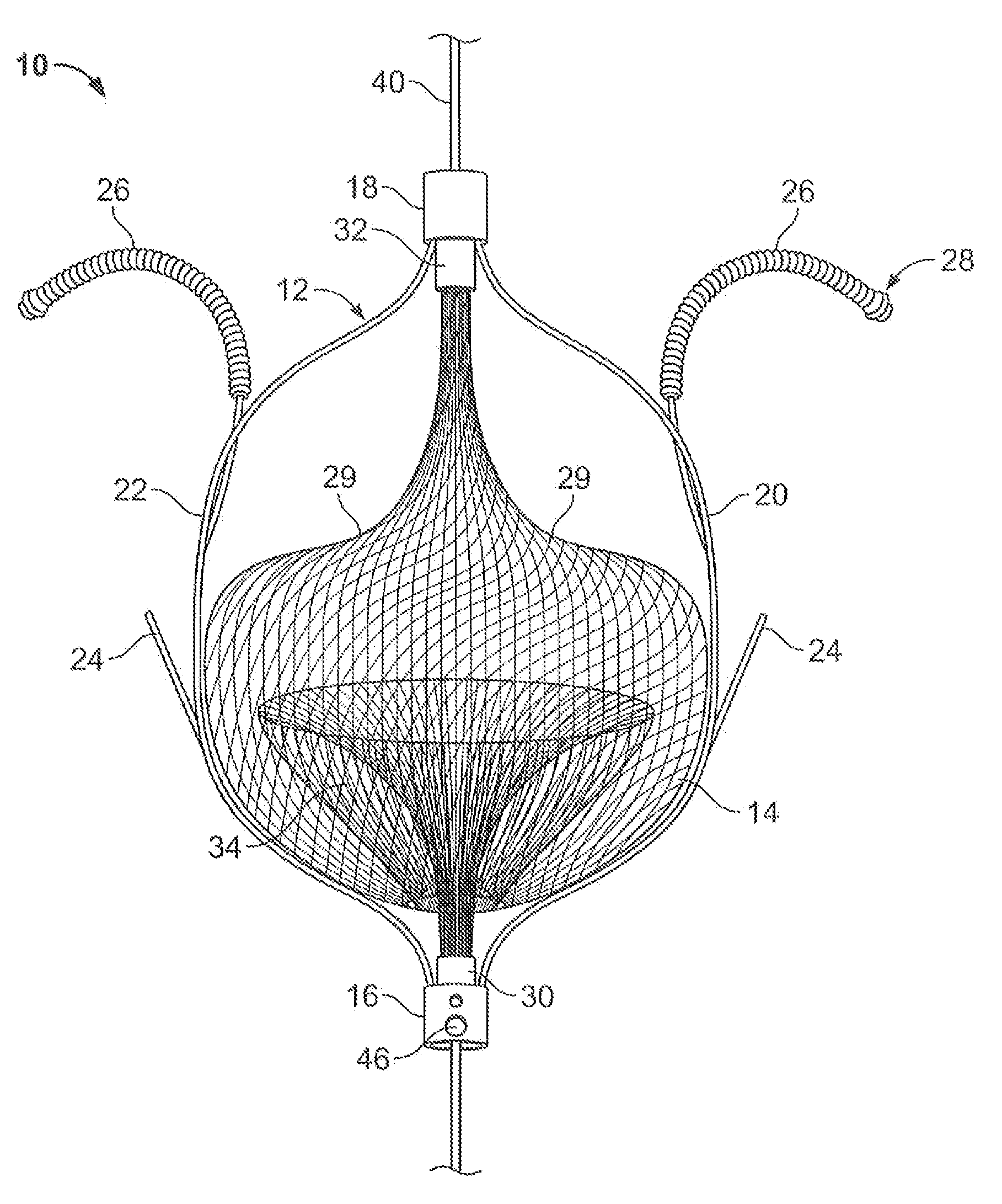
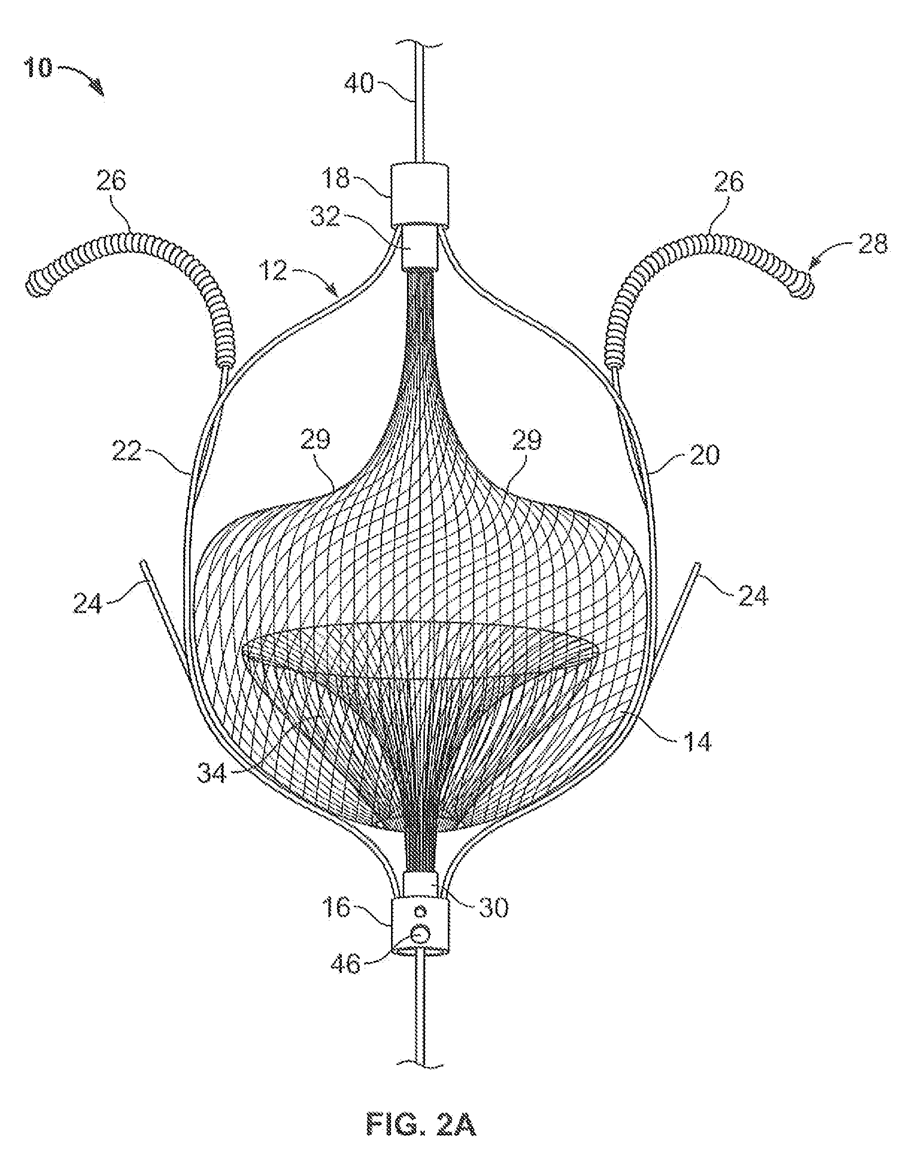
[0040]FIG. 2A illustrates an embolic filtering device 10 comprising a frame 12 and an embolic filter 14 comprising a mesh of stranded fabric, wire, or combination thereof. Any and / or all elements of the embolic filtering device 10, including the frame 12 and the embolic filter 14, can be entirely or partially biodegradable and / or bio-inert (e.g., non-biodegrading). After being deployed in the patient, the embolic filterin...


PUM


Abstract

The present invention relates generally to a device and method for preventing the undesired passage of emboli from a venous blood pool to an arterial blood pool. The invention relates especially to a device and method for treating certain cardiac defects, especially patent foramen ovales and other septal defects, through the use of an embolic filtering device capable of instantaneously deterring the passage of emboli from the moment of implantation. The device consists of a frame, and a braided mesh of sufficient dimensions to prevent passage of emboli through the mesh. The device is preferably composed of shape memory allow, such as Nitinol, which conforms to the shape and dimension of the defect to be treated.

Description

CROSS-REFERENCE TO RELATED APPLICATIONS[0001]This application claims the benefit of U.S. Provisional Application Nos. 60 / 860,393, filed 20 Nov. 2006; and 60 / 866,847, filed 21 Nov. 2006, which are incorporated by reference herein in their entireties.BACKGROUND OF THE INVENTION[0002]1. Field of the Invention[0003]The present invention relates generally to a device and method for preventing the undesired passage of emboli from a venous blood pool to an arterial blood pool. The invention relates especially to a device and method for treating certain cardiac defects, especially patent foramen ovales and other septal defects through the use of an embolic filtering device capable of instantaneously deterring the passage of emboli from the moment of implantation.[0004]2. Description of Related Art[0005]The fetal circulation is vastly different than the normal adult circulation. The blood circulating in a fetus is oxygenated by the placenta, not the developing lungs. Therefore, the fetal cir...

Claims


Application Information

Patent Timeline
Patent Type & Authority Applications(United States)
IPC IPC(8): A61M29/00
CPCA61F2230/0076A61F2230/0006A61F2230/008A61B17/0057A61B17/12022A61B17/12122A61B17/12168A61B17/12172A61B17/12177A61B17/221A61B2017/00575A61B2017/00632A61B2017/00867A61B2017/12054A61F2/01A61F2002/018A61F2230/0078
Inventor GREENHALGH, E. SKOTTKLESHINSKI, STEPHEN J.
Owner SEPTRX
Why Eureka
Try Eureka