Analysis of rare cell-enriched samples

a cell enrichment and sample technology, applied in the field of rare cell enrichment sample analysis, can solve the problems of unreliable testing, many cancers continue to go undetected, and the amniocentesis and chorionic villus sampling (cvs) are potentially harmful to the mother and the fetus

Inactive Publication Date: 2008-05-29
GPB SCI +2
View PDF99 Cites 169 Cited by
  • Summary
  • Abstract
  • Description
  • Claims
  • Application Information

AI Technical Summary

Problems solved by technology

However, the available methods today, amniocentesis and chorionic villus sampling (CVS) are potentially harmful to the mother and to the fetus.
For example, maternal serum alpha-fetoprotein, and levels of unconjugated estriol and human chorionic gonadotropin are used to identify a proportion of fetuses with Down's syndrome, however, these tests are not one hundred percent accurate.
Moreover, despite decades of advances in cancer diagnosis and therapy, many cancers continue to go undetected until late in their development.
However, fetal cells and cancer cells are rare as compared to the presence of other cells in the blood.
Enriching fetal cells from maternal peripheral blood and cancer cells from patient's blood is challenging, time intensive and any analysis derived there from is prone to error.

Method used


Image

  • Analysis of rare cell-enriched samples
  • Analysis of rare cell-enriched samples
  • Analysis of rare cell-enriched samples

Examples


example 1

Separation of Fetal Cord Blood

[0159]FIGS. 14A-14D shows a schematic of the device used to separate nucleated cells from fetal cord blood.

[0160]Dimensions: 100 mm×28 mm×1 mm

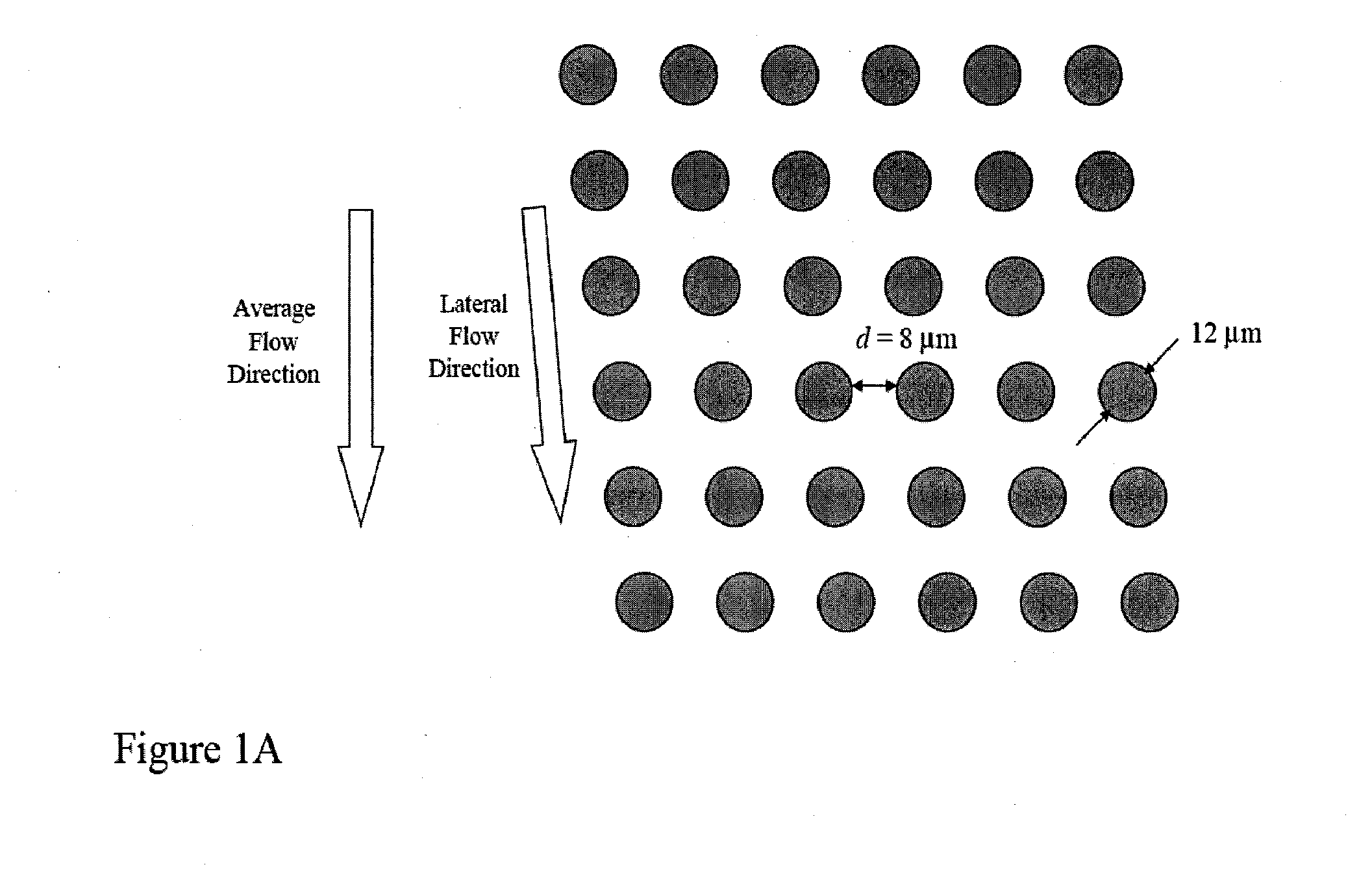
[0161]Array design: 3 stages, gap size=18, 12 and 8 μm for the first, second and third stage, respectively.

[0162]Device fabrication: The arrays and channels were fabricated in silicon using standard photolithography and deep silicon reactive etching techniques. The etch depth is 140 μm. Through holes for fluid access are made using KOH wet etching. The silicon substrate was sealed on the etched face to form enclosed fluidic channels using a blood compatible pressure sensitive adhesive (9795, 3M, St Paul, Minn.).

[0163]Device packaging: The device was mechanically mated to a plastic manifold with external fluidic reservoirs to deliver blood and buffer to the device and extract the generated fractions.

[0164]Device operation: An external pressure source was used to apply a pressure of 2.0 PSI to the buffer and blood r...

example 2

Isolation of Fetal Cells from Maternal Blood

[0168]The device and process described in detail in Example 1 were used in combination with immunomagnetic affinity enrichment techniques to demonstrate the feasibility of isolating fetal cells from maternal blood.

[0169]Experimental conditions: blood from consenting maternal donors carrying male fetuses was collected into K2EDTA vacutainers (366643, Becton Dickinson, Franklin Lakes, N.J.) immediately following elective termination of pregnancy. The undiluted blood was processed using the device described in Example I at room temperature and within 9 hrs of draw. Nucleated cells from the blood were separated from enucleated cells (red blood cells and platelets), and plasma delivered into a buffer stream of calcium and magnesium-free Dulbecco's Phosphate Buffered Saline (14190-144, Invitrogen, Carlsbad, Calif.) containing 1% Bovine Serum Albumin (BSA) (A8412-100ML, Sigma-Aldrich, St Louis, Mo.). Subsequently, the nucleated cell fraction was ...

example 3

Amplification and Sequencing of STRs for Fetal Diagnosis

[0172]Fetal cells or nuclei can be isolated as describe in the enrichment section or as described in example 1 and 2. DNA from the fetal cells or isolated nuclei from fetal cells can be obtained using any methods known in the art. STR loci can be chosen on the suspected trisomic chromosomes (X, 13, 18, or 21) and on other control chromosomes. These would be selected for high heterozygosity (variety of alleles) so that the paternal allele of the fetal cells is more likely to be distinct in length from the maternal alleles, with resulting improved power to detect. Di-, tri-, or tetra-nucleotide repeat loci can be used. The STR loci can then be amplified according the methods described in the amplification section.

[0173]For instance, the genomic DNA from the enriched fetal cells and a maternal control sample can be fragmented, and separated into single strands. The single strands of the target nucleic acids would be bound to beads...


PUM

PropertyMeasurementUnit
sizeaaaaaaaaaa
sizeaaaaaaaaaa
timeaaaaaaaaaa

Abstract

The present invention relates to methods for detecting, enriching, and analyzing rare cells that are present in the blood, e.g., epithelial cells. The invention further features methods of analyzing rare cell(s) to determine the presence of an abnormality, disease or condition in a subject by analyzing a cellular sample from the subject.

Description

CROSS REFERENCE TO RELATED APPLICATIONS[0001]This application claims priority, under 35 U.S.C. §119, to U.S. provisional patent application Nos. 60 / 804,819 and 60 / 804,817 both filed on Jun. 14, 2006 and incorporated herein by reference in their entirety.BACKGROUND OF THE INVENTION[0002]Analysis of specific cells can give insight into a variety of diseases. These analyses can provide non-invasive tests for detection, diagnosis and prognosis of diseases, thereby eliminating the risk of invasive diagnosis. For instance, social developments have resulted in an increased number of prenatal tests. However, the available methods today, amniocentesis and chorionic villus sampling (CVS) are potentially harmful to the mother and to the fetus. The rate of miscarriage for pregnant women undergoing amniocentesis is increased by 0.5-1%, and that figure is slightly higher for CVS. Because of the inherent risks posed by amniocentesis and CVS, these procedures are offered primarily to older women, i...

Claims


Application Information

Patent Timeline
Patent Type & Authority Applications(United States)
IPC IPC(8): C12Q1/68G01N33/574C12M1/34
CPCG01N33/5091G01N33/57484G01N2015/1087G01N33/5005G01N2015/1006G01N2800/385C12Q1/6886C12Q2600/156G01N1/405
Inventor FUCHS, MARTINKAPUR, RAVITONER, MEHMETWANG, ZIHUA
Owner GPB SCI
Why Eureka
Try Eureka