Method of Detecting Hepatitis B Virus S Antigen

Active Publication Date: 2008-08-14
ADVANCED LIFE SCI INST
View PDF2 Cites 6 Cited by
  • Summary
  • Abstract
  • Description
  • Claims
  • Application Information

AI Technical Summary

Benefits of technology

[0029]By the probe of the present invention and a method of detecting HBV or HBs antigen by using the same, an escape mutant etc. having a mutation on common “a” determinant of HBs antigen, which cannot be detected by the conventional HBs antigen detection method, can be highly sensitively detected thereby highly reliably judging HBV infection. Even if a patient's antibody to the common “a” determinant of HBs antigen inhibits detection of HBs antigen, HBV infection can be reliably judged by the detection me

Problems solved by technology

One problem associated with occurrence of the escape mutant is that this mutant can keep persistent infection because it can escape from an antibody induced by inoculation with a vaccine utilizing the “a” determinant before mutation.
Another problem is that this escape mutant cannot be detected in conventional HBs antigen examination methods.
Generally, the escape mutant having a mutati

Method used


Image

  • Method of Detecting Hepatitis B Virus S Antigen
  • Method of Detecting Hepatitis B Virus S Antigen

Examples


Example

Example 1

Expression and Purification of TrpE-HBs (26 to 80) Antigen

(A) Construction of TrpE-HBs (26 to 80) Antigen-Expressing Plasmid

[0053]An expression plasmid for HBs (26 to 80) region was constructed by the following method. 100 μl serum from an HBV patient was mixed with 100 μl DNA extract [10 μl of 1 M Tris-HCl (pH 8.4), 8 μl of 250 mM EDTA, 40 μl of 10% SDS, 8 μl of 5 M NaCl, 10 μl of 20 mg / ml. Proteinase K, 1 μl tRNA (5 μg / μl), and 23 μl sterilized water] and incubated at 54° C. for 30 minutes. The sample was mixed with 200 μl phenol / chloroform (1 / 1) solution and then centrifuged at 15 Krpm for 5 minutes to give a supernatant, and 150 μl isopropanol and 7 μl of 5 M NaCl were added to the supernatant and left at −20° C. for 1 hour. After centrifugation at 15 Krpm at 4° C. for 5 minutes, the precipitates were rinsed with 70% ethanol and then centrifuged again at 15 Krpm at 4° C. for 5 minutes. The precipitates were air-dried and dissolved in 20 μl sterilized water to give an HB...

Example

Example 2

Preparation of Hybridoma

[0061]The polypeptide [trpE-HBs (26 to 80)] prepared by the method described above was dissolved with 6M urea and then diluted at a final concentration of 0.2 to 1.0 mg / ml in 10 mM phosphate buffer (pH 7.3) containing 0.15 M NaCl (PBS), then mixed with an equal volume of Freund's adjuvant, and administered intraperitoneally in a dose of 10 to 20 μg to a 4- to 6-week-old BALB / c mouse.

[0062]Booster was carried out every 2 to 4 weeks in the same manner as above, and for final immunization, 10 μg HBs dissolved in PBS was administered to the caudal vein.

[0063]At three days after the final immunization, the spleen was aseptically removed from the mouse, then broken into individual cells with scissors and a metallic mesh and washed 3 times with RPMI-1640 medium. Mouse myeloma cell strain Sp2 / OAg14 at the logarithmic growth phase was washed 3 times with RPMI-1640 medium, and the cells were mixed with the spleen cells at a ratio of 1:5. After centrifugation a...

Example

Example 3

Preparation and Analysis of Monoclonal Antibody

[0066]Each of the hybridomas obtained by the method described in Example 2 was transplanted in a BALB / c mouse abdominal cavity previously administered with pristane, and the monoclonal antibody produced in the ascites was obtained.

[0067]The IgG fraction containing the monoclonal antibody was purified by affinity chromatography on a protein A Sepharose column.

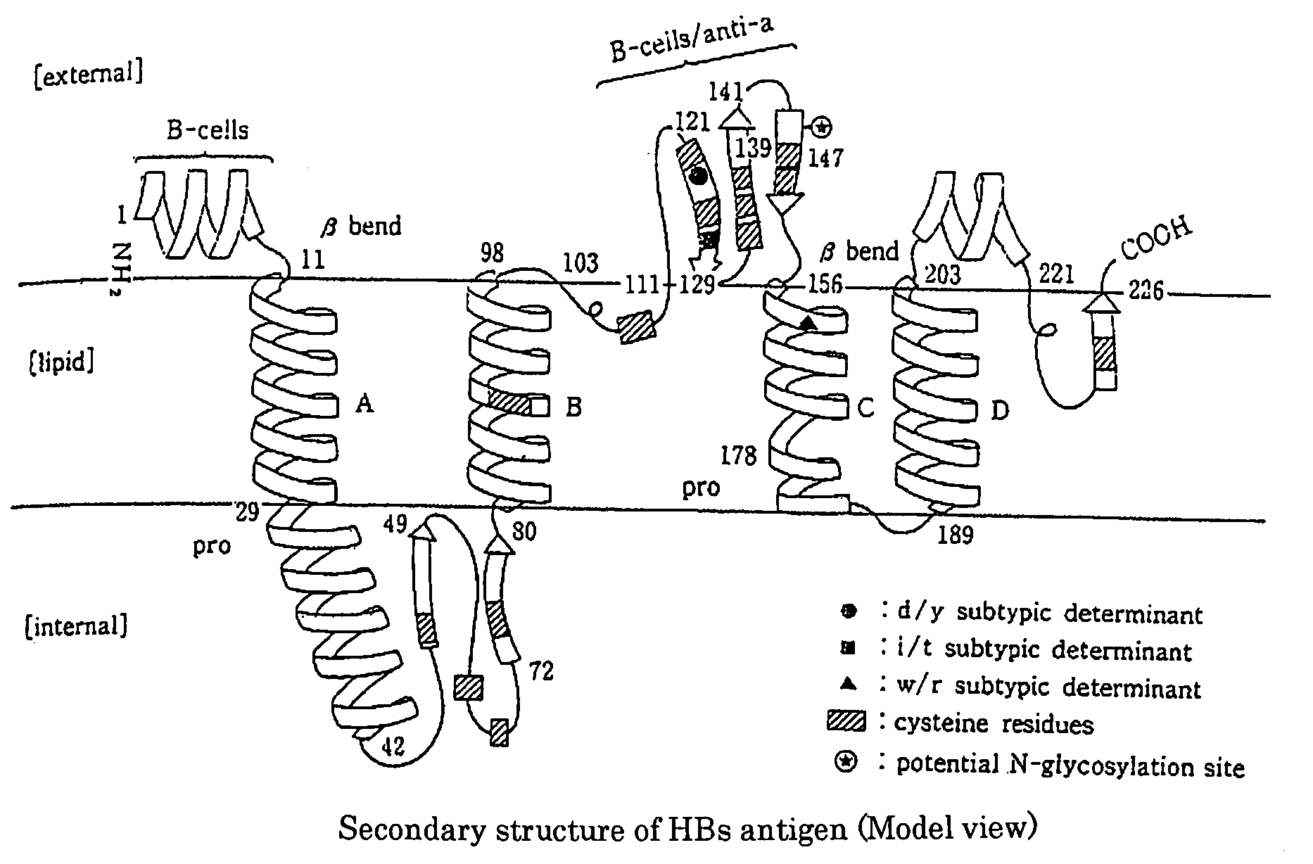
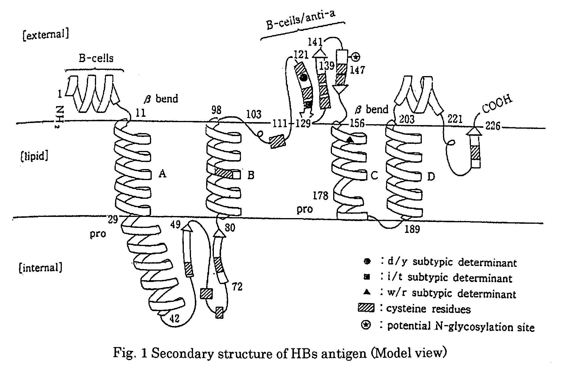
[0068]The respective obtained monoclonal antibodies were analyzed for their target epitope by using the TrpE-HBs (26 to 80) antigen and synthetic peptides each consisting of 20 amino acids synthesized on the basis of a sequence derived from the HBs region, and as a result, it was found that as shown in Table 1, these monoclonal antibodies recognize an epitope (amino acid numbers: 26 to 80) of the HBs antigen, which is located in the inside of a lipid bilayer.

[Table 1]

[0069]

TABLE 1(Poly)peptideAmino acidMonoclonal antibody namenamenumber4A36G61C10HBS-1 1-20−−−HBS-211-30−−−HB...


PUM

PropertyMeasurementUnit
Volumeaaaaaaaaaa
Volumeaaaaaaaaaa
Volumeaaaaaaaaaa

Abstract

[PROBLEMS] To provide a probe useful in the detection of HBV or HBs antigen by which an escape mutant of hepatitis B virus (HBV) possibly occurring in a specimen can be detected; and a method of using the same.
[MEANS FOR SOLVING PROBLEMS]A probe capable of recognizing an epitope located on a peptide comprising the amino acid sequence of SEQ ID NO:1; and a method of detecting hepatitis B virus or hepatitis B virus s antigen by using this probe.

Description

TECHNICAL FIELD[0001]The present invention relates to a probe recognizing a novel epitope of hepatitis B virus (HBV) s antigen (HBs antigen) and a method of detecting HBV, or HBs antigen, by using the probe.BACKGROUND ART[0002]Diagnosis of viral infection is carried out mainly by a method of detecting a virus or a virus-related component (protein or nucleic acid) or by a method of detecting a specific antibody produced by the living body upon viral infection.[0003]Usually, infection with HBV can be known by confirming the presence of HBs antigen or HBc antibody. As an indicator not only for infection with HBV but also for a clinical state of an HBV carrier or judgment of prognosis and treatment efficacy, the amount of hepatitis B virus e antigen (HBe antigen), an antibody to the antigen, or DNA of HBV (HBV-DNA) is measured.[0004]Among antigens constituting HBV viral particles (HBV particles), HBs antigen is a major constitutional envelope protein on the surface of infectious HBV par...

Claims


Application Information

Patent Timeline
IPC IPC(8): C12Q1/70C07H21/04
CPCC07K14/005G01N33/5761C12N2730/10122C07K16/082C07K14/02C12N15/02C12P21/00G01N33/531
Inventor MAKI, NOBORUFUKUDA, YASUYUKIKIMURA, TATSUJIODA, YOKOOHUE, CHIHARUKUSANO, OSAMU
Owner ADVANCED LIFE SCI INST
Why Eureka
Try Eureka