
Biocompatible fluorescent metal oxide nanoparticles

a metal oxide nanoparticle, biocompatible technology, applied in the direction of instruments, applications, drug compositions, etc., can solve the problems of limited target to background ratio, limited biocompatibility and stability, and limited development of combined optical/mri agents for molecular imaging applications

Active Publication Date: 2008-09-18
VISEN MEDICAL INC
View PDF3 Cites 77 Cited by
  • Summary
  • Abstract
  • Description
  • Claims
  • Application Information

AI Technical Summary

Benefits of technology

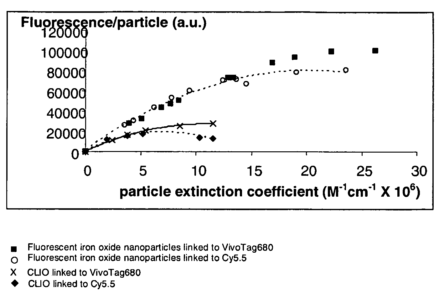
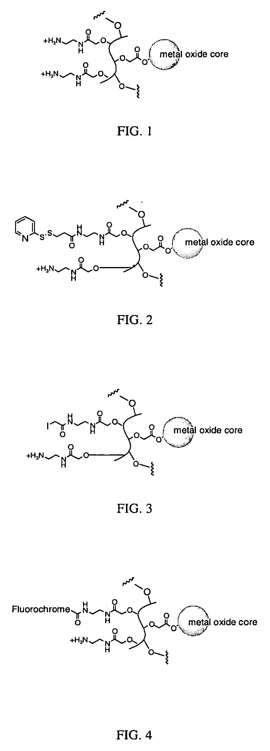
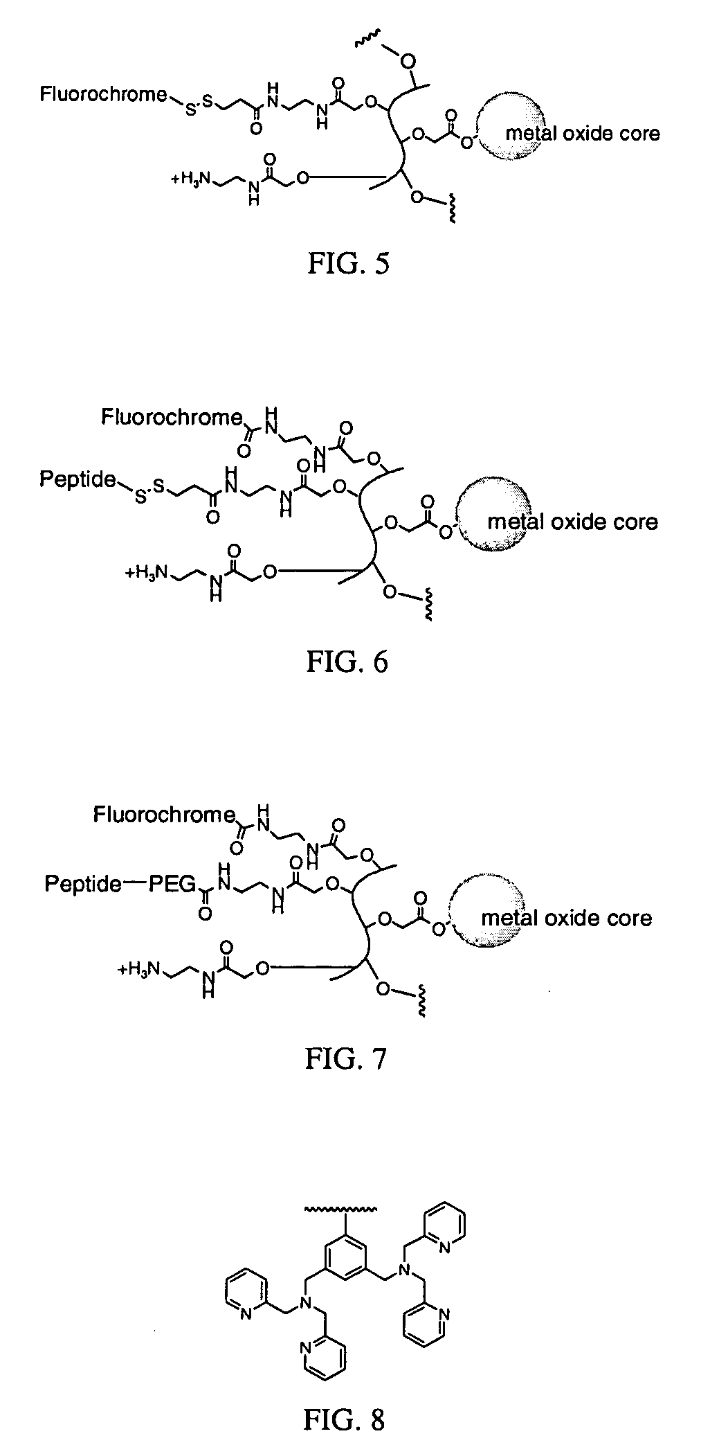
[0012]Specifically, the design of the agents of the present invention provide fluorescent metal oxide nanoparticles with one or more of the following features: (1) a polymer coating suitable for attaching a plurality of fluorochromes thereby achieving large extinction coefficients (in excess of 1,000,000 M−1cm−1), (2) a non-crosslinked polymer coating suitable for attaching from about 10 to about 300 fluorochromes per particle, (3) a polymer coating suitable for attaching a plurality of fluorochromes in a manner that does not significantly compromise the quantum yield of the fluorochromes (e.g., the nanoparticles retain at least 50% of the fluorescent signal that is created by substantially the same number of free fluorochromes when tested under the same conditions), and (4) a polymer coating that is amenable to efficient chemical linking of biomolecules with retention of their biological properties to yield molecular imaging agents. The fluorescent metal oxide nanoparticles are highly stable molecular imaging agents in vitro, both before and after chemical linking of fluorochromes and biomolecules, but yet are labile and / or degradable in vivo.

Problems solved by technology

Unfortunately, most current imaging techniques report primarily on anatomical or physiological information (e.g., magnetic resonance imaging (MRI), computed tomography (CT), ultrasound).
To date however, the development of optical, MRI, and combined optical / MRI agents for molecular imaging applications has been limited by: 1) delivery barriers standing between molecularly targeted agents and their intended molecular target, 2) the fact that many monovalent ligands (antibodies, proteins, peptides) lack the affinity needed for efficient agent targeting, 3) limited target to background ratio due to sensitivity issues related to the signal strength of the individual agent (e.g., fluorescence or fluorescent brightness) of the individual agent, especially with regards deep tissue imaging (for optical agents) and low target abundance (MRI and optical agents), and 4) the limited biocompatibility and stability of some MRI (Palmacci and Josephson, U.S. Pat. No. 5,262,176) and optical agents (Bhatia et al., Nano Lett.

Method used


Image

  • Biocompatible fluorescent metal oxide nanoparticles
  • Biocompatible fluorescent metal oxide nanoparticles
  • Biocompatible fluorescent metal oxide nanoparticles

Examples


example 1

Synthesis of an Exemplary Fluorescent Metal Oxide Nanoparticle

[0162]Polyvinyl alcohol (6 kDa, Polysciences, 30 g) was dissolved in 140 mL of water by heating to 90° C. The solution was cooled and transferred to a 500 mL jacketed beaker equilibrated to 15° C. 100 mL of 50% NaOH was added slowly with vigorous stirring using an overhead stirrer until a smooth, relatively homogeneous suspension formed. Bromoacetic acid (100 g) was dissolved in 50 mL of water and added slowly with vigorous stirring, making sure the temperature stayed below 35° C. After complete addition, the mixture was stirred for 15 hours at room temperature, and then neutralized with 6 M HCl to pH ˜7. Carboxymethyl-polyvinylalcohol was precipitated by addition of cold ethanol, filtered, washed with 70% ethanol, pure ethanol, and ether and then dried under vacuum. The carboxyl content (in μmol / g) of the polymer was determined by titration to neutrality of an acidified sample of the polymer. A range of 450-850 μmol / g wa...

example 2

Synthesis of an Exemplary Fluorescent Metal Oxide Nanoparticle with Polyethyleneglycol

[0166]The nanoparticle of Formula I from Example 1 (500 μL, 6.8 mg / mL iron) and 50 μL 0.1 M HEPES, pH 7, was combined with 8 mg of mPEG-SPA (5 kDa) and allowed to react at room temperature overnight. The pegylated particles were purified by gel filtration using Biorad Biogel P-60 eluting with 1×PBS to yield a nanoparticle of Formula II.

example 3

Synthesis of Exemplary Succinylated Fluorescent Metal Oxide Nanoparticles

[0167]Nanoparticles of Formula I from Example 1 (600 μL, 2.5 mg / mL iron) and 50 μL 0.1 M HEPES, pH 7, was combined with succinic anhydride (5 mg, 50 μmol) and reacted at room temperature for 5 hours. The succinylated particles were purified by gel filtration using Biorad Biogel P-60 eluting with 1×PBS to yield nanoparticles of Formula III.


PUM

PropertyMeasurementUnit
diameteraaaaaaaaaa
wavelengthsaaaaaaaaaa
diameteraaaaaaaaaa

Abstract

The invention relates to highly fluorescent metal oxide nanoparticles to which biomolecules and other compounds can be chemically linked to form biocompatible, stable optical imaging agents for in vitro and in vivo applications. The fluorescent metal oxide nanoparticles may also be used for magnetic resonance imaging (MRI), thus providing a multi modality imaging agent.

Description

RELATED APPLICATIONS[0001]This application claims the benefit and priority to U.S. Provisional Patent Application Ser. No. 60 / 753,805, filed Dec. 22, 2005, and U.S. Provisional Patent Application Ser. No. 60 / 783,460, filed Mar. 16, 2006, the disclosures of which are incorporated by reference herein.GOVERNMENT SUPPORT[0002]The invention was supported, in whole or in part, by grant 1 R44RR020233-01 from the National Institutes of Health. The Government has certain rights in the invention.BACKGROUND[0003]Current approaches to the assessment of molecular endpoints in disease usually require tissue and blood sampling, surgery, and in the case of experimental animals, sacrifice at different time points. Despite improvement in noninvasive imaging, more sensitive and specific imaging agents and methods are urgently needed. Imaging techniques capable of visualizing specific molecular targets and / or entire pathways would significantly enhance our ability to diagnose and assess treatment effic...

Claims


Application Information

Patent Timeline
Patent Type & Authority Applications(United States)
IPC IPC(8): A61K49/00G01N33/53A61P43/00
CPCA61K49/0002A61K49/0032A61K49/0056B82Y5/00A61K49/1833A61K49/186A61K49/1866A61K49/0093A61P43/00
Inventor GROVES, KEVINRAJOPADHYE, MILIND
Owner VISEN MEDICAL INC
Learn More