Systems for monitoring and applying electrical currents in an organ perfusion system

an organ perfusion system and electrical current technology, applied in the field of organ perfusion system monitoring and applying electrical currents, can solve the problems of coronary vasomotor dysfunction, unsatisfactory protection of the heart from myocardial damage, and particularly undesirable injuries, and achieve better electrical connection, stable position, and stable ecg signals.

Active Publication Date: 2008-09-25
TRANSMEDICS
View PDF101 Cites 56 Cited by
  • Summary
  • Abstract
  • Description
  • Claims
  • Application Information

AI Technical Summary

Benefits of technology

[0009]An advantage of this configuration allows an electrode to be held against the right atrium of the explanted heart under the heart's own weight, which reduces the likelihood that the electrode will shift during transport of the heart due to vibrations or the beating of the heart itself. As well, placing the electrode epicardially allows the electrode to be manipulated to ensure better electrical connection as well as adjustments for differently shaped and sized hearts.
[0010]Further, placement of an electrode in the aortic bloodpath supplies a more stable position for the sensing and detection of electrocardiogram (ECG) signals from the heart. This configuration provides an electrical connection for sensing and detecting ECG signals from the electrode in the aortic bloodpath, through the blood and heart muscle to the electrode, placed epicardially, on the right atrium. This electrode configuration has been shown to provide more stable ECG signals than two electrodes placed epicardially on the heart.
[0011]In addition to sensing and detecting ECG signals, the right atrial electrode, in combination with a left ventricle electrode, is used to deliver defibrillation energy and / or pacing signals to the explanted heart after being placed in a perfusion system to ensure the heart is beating normally before the organ chamber is sealed. After the heart is beating normally, the left ventricle electrode may be moved aside, such that fewer elements are in contact with the heart that may cause irritation to the tissue. However, it is envisioned that in some embodiments, the left ventricle electrode may be left in place after a normal heart beat is achieved so defibrillation energy and / or pacing signals may be delivered to the heart after the organ chamber is sealed without the need for further manipulating the electrode through the membrane.
[0012]A perfusion system for maintaining an organ ex-vivo may include a housing comprising an outer lid and an intermediate lid. The intermediate lid covers an opening to the housing for substantially enclosing the organ within the housing, and includes a frame and a flexible membrane suspended within the frame. The flexible membrane includes sufficient excess membrane material to contact an organ contained within the chamber, which enables a medical operator to touch / examine the organ indirectly through the membrane or manipulate one or more electrodes contained within the organ chamber while still maintaining sterility of the system and the organ. The outer lid opens and closes over the intermediate lid independently from the intermediate lid. Preferably, the outer lid is rigid enough to protect the organ from physical contact, indirect or direct, and provide structural integrity to the organ chamber assembly.

Problems solved by technology

These techniques utilize a variety of cardioplegic and cold preservation solutions, none of which sufficiently protect the heart from myocardial damage resulting from ischemia.
Such injuries are particularly undesirable when an organ, such as a heart, is intended to be transplanted from a donor into a recipient.
In addition to myocardial damage resulting from ischemia, reperfusion of a heart may exacerbate the myocardial injury and may cause coronary vascular endothelial and smooth muscle injury, which may lead to coronary vasomotor dysfunction.
This relatively brief time period limits the number of recipients who can be reached from a given donor site, thereby restricting the recipient pool for a harvested heart.
Even within the 4-6 hour time limit, the heart may nevertheless be significantly damaged.
A significant issue is that there may not be any apparent indication of the damage.
Compounding the effects of cold ischemia, current cold preservation techniques preclude the ability to evaluate and assess an organ ex-vivo.
Because of this, less-than-optimal organs may be transplanted, resulting in post-transplant organ dysfunction or other injuries.
Current transplantation techniques rely mainly on matching donor and recipient blood types, which by itself is not a foolproof indicator of whether or not the organ will be rejected by the recipient.
A more complete test for organ compatibility is a Human Leukocyte Antigen (HLA) matching test, but current cold ischemic organ preservation approaches preclude the use of this test, which can often require twelve hours or more to complete.
There are a number of issues associated with these electrodes, such as their size, which makes them difficult to position and may cause them to come in contact with each other resulting in erroneous signals, particularly on smaller hearts.
In addition, these electrodes require wetting with blood to establish electrical contact with the heart, have a tendency to move around due to vibration during transport and beating of the heart resulting in a loss of signal fidelity, have biocompatibility issues, and are incompatible with the sterilization method (ETO) used to sterilize components of the perfusion systems.

Method used


Image

  • Systems for monitoring and applying electrical currents in an organ perfusion system
  • Systems for monitoring and applying electrical currents in an organ perfusion system
  • Systems for monitoring and applying electrical currents in an organ perfusion system

Examples


Embodiment Construction

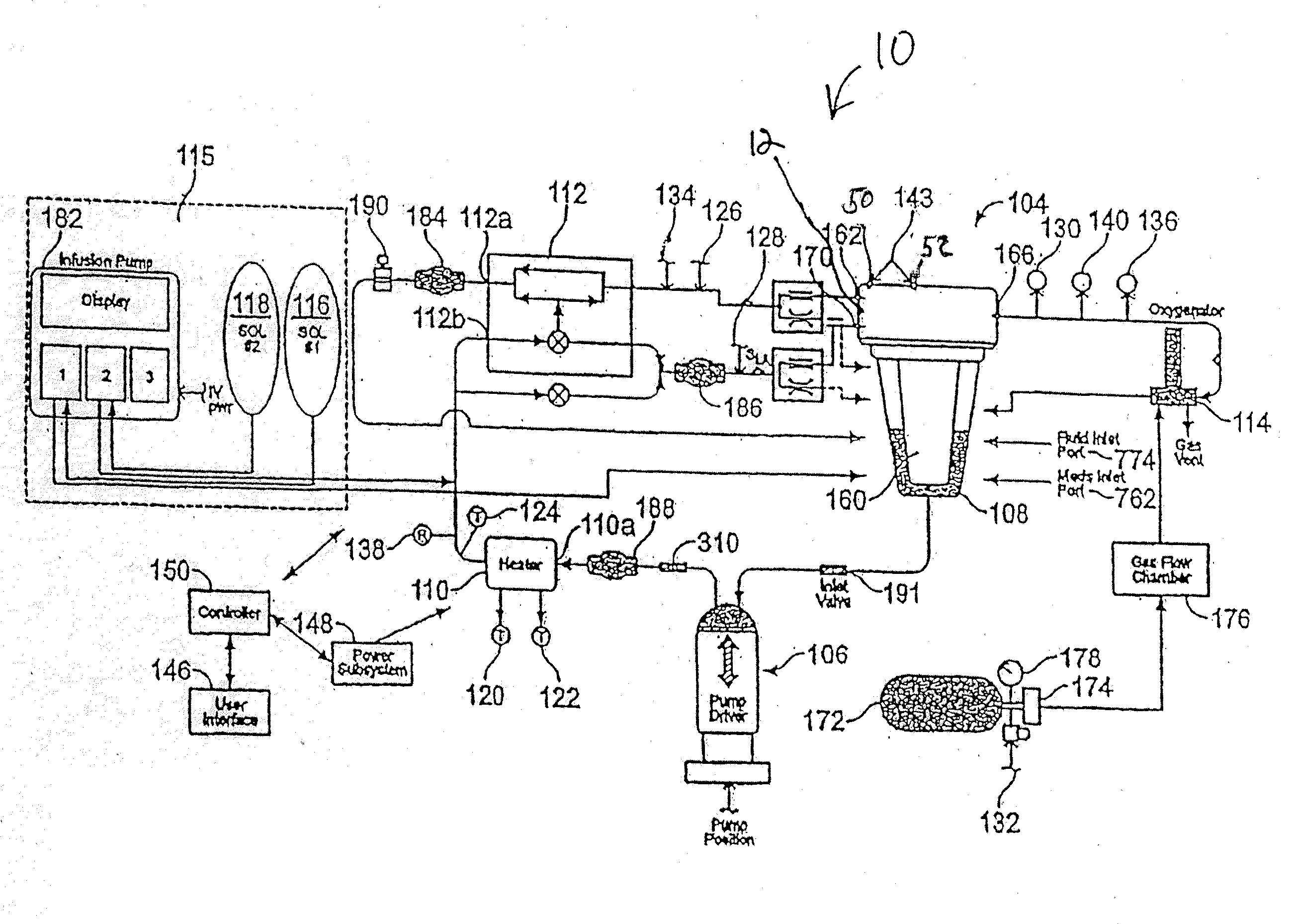
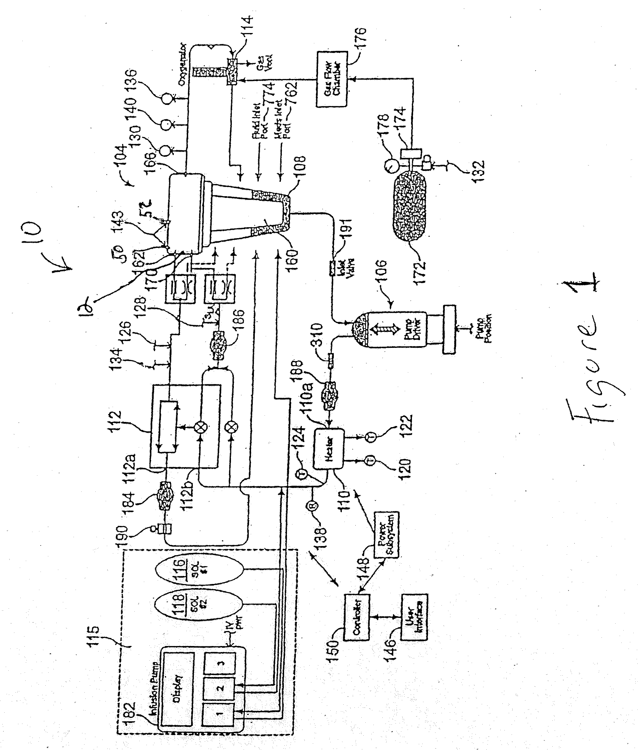
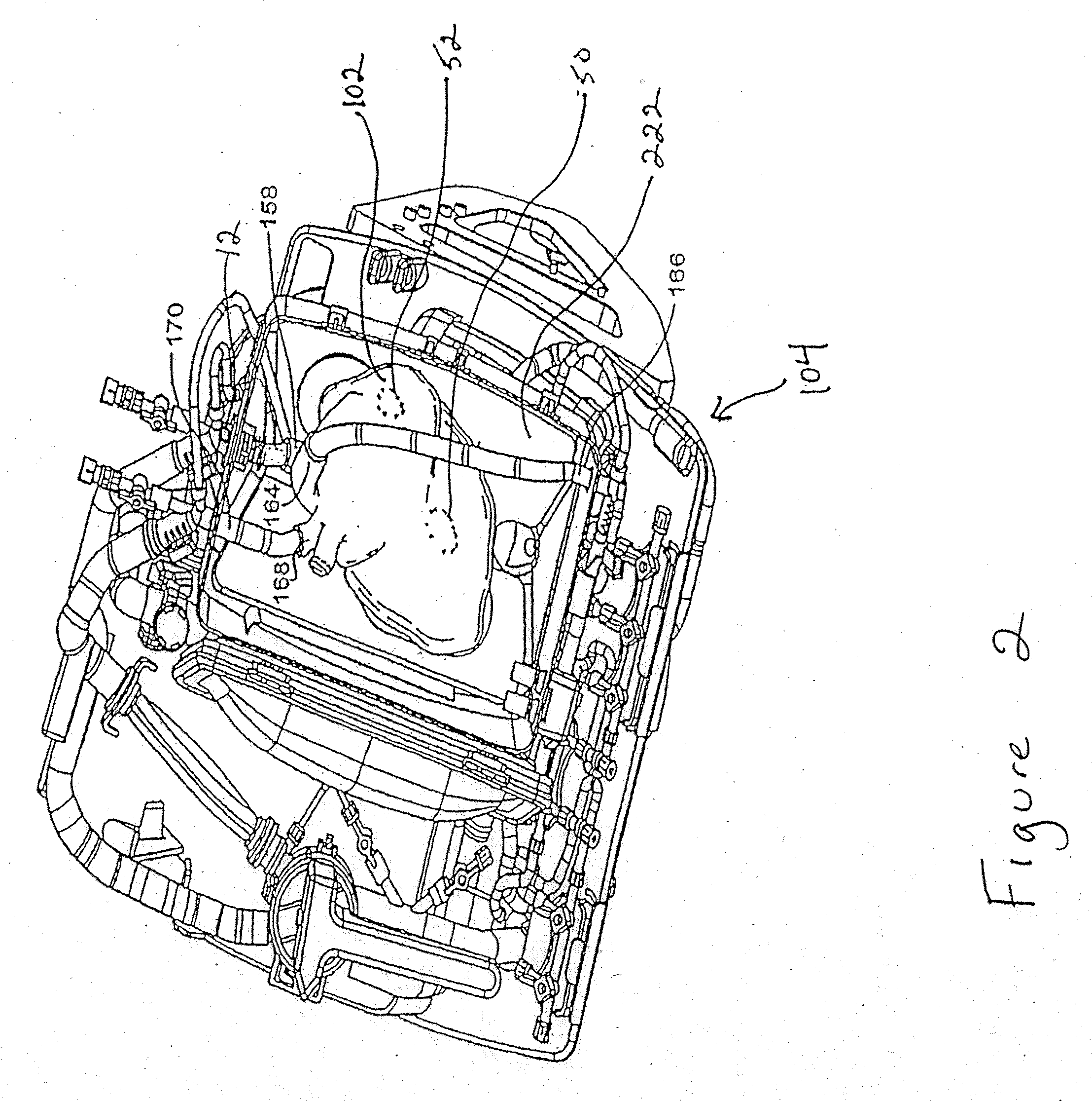
[0008]Electrode systems have been developed for use in perfusion systems to measure the electrical activity of an explanted heart and to provide defibrillation energy as necessary. The perfusion systems maintain the heart in a beating state at, or near, normal physiologic conditions; circulating oxygenated, nutrient enriched perfusion fluid to the heart at or near physiologic temperature, pressure and flow rate. These systems include a pair of electrodes that are placed epicardially on the right atrium and left ventricle of the explanted heart, as well as an electrode placed in the aortic blood path.

[0009]An advantage of this configuration allows an electrode to be held against the right atrium of the explanted heart under the heart's own weight, which reduces the likelihood that the electrode will shift during transport of the heart due to vibrations or the beating of the heart itself. As well, placing the electrode epicardially allows the electrode to be manipulated to ensure bett...


PUM


Abstract

Electrode systems have been developed for use in perfusion systems to measure the electrical activity of an explanted heart and to provide defibrillation energy as necessary. The perfusion systems maintain the heart in a beating state at, or near, normal physiological conditions; circulating oxygenated, nutrient enriched perfusion fluid to the heart at or near physiological temperature, pressure and flow rate. These systems include a pair of electrodes that are placed epicardially on the right atrium and left ventricle of the explanted heart, as well as an electrode placed in the aortic blood path.

Description

RELATED APPLICATIONS[0001]This application is related to Application Ser. No. 60 / 919,306, titled “Systems for Monitoring Organ Electrical Activity in a Perfusion System,” filed Mar. 20, 2007, which is incorporated herein by reference in its entirety.BACKGROUND OF THE INVENTION[0002]Organ preservation techniques typically involve hypothermic storage of the organ in a chemical perfusate solution on ice. In the case of a heart, it is typically arrested, and cooled with a cardioplegic solution until it reaches a hypothermic, non-functioning state and then is stored in or perfused with a cold preservation solution. These techniques utilize a variety of cardioplegic and cold preservation solutions, none of which sufficiently protect the heart from myocardial damage resulting from ischemia. Such injuries are particularly undesirable when an organ, such as a heart, is intended to be transplanted from a donor into a recipient. In addition to myocardial damage resulting from ischemia, reperfu...

Claims


Application Information

Patent Timeline
Patent Type & Authority Applications(United States)
IPC IPC(8): A61N1/04
CPCA61N1/046A61N1/0492A61N1/0488A01N1/0247A01N1/0294
Inventor HASSANEIN, WALEEDELBETANONY, AHMEDBRINGHAM, RICHARDHAVENER, ROBERTLAMBERT, VINCENTOCHS, BURT
Owner TRANSMEDICS
Why Eureka
Try Eureka