Depletion of plasma proteins

Inactive Publication Date: 2009-02-05
HEALTHLINX LTD
View PDF10 Cites 3 Cited by
  • Summary
  • Abstract
  • Description
  • Claims
  • Application Information

AI Technical Summary

Benefits of technology

[0012]We have now surprisingly found that a simple immuno-affinity procedure, combined with the use of existing solid phase affinity capture supports, can

Problems solved by technology

In routine diagnostic laboratories, identification of specific low abundant disease-associated proteins in serum relies heavily on time-consuming and expensive radiolabelled or enzyme-linked immunoassay methods (RIA or ELISA) which only have the ability to evaluate a single protein component at a time.
Unfortunately, the analysis by 2DE gels of proteins in samples of biological fluids such as serum and plasma is very difficult.
This is because of the limited amount of protein able to be resolved by a gel, and the great variation in the concentration of proteins in many samples.
Thus the application of current proteomic technologies is limited by the presence of high abundance “housekeeping” proteins like albumin and immunoglobulins, which constitute approximately 60-97% of the total serum protein (Georgiou et al, 2001).
Such proteins hinder the detection of hundreds of low abundance proteins, some of whi

Method used


Image

  • Depletion of plasma proteins
  • Depletion of plasma proteins
  • Depletion of plasma proteins

Examples


Example

EXAMPLE 1

Production of Polyclonal Antibodies to Human Plasma

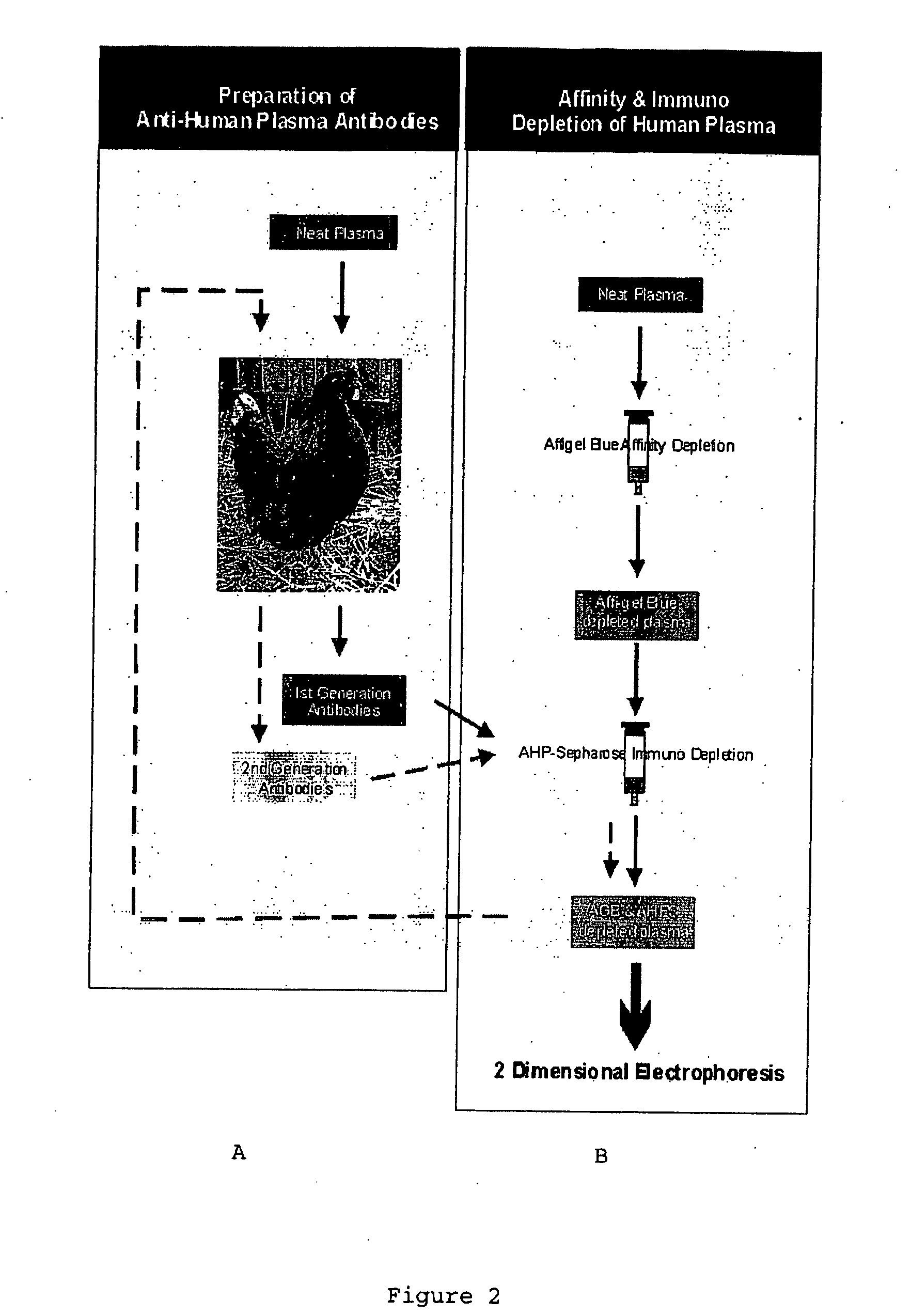
[0080]First generation polyclonal antibodies to human plasma were produced in female chickens. The procedure is summarised in FIG. 2. Chickens (14 week old White Leghorn / Rhode Island Red cross) were immunised according to the recommendations of the 21st European Centre for the Validation of Alternative Methods (ECVAM) workshop, using 1 mg plasma proteins / bird (12.5 μl of 80 mg / ml) suspended in saline (87.5 μl) / Freund's Incomplete Adjuvant (100 μl). 100 μl of total plasma proteins were injected subcutaneously over the pectoralis major muscle, using a 25-gauge needle at four sites (i.e. 50 μl per site).

[0081]Birds received three booster injections as described above, 4, 8 and 12 weeks later. Eggs were collected prior to immunization and the yolks stored at −20° C. Eggs were collected daily during the immunization schedule, up to—30 days after the last booster injection and the yolks extracted as described in Example 2.

Example

EXAMPLE 2

Extraction of IgY

[0082]Egg yolks (10 per batch) were separated and then suspended in 2 volumes of 100 mM phosphate buffer (pH7.6) in a glass beaker. An equal volume of chloroform was added and then stirred for 5 min at room temperature. The resultant emulsion was then transferred to 100 ml glass centrifuge tubes and centrifuged at 2000 g for 1 h at 4° C. The supernatant was collected and its volume determined. PEG 6000 (Sigma Chemical Company, St Louis, USA) was dissolved in the supernatant to final concentration of 12% w / v, incubated for 10 min at room temperature and then centrifuged at 2000 g for 1 h at 4° C. The supernatant was discarded and the pellet resuspended in 100 mM phosphate buffer pH7.6 (⅙ original yolk volume) and stored at −20° C. as 1 ml aliquots.

[0083]Egg yolks were collected for four weeks following the final immunization, pooled and extracted as described above.

[0084]The binding characteristics of the extracted antibodies were determined by 2DE Western B...

Example

EXAMPLE 3

Coupling of IgY to Sepharose 4B

[0085]PEG 6000 was dissolved in 2 ml IgY solution (17.3 mg protein / ml), incubated for 10 min at room temperature and then centrifuged at 2000 g for 1 h. The pellet was resuspended in coupling buffer (0.1M NaHCO3 pH 8.3, containing 0.5M NaCl) to a final concentration of 7.5 mg protein / ml.

[0086]CNBr-activated Sepharose 4B (Pharmacia; 1 g) was suspended in 20 ml of 1 mM HCl. The suspension was then washed with 200 ml 1 mM HCl on a sintered glass filter. The washed gel was resuspended in the IgY solution, and mixed on a rotary mixer for 18 h at 4° C. The gel was then washed with 5 volumes of coupling buffer and incubated in 0.1M Tris-HCl buffer, pH8.0 for 2 h at 4° C. The gel was washed 3 times alternately with 5 volumes 0.1M acetate buffer pH 4.0 containing 0.5M NaCl, and then 0.1M Tris HCl pH 8.0 containing 0.5M NaCl. The anti-human plasma antibody-Sepharose 4B (AHP-Sepharose) gel was then stored at 4° C. in 0.01 M phosphate-buffered saline, pH7...


PUM

PropertyMeasurementUnit
Timeaaaaaaaaaa
Timeaaaaaaaaaa
Affinityaaaaaaaaaa

Abstract

This invention relates to methods of analysis, and in particular to methods for the preliminary fractionation of samples in which low abundance molecules of interest, for example proteins, polysaccharides or fatty acids, are present together with more abundant molecules of little or no interest. In particular, the invention relates to methods of depletion of high abundance proteins from biological samples. Products and kits for use in the method are also disclosed, and form part of the invention.
In one aspect, the invention provides a method of depleting a high-abundance molecule from a biological sample, comprising the steps of
    • a) subjecting the sample to affinity depletion using an affinity support with high affinity for a high abundance molecule, and/or
    • b) immunodepletion using an affinity support coupled to an antibody directed against whole or previously fractionated plasma or serum.

Description

[0001]This invention relates to methods of analysis, and in particular to methods for the preliminary fractionation of samples in which low abundance molecules of interest, for example proteins, polysaccharides or fatty acids, are present together with more abundant molecules of little or no interest. In particular, the invention relates to methods of depletion of high abundance proteins from biological samples. The method is particularly applicable to samples of human biological fluids such as serum, plasma, tears, saliva, cerebrospinal fluid, uterine washings, amniotic fluid, cervico-vaginal fluid or urine. It is contemplated that the method of the invention will be especially useful for proteomic applications involving biomarker discovery. Products and kits for use in the method are also disclosed, and form part of the invention.BACKGROUND OF THE INVENTION[0002]All references, including any patents or patent applications, cited in this specification are hereby incorporated by ref...

Claims


Application Information

Patent Timeline
IPC IPC(8): C12M1/00C07K1/00C07K16/18C07K1/22G01N30/00G01N30/14G01N33/68
CPCC07K1/22G01N30/14G01N33/6854G01N2030/009B01D15/3804
Inventor RICE, GREGORY E.BAKER, MARK S.QUINN, MICHAEL
Owner HEALTHLINX LTD
Why Eureka
Try Eureka