
Method for the production of bio-imaging nanoparticles with high yield by early introduction of irregular structure

a bio-imaging and nanoparticle technology, applied in the field of bioimaging nanoparticle preparation, can solve the problems of large loss of nanoparticles, no further studies, and increased aggregation and precipitation, and achieve high yield and high dispersibility and stability

Inactive Publication Date: 2009-04-30
KOREA INST OF SCI & TECH
View PDF16 Cites 23 Cited by
  • Summary
  • Abstract
  • Description
  • Claims
  • Application Information

AI Technical Summary

Benefits of technology

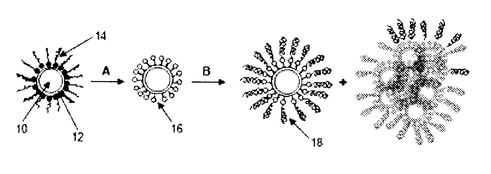
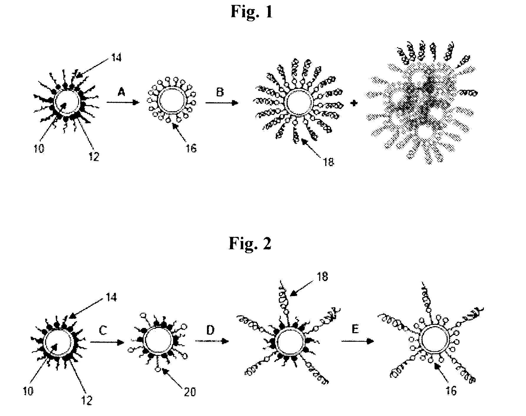
[0019]An object of the present invention is to provide a method of preparing bio-imaging nanoparticles by early introduction of an irregular structure into the surface of nanoparticles via partial surface modification, allowing the structural hinderance of the aggregation and precipitation phenomena due to internanoparticle hydrogen bonding attraction, thereby preparing bio-imaging nanoparticles having high dispersibility and stability in a nonpolar organic solvent or an aqueous solution with high yield without the loss of nanoparticles.
[0020]It is another object of the present invention to provide bio-imaging nanoparticles having high dispersibility and stability prepared according to the method of the present invention.

Problems solved by technology

However, because the hydroxy group or amine group easily aggregates and precipitates in a neutral or near neutral solution, no further studies have been made.
In particular, the aggregation and precipitation are more severe in the case of magnetic nanoparticles.
Since these precipitates must eventually be removed and only the well-dispersed portion of the solution layer is used, there is a serious problem in that a large quantity of nanoparticles is lost.
However, the disclosed method still suffers from the problem of low reaction yield, since the hydrophilic thiol group that is linked by a long hydrocarbon chain has to penetrate into the surface of nanoparticles surrounded with surfactants.

Method used


Image

  • Method for the production of bio-imaging nanoparticles with high yield by early introduction of irregular structure
  • Method for the production of bio-imaging nanoparticles with high yield by early introduction of irregular structure
  • Method for the production of bio-imaging nanoparticles with high yield by early introduction of irregular structure

Examples


example 1

Preparation of Semiconductor Nanoparticles CdSe / CdS-DA by Surface Ligand Exchange

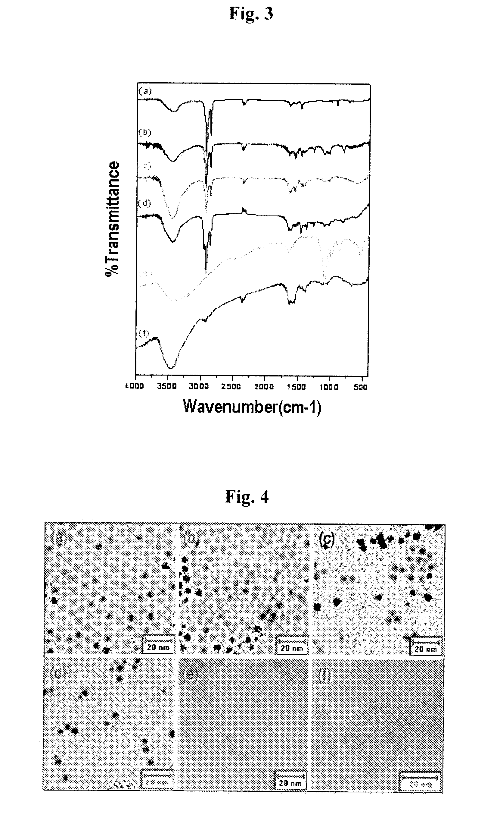
[0064]5 ml of a hydrophobic CdSe / CdS-ODA quantum dot solution (8×10⁻⁵ M) was subjected to vacuum evaporation to remove the solvent and dispersed in 20 ml of chloroform. To the dispersion was added 1000 equivalents of decylamine (DA), and the mixture was stirred for 2 days in a dark inert atmosphere. The resulting solution was mixed with acetone and centrifuged to separate the precipitate. Thus separated precipitate was dispersed in chloroform to prepare 20 ml of a CdSe / CdS-DA solution (2×10⁻⁵ M). The CdSe / CdS-DA sample was analyzed with an infrared spectrophotometer and a transmission electron microscope (TEM), where the results are shown in FIG. 3(b) and FIG. 4(b), respectively. The occurrence of surface ligand exchange was confirmed by the shorter distance between CdSe / CdS-DA quantum dots than CdSe / CdS-ODA in the TEM image.

example 2

Preparation of Partially Surface Modified Semiconductor Nanoparticles CdSe / CdS(-DA)ex(-MUA)5

[0065]To 17 ml of the CdSe / CdS-DA solution prepared in Example 1 was added 5 equivalents of mercaptoundecanoic acid (MUA) and stirred for 19 hours in a dark inert atmosphere. The resulting solution was concentrated, mixed with acetone, and centrifuged to separate the precipitate. Thus separated precipitate was dispersed in chloroform to prepare 17 ml of a CdSe / CdS(-DA)ex(-MUA)5 solution (2×10⁻⁵ M). The resulting sample was analyzed with an infrared spectrophotometer and a transmission electron microscope (TEM), where the results are shown in FIG. 3(c) and FIG. 4(c), respectively. The TEM image confirmed that self-assembly of the nanoparticles did not occur any longer, due to the destruction of their uniform structure caused by a partial replacement of MUA, as in step C of FIG. 2.

example 3

Preparation of Targeting Hydrophobic Semiconductor Nanoparticles CdSe / CdS(-DA)ex(-MUA-en-FA)5

[0066]2 ml of the CdSe / CdS(-DA)ex(-MUA)5 solution prepared in Example 2 was diluted with chloroform to a final volume of 10 ml. After 5 equivalents of dicyclohexylcarbodiimide (DCC) was added to the diluent and stirred for 3 hours in a dark inert atmosphere, 50 equivalents of en-FA prepared as follows were added thereto and further stirred for 2 hours. The resulting solution was concentrated, mixed with acetone, and centrifuged to separate the precipitate. Thus separated precipitate was dispersed in chloroform, to prepare 10 ml of a CdSe / CdS(-DA)ex(-MUA-en-FA)5 solution (4×10⁻⁶ M). The bonding of en-FA to MUA was analyzed with an infrared spectrophotometer and a TEM, where the results are shown in FIG. 3(d) and FIG. 4(d), respectively.

Preparation of a Complex en-FA of a Targeting Molecule Folic Acid (FA) and Ethylenediamine (en)

[0067]441 mg (1 mmol) of folic acid was added to 10 ml of dry-d...


PUM

PropertyMeasurementUnit
Equivalent massaaaaaaaaaa
Hydrophilicityaaaaaaaaaa
Biocompatibilityaaaaaaaaaa

Abstract

Methods of preparing bio-imaging nanoparticles having high dispersibility in an aqueous solution, biocompatibility, and targetability with high yield, by early introduction of an irregular structure are disclosed.

Description

CROSS REFERENCE TO RELATED APPLICATIONS[0001]This application claims priority to Korean Patent Application No. 10-2007-110333, filed Oct. 31, 2007. The entire contents of that application are hereby incorporated by reference.FIELD OF THE INVENTION[0002]The present invention relates to a method of preparing bio-imaging nanoparticles having high dispersibility in an aqueous solution, biocompatibility, and targetability with high yield by early introduction of an irregular surface structure.BACKGROUND OF THE INVENTION[0003]Since the establishment of a method of chemically synthesizing hydrophobic inorganic nanoparticles having homogeneous size distribution in an organic solvent including a surfactant, various attempts have been made to put the method to practical use. In particular, since the nanoparticles prepared in an aqueous solution show a much more heterogeneous size distribution than those prepared in an organic solvent and water is the cheapest, most environmentally friendly, a...

Claims


Application Information

Patent Timeline
IPC IPC(8): A61K49/18B82B1/00
CPCA61K49/0052A61K49/0067B82Y5/00A61K49/1839A61K49/186A61K49/1836B82B3/00B82Y30/00
Inventor WOO, KYOUNGJAMOON, JIHYUNG
Owner KOREA INST OF SCI & TECH
Learn More