
Methods for detection of pathogenic prion proteins associated with prion diseases, using conjugated polyelectrolytes

a technology of conjugated polyelectrolytes and prion proteins, which is applied in the field of pathogenic prion proteins associated with prion diseases, can solve the problems of limited early stage therapeutic interventions for misfolding diseases, and achieve the effect of facilitating a greater understanding of the conformational phenotype encoded

Inactive Publication Date: 2009-08-06
BIOCHROMIX
View PDF1 Cites 4 Cited by
  • Summary
  • Abstract
  • Description
  • Claims
  • Application Information

AI Technical Summary

Benefits of technology

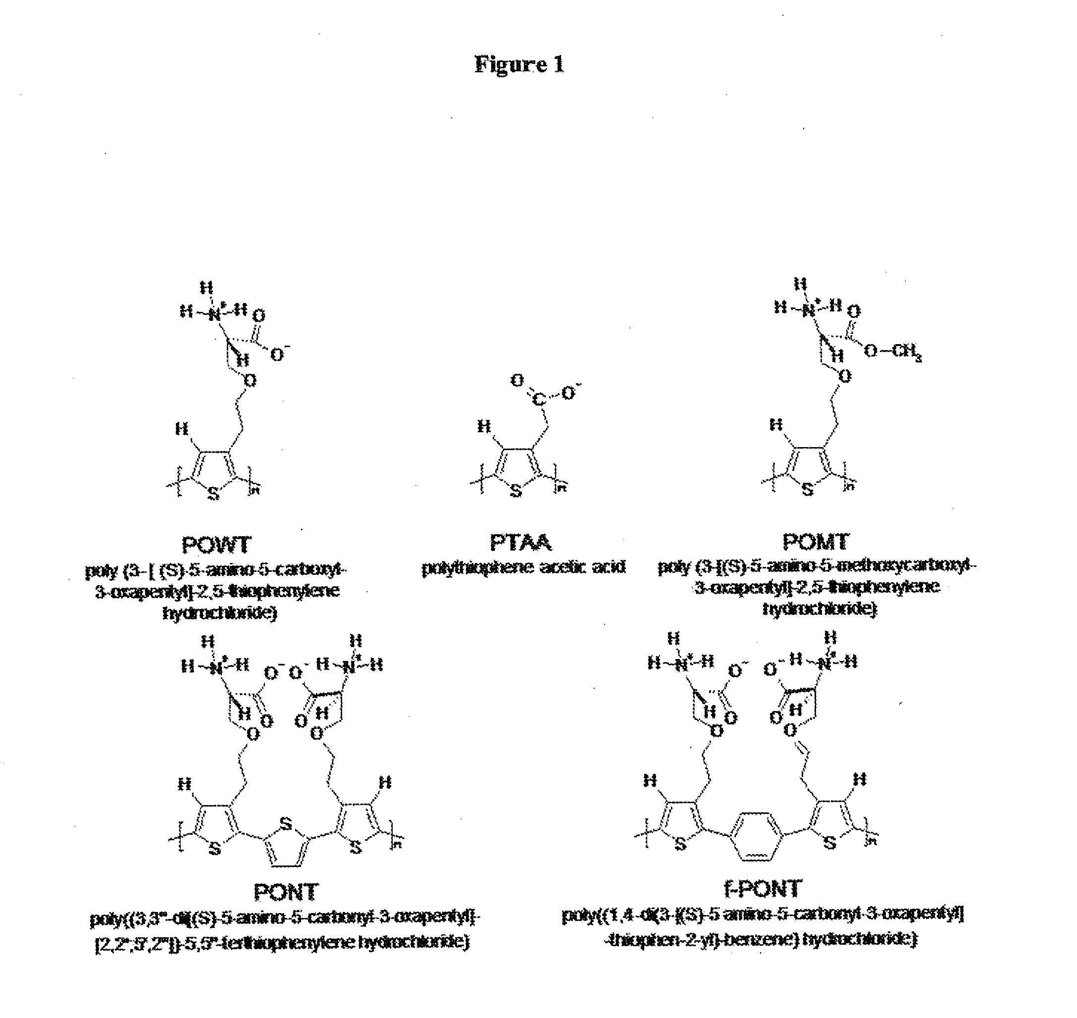
[0008]Conjugated polyelectrolytes, such as poly (thiophene) and poly (pyrrole) can be used to record conformational changes of proteins into observable responses. Sensors based on conjugated polyelectrolytes are sensitive to very minor perturbations, due to amplification by a collective system response and therefore offer a key advantage compared to small-molecule based sensors of the prior art and the polyelectrolytes also offers a direct detection of the pathogenic prion protein. This direct detection of the prion protein is an advantage compared to immunohistological techniques as these methods often requires the use of a secondary antibody for visualization of the pathogenic prion protein. The possibility to use conjugated polyelectrolytes as detecting elements for biological molecules requires that polymers are compatible with an aqueous environment and this has been accomplished by making conjugated luminescent polyelectrolytes.

Problems solved by technology

Currently, early stage therapeutic interventions of the misfolding diseases are limited by the absence of sensitive diagnostic tests for the pathogenic misfolded prion protein.

Method used


Image

  • Methods for detection of pathogenic prion proteins associated with prion diseases, using conjugated polyelectrolytes
  • Methods for detection of pathogenic prion proteins associated with prion diseases, using conjugated polyelectrolytes
  • Methods for detection of pathogenic prion proteins associated with prion diseases, using conjugated polyelectrolytes

Examples


example 1

Histological Staining of Scrapie Infected Nervous Tissue from Sheep

[0060]Sections (5 μm) from formaldehyde-fixed, paraffin-embedded amyloid-containing tissue were placed on plus-slides and deparaffinized with xylene (60 min), absolute alcohol (15 min), 95% alcohol (15 min) and 70% alcohol (10 min) and finally rinsed in distilled water for a couple of minutes. The sections were equilibrated in incubation buffer solution, 100 mM Na-Carbonate pH 10, for 10 min. PTAA were mixed with the same buffer used for equilibration (5 μg probe in 100 μl) and added to the sections. The incubation took place in a humidity chamber for 1 hour and superfluous probe solution was washed away with incubation buffer. When PTAA binds to or interacts with the misfolded prion protein (PrPSc), the misfolded pathogenic prion protein is associated with Scrapie disease normally seen in sheep, it can be detected by electromagnetic radiation or absorption, preferably between UV and IR range, optimal in the visible ...

example 2

Histological Staining of Non-Infected Nervous Tissue with PTAA

[0061]Sections (5 μm) from formaldehyde-fixed, paraffin-embedded amyloid-containing tissue were placed on plus-slides and deparaffinized with xylene (60 min), absolute alcohol (15 min), 95% alcohol (15 min) and 70% alcohol (10 min) and finally rinsed in distilled water for a couple of minutes. The sections were equilibrated in incubation buffer solution, 100 mM Na-Carbonate pH 10, for 10 min. PTAA were mixed with the same buffer used for equilibration (5 μg probe in 100 μl) and added to the sections. The incubation took place in a humidity chamber for 1 hour and superfluous probe solution was washed away with incubation buffer. The fluorescence from the tissues samples can be recorded with an epifluorescence microscope (Zeiss Axiovert inverted microscope A200 Mot) equipped with a CCD camera (Axiocam HR), using a 405 / 30 nm bandpass filter (LP450), a 470 / 40 nm bandpass filter (LP515) and a 546 / 12 nm bandpass filter (LP590)....

example 3

Histological Staining of Chronic Wasting Disease (CWD) Infected Mouse Tissue with PTAA

[0062]Frozen sections from CWD infected mouse brain were fixed in ice cold ethanol for 10 minutes and washed with buffer solution, 100 mM Na-Carbonate pH 10. The sections were equilibrated in incubation buffer solution, 100 mM Na-Carbonate pH 10, for 10 min. PTAA were mixed with the same buffer used for equilibration (5 μg probe in 100 μl) and added to the sections. The incubation took place in a humidity chamber for 1 hours and superfluous probe solution was washed away with incubation buffer. When PTAA binds to or interacts with the misfolded prion protein (PrPSc), the misfolded pathogenic prion protein is associated with chronic wasting disease normally seen in deer, it can be detected by electromagnetic radiation or absorption, preferably between UV and IR range, optimal in the visible range. In the visible range it is normally seen as a change of the color and the intensity of the emitted ligh...


PUM

PropertyMeasurementUnit
wavelengthsaaaaaaaaaa
pHaaaaaaaaaa

Abstract

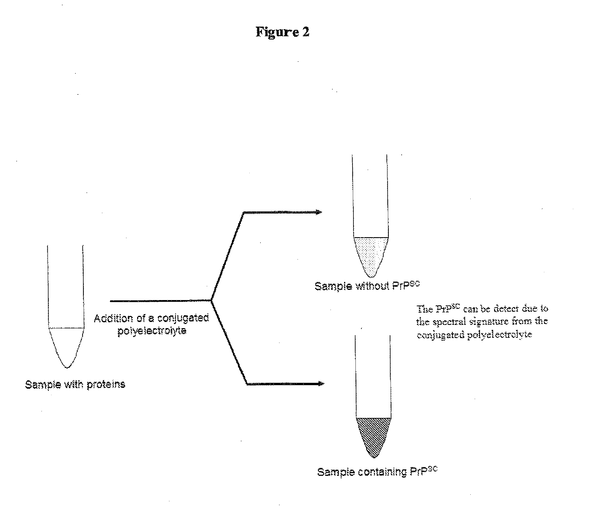
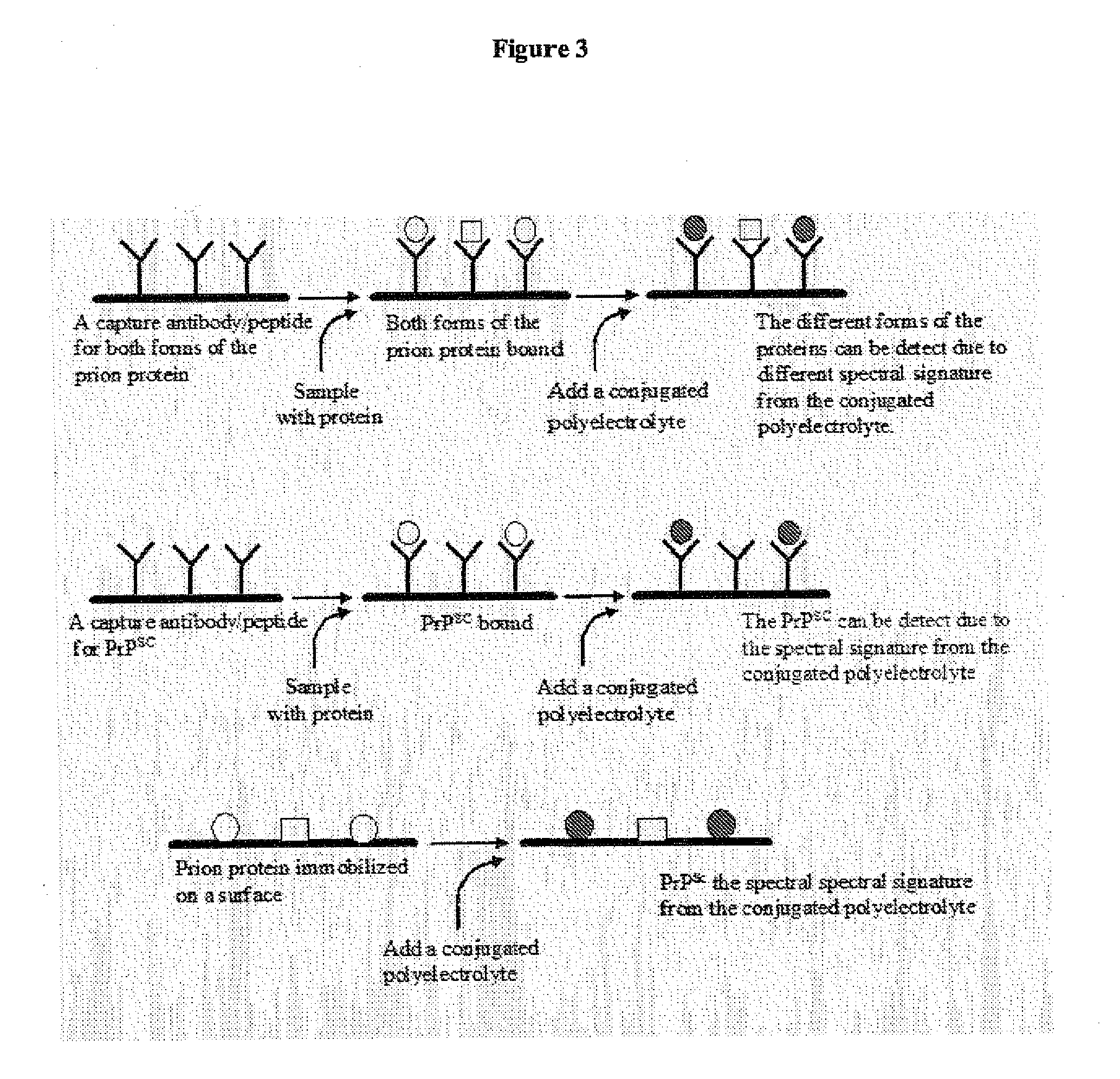
The present invention relates to a method for detecting the presence of a pathogenic prion species in a sample comprising the steps—bringing the sample in contact with at least one conjugated polyelectrolyte (CPE)—irradiating the CPE with electromagnetic radiation—measuring the radiation emitted or absorbed by the CPE at at least one wavelength, and—comparing the measured emitted or absorbed radiation to at least one reference value corresponding to the CPE interacting with a known prion species. Optionally, the emitted or absorbed radiation is measured at least two wavelengths and a ratio is formed of the values of the measured radiation. The method facilitates differentiation between different strains of pathogenic prion species. The invention also relates to devices for performing the method.

Description

FIELD OF THE INVENTION[0001]The present invention relates to methods for detection of pathogenic prion proteins associated with prion diseases, using conjugated polyelectrolytes.BACKGROUND OF THE INVENTION[0002]The development of materials that are capable of selectively detecting pathogenic prion proteins have received increasing attention, owing to their large potential for being used as analytic tools for pre-clinical diagnosis of prion diseases. Prion diseases are normally diagnosed by immunohistological techniques using antibodies, or by methods utilizing small molecule dyes, such as Congo red and thioflavin T, if the prion protein has an amyloid like structure.[0003]Natural biopolymers, such as proteins, frequently have ordered conformations, such as alpha-helix and beta-sheets, which contribute to the three-dimensional ordered structure and the specific function of the biopolymer. Proteins frequently alter their conformation due to different external stimuli and the importanc...

Claims


Application Information

Patent Timeline
Patent Type & Authority Applications(United States)
IPC IPC(8): G01N33/50
CPCG01N21/64G01N2800/2828G01N33/6896
Inventor NILSSON, PETERHAMMARSTROM, PER
Owner BIOCHROMIX
Learn More