
Eureka
For R&D, Eureka makes reading and utilizing patents & technical documents easy.
Eureka AIR
Designed for self-driven R&D workflows. Generate viable solutions, solve complex R&D challenges, empower your innovation with AI.

Eureka Materials
Designed for material experts only. Revolutionize your material R&D, from search, analyze, to developing new materials.

TechResearch
Generate reliable direction feasibility study reports for your R&D in just a few steps.

TechSeek
Discover and master advanced knowledge NOW. Basics, ideas, possibilities, all at once.

TechMind
As an expert in R&D Theories, TechMind can generates customized viable solutions instantly.

TechRisk
Analyze your overall solution with one click, know your potential R&D risks in advance.

TechMonitor
Get weekly tech updates, stay abreast of the latest tech innovations and key insights.
Encapsulated nanoparticles for computed tomography imaging
- Summary
- Abstract
- Description
- Claims
- Application Information
AI Technical Summary
Benefits of technology
Problems solved by technology
Method used
Image

Examples
example 1
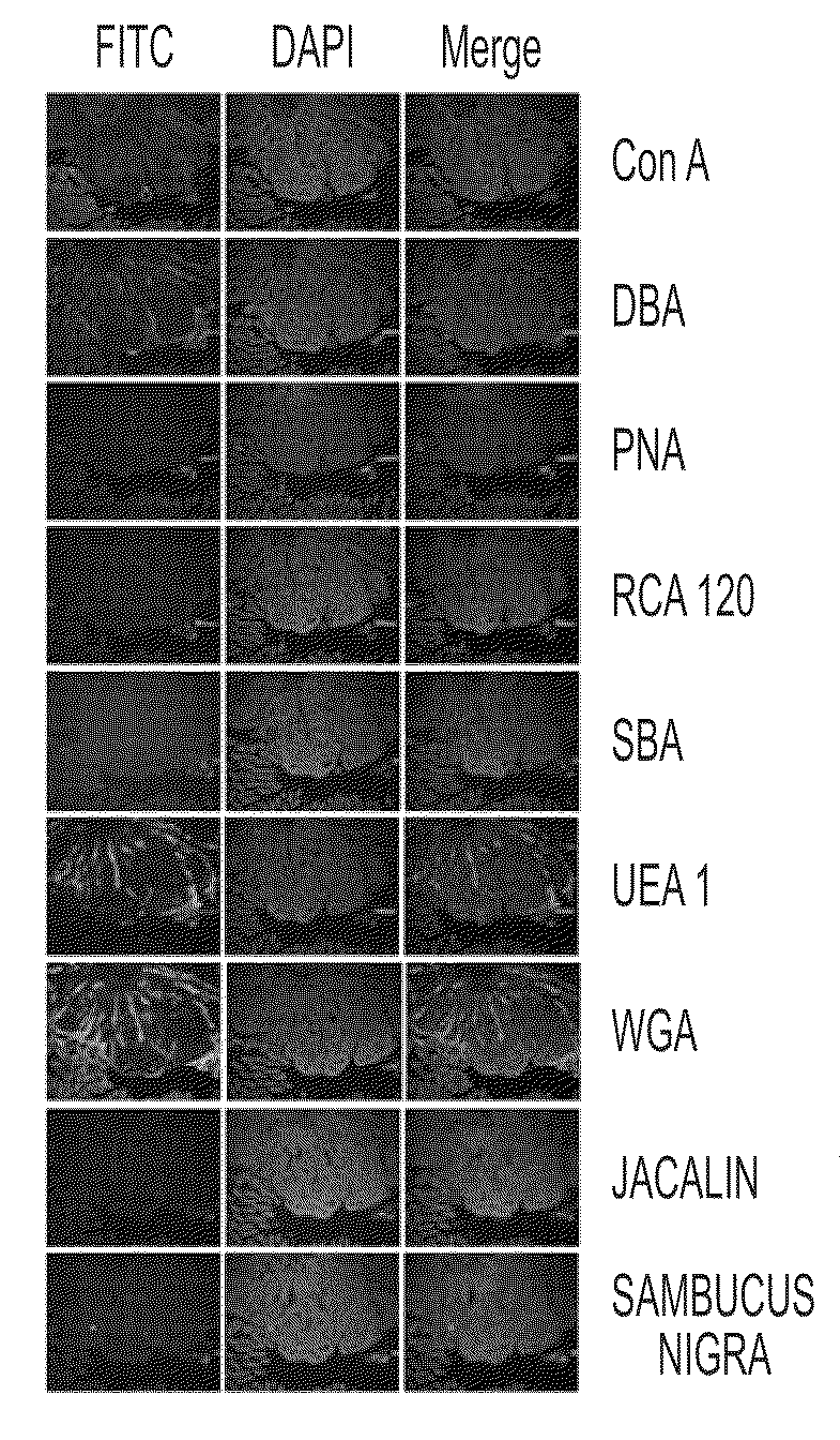
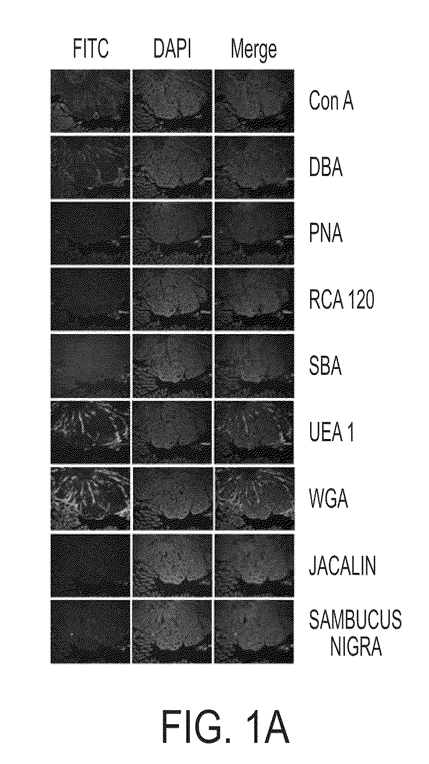
FITC Lectin Histochemistry
[0053]Nine different fluorescein (FITC)-labeled lectins were commercially purchased from Vector Laboratories (Burlingame, Calif., USA). The FITC-lectins used included: Concavalin A (ConA), Dolichos biflorus agglutinin (DBA), Peanut agglutinin (PNA), Ricinus communis agglutinin (RCA), Soybean agglutinin (SBA), Ulex europaeus agglutinin (UEA), Wheat germ agglutinin (WGA), Jacalin (Jac), and Sambucus nigra agglutinin (SNA).
[0054]Parafinized intestinal tissue sections (N=13, containing adenomatous polyps) were obtained from 8 weeks old heterozygous male mice of the strain C57BL / 6J APCMin / + (The Jackson Laboratory, Bar Harbor, Me.,) with 10% high fat diet. Paraffin sections were deparaffinized in three changes each of xylene, 100% ethanol (EtOH), 75% EtOH, 50% EtOH, and water. The final wash solvent was PBS. The sections were incubated in two antibody blocking solutions for 10 mins and 30 mins, respectively. These bowels excised from C57BL / 6J APCMin / + mice were ...
example 2
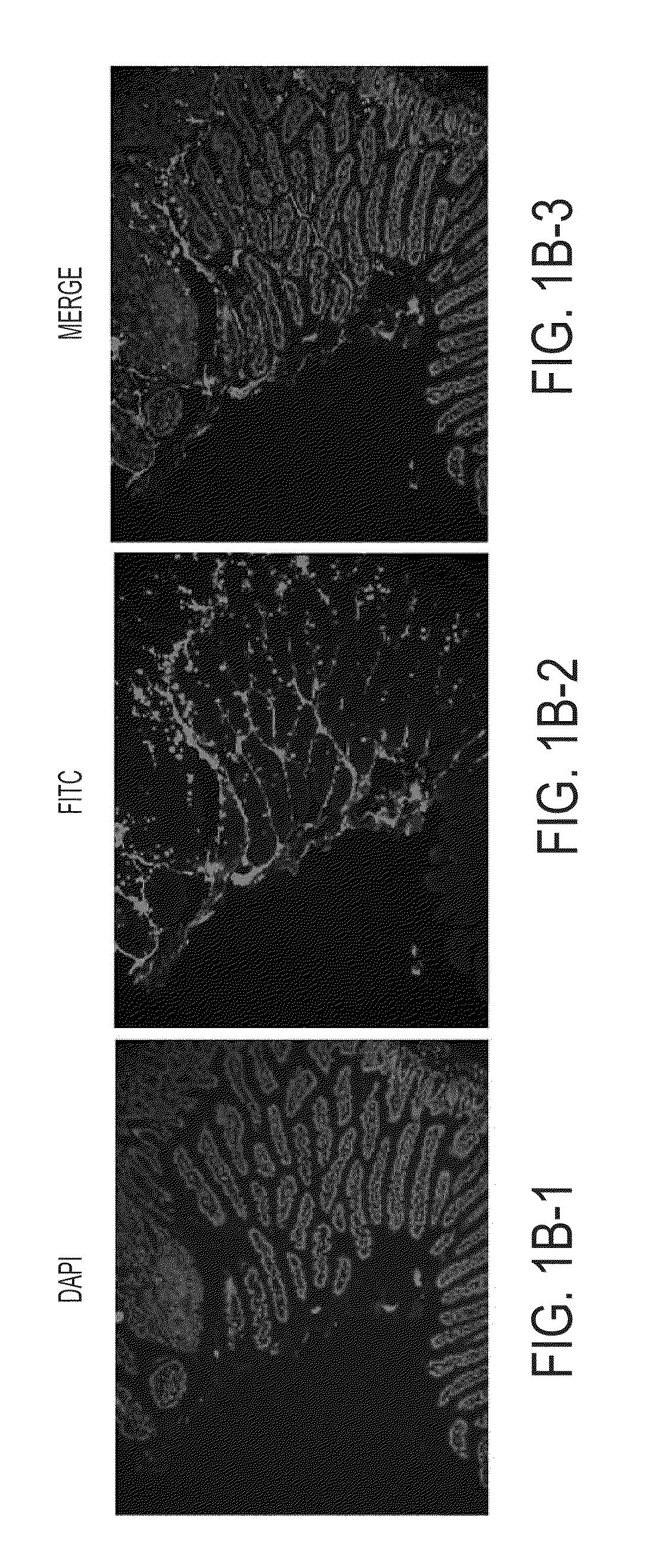
Ex Vivo Multispectral Optical Imaging
[0059]Excised small and large bowels (N=4) of male C57BL / 6J APCMin / + mice were commercially purchased from The Jackson Laboratory (8 wks old with 10% high fat diet) and stored at −80° C. until use. The bowels were thawed to room temperature (RT, 15-20 mins), cut along the longitudinal axis, and preserved in formaldehyde (RT, 15 mins) before the commencement of staining.
[0060]The bowels were incubated in 15 mL conical tubes in two antibody blocking solutions for 10 mins and 30 mins, respectively. After blocking, the bowels were incubated, with constant gentle shaking, in 5 μg / mL FITC-UEA-1 for 30 mins and followed by 3 times 5 mins PBS washing. Optical imaging was performed by using Maestro™ In-Vivo Multispectral Imaging System (CRI, Inc., Woburn, Mass.). During image acquisition, FITC was characteristically excited at 494 nm although emission data were collected from 500 to 950 nm including data from FITC's characteristic emission at 518 nm. The ...
example 3
CT Detection Agent: UEA-1 Conjugated Polymerized Liposomes
[0063]In an embodiment, a computed tomography (CT) detection agent includes Ulex europaceus agglutinin 1 (UEA-1) conjugated to a liposome, with the liposome including a radiologic contrast agent. To produce the CT detection agent, total lipid in chloroform was dried to form a thin lipid film, hydrated with iodinated contrast solution, extruded and crosslinked to form polymerized liposomes. UEA-1 protein was then conjugated to the liposomes.
[0064]The polymerizable diacetylene phospholipid DAPC (Avanti Polar Lipids, Alabaster, Ala.) was mixed with the saturated spacer lipid DNPC (Lipoid, Newark, N.J.) and DMPE-NHS (NOF) with 1:1 ratio together with 1% 18:1 Lissamine Rhodamine PE (Avanti Polar Lipids). The lipids in chloroform were added to a round bottom flask in the dark and protected with aluminum foil. The solvent was removed under vacuum using BUCHI Rotavapor for 1-2 hrs and further removed by leaving the mixture under high...
PUM
| Property | Measurement | Unit |
|---|---|---|
| Concentration | aaaaa | aaaaa |
Abstract
Description
Claims
Application Information
 - R&D Engineer
- R&D Manager
- IP Professional
- Industry Leading Data Capabilities
- Powerful AI technology
- Patent DNA Extraction
Browse by: Latest US Patents, China's latest patents, Technical Efficacy Thesaurus, Application Domain, Technology Topic, Popular Technical Reports.
© 2024 PatSnap. All rights reserved.Legal|Privacy policy|Modern Slavery Act Transparency Statement|Sitemap|About US| Contact US: help@patsnap.com