

Method for Cell Implantation
a cell and hyaln technology, applied in the field of cell implantation, can solve the problems of poor fixation of the scaffold to the defect, higher likelihood of the scaffold being damaged, etc., and achieve the effects of high quality repair, high efficiency and robustness, and efficient preparation of high quality ‘hyalln’
- Summary
- Abstract
- Description
- Claims
- Application Information
AI Technical Summary
Benefits of technology
Problems solved by technology
Method used
Image

Examples
example 1
[0219]A hydrophilic scaffold containing human cells embedded in a hydrogel according to the invention was prepared in the following manner.
[0220]The MPEG-PLGA scaffold (prepared according to the examples from DK application PA2006 00337 which are provided for reference below) was cut out with a sterile scalpel to a circular shape of 10 mm in diameter.
[0221]In regards to the species of thrombin used in the example in this context, was bovine thrombin used as thrombin “In general” for the examples, but the resulting gelation would be the same with human thrombin. Although, as discussed herein, the use of bovine thrombin should be avoided in a composition or kit according to the present invention, the thrombin used in the examples is merely to demonstrate the gelating effect thrombin with no respect to the origin of the thrombin. This thrombin was prepared by dissolving the thrombin in sterile H2O containing 0.1% BSA.
[0222]In regards to the species of fibrinogen used in the example in ...
example 2
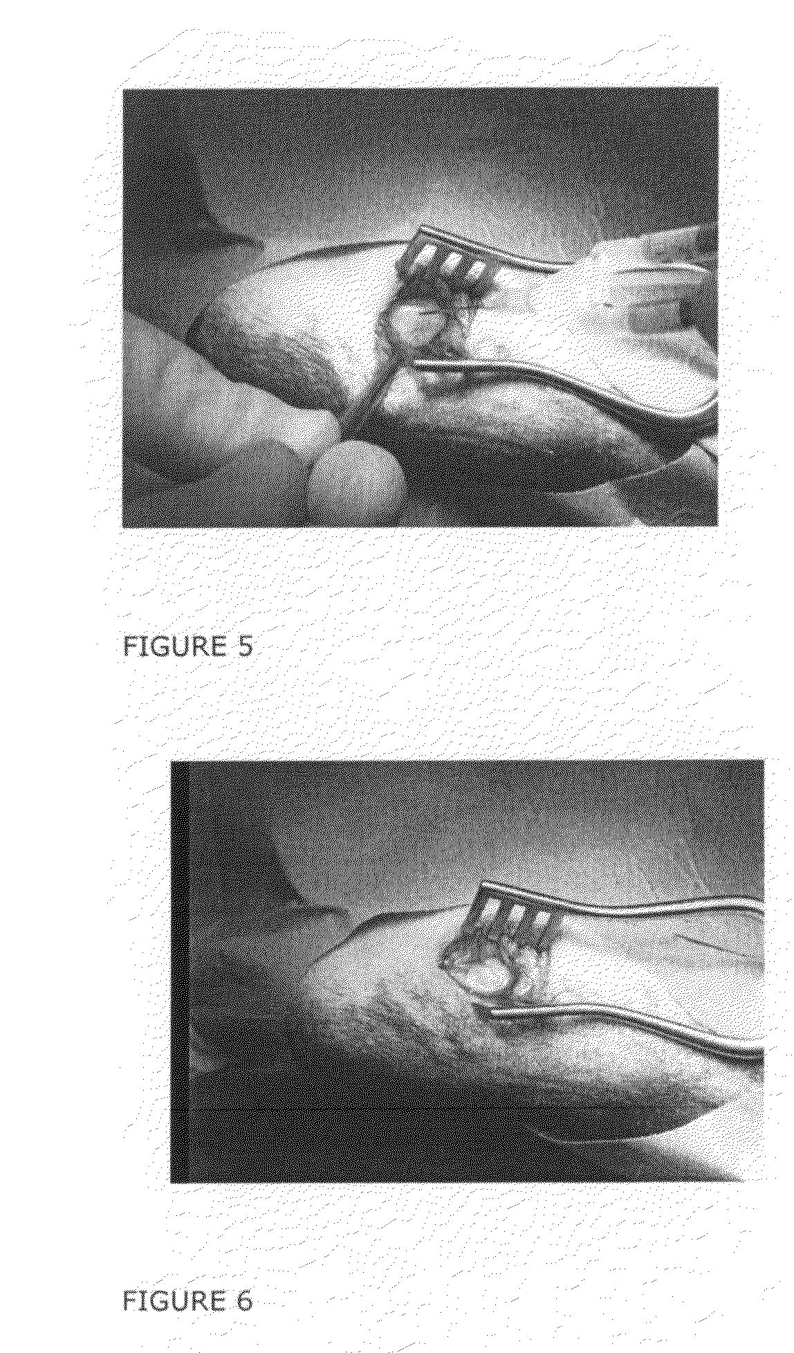
[0227]A hydrophilic scaffold containing human cells embedded in a hydrogel according to the invention was prepared in the following manner. The Trufit® from Osteobiologics, Inc., for pre-clinical studies called PolyGraft® BGS, Lot # X41053, to be used for pre-clinlcal studies only, was measuring 5.3×3 mm, and packed in a sterile package consisting of aluminum foil.
[0228]In regards to the species of thrombin used in this example and in this context, similar to the above described Example 1, was bovine thrombin used as thrombin “in general” for the examples, but the resulting gelation would be the same with human thrombin. Although, as discussed herein, the use of bovine thrombin should be avoided in a composition or kit according to the present invention, the thrombin used in the examples is merely to demonstrate the gelating effect thrombin with no respect to the origin of the thrombin. This thrombin was prepared by dissolving the thrombin In sterile H2O containing 0.1% BSA.
[0229]In...
example 3
[0233]Human articular chondrocytes (hACs) were cultured as a monolayer cell culture and then released from the cell culture flask using trypsin—EDTA.
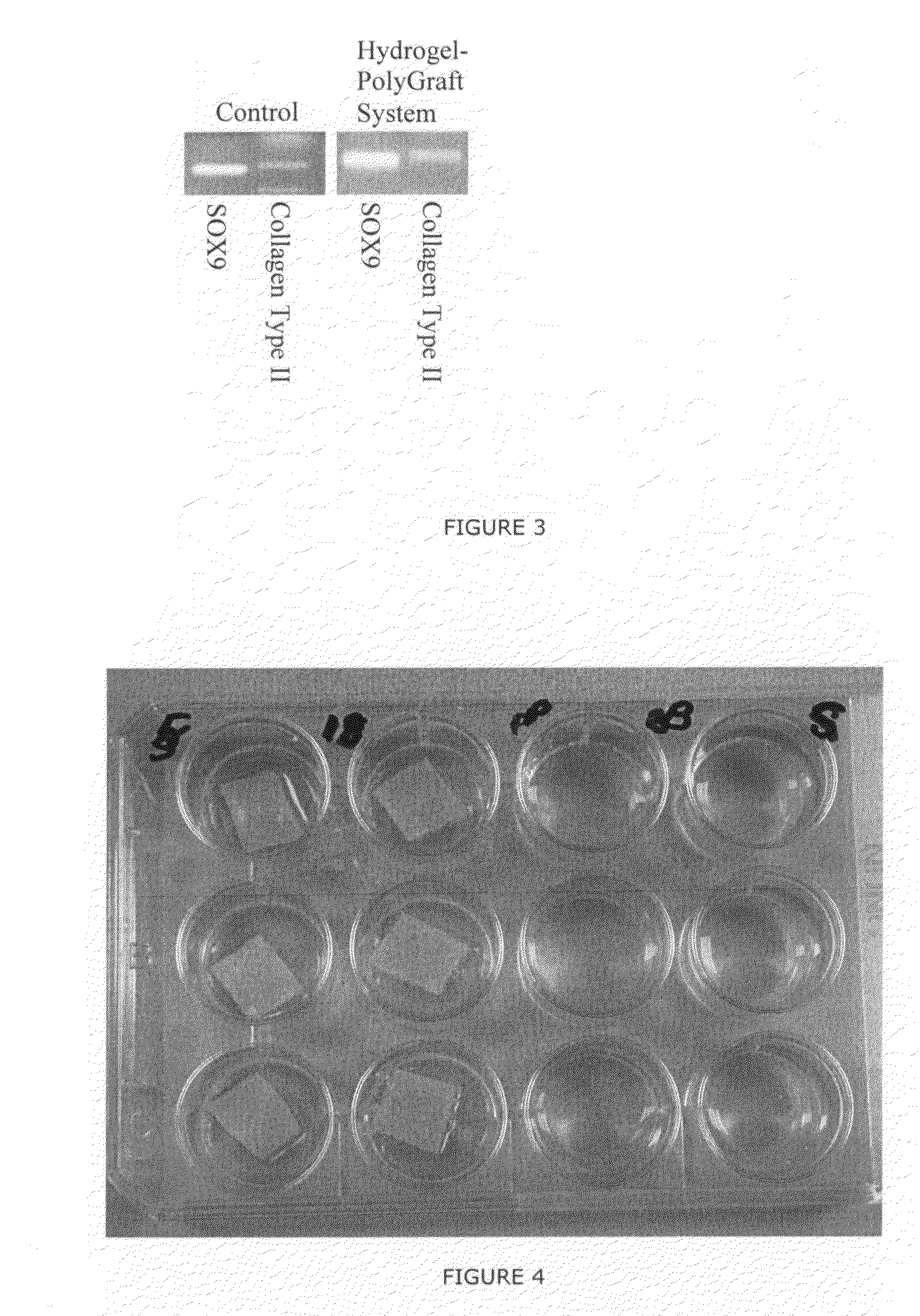
[0234]0.5×10⁶ cells were combined with the hydrogel composed of chondrocytes and fibrinogen / thrombin, and subsequently applied to the special Trufit (called Poly-Graft Top Phase from Osteobiologics, San Antonio, Tex.) in a Petri dish. After 5 minutes the “Hydrogel-PolyGraft System was placed in a 24 well plate and growth medium was applied to the wells. 6 (six) samples were cultured at 37° C. In a CO2 incubator; growth medium were changed around twice a week. 2 (two) samples were kept for 14 days to get a “14 day” time point analysis, 2 (two) for 2 months analysis and 2 for 4 months analysis.
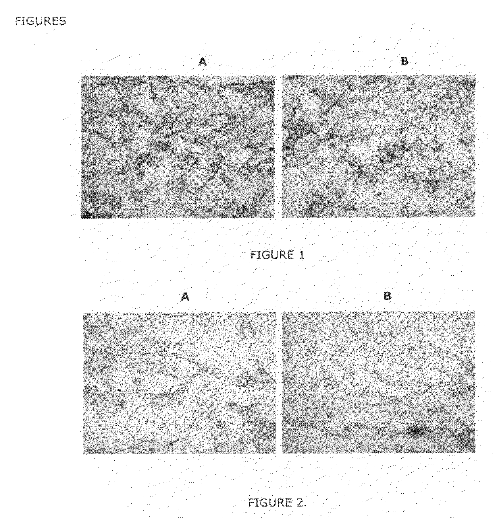
[0235]After each time point the following analysis were conducted:[0236]Cryo-section followed by histology (Toluldine Blue Staining and Safranin O staining) and immunocytochemistry (monoclonal antibodies against Aggrecan and Collagen type II)[0237]R...
PUM
| Property | Measurement | Unit |
|---|---|---|
| Concentration | aaaaa | aaaaa |
| Porosity | aaaaa | aaaaa |
| Molar mass | aaaaa | aaaaa |
Abstract
Description
Claims
Application Information