

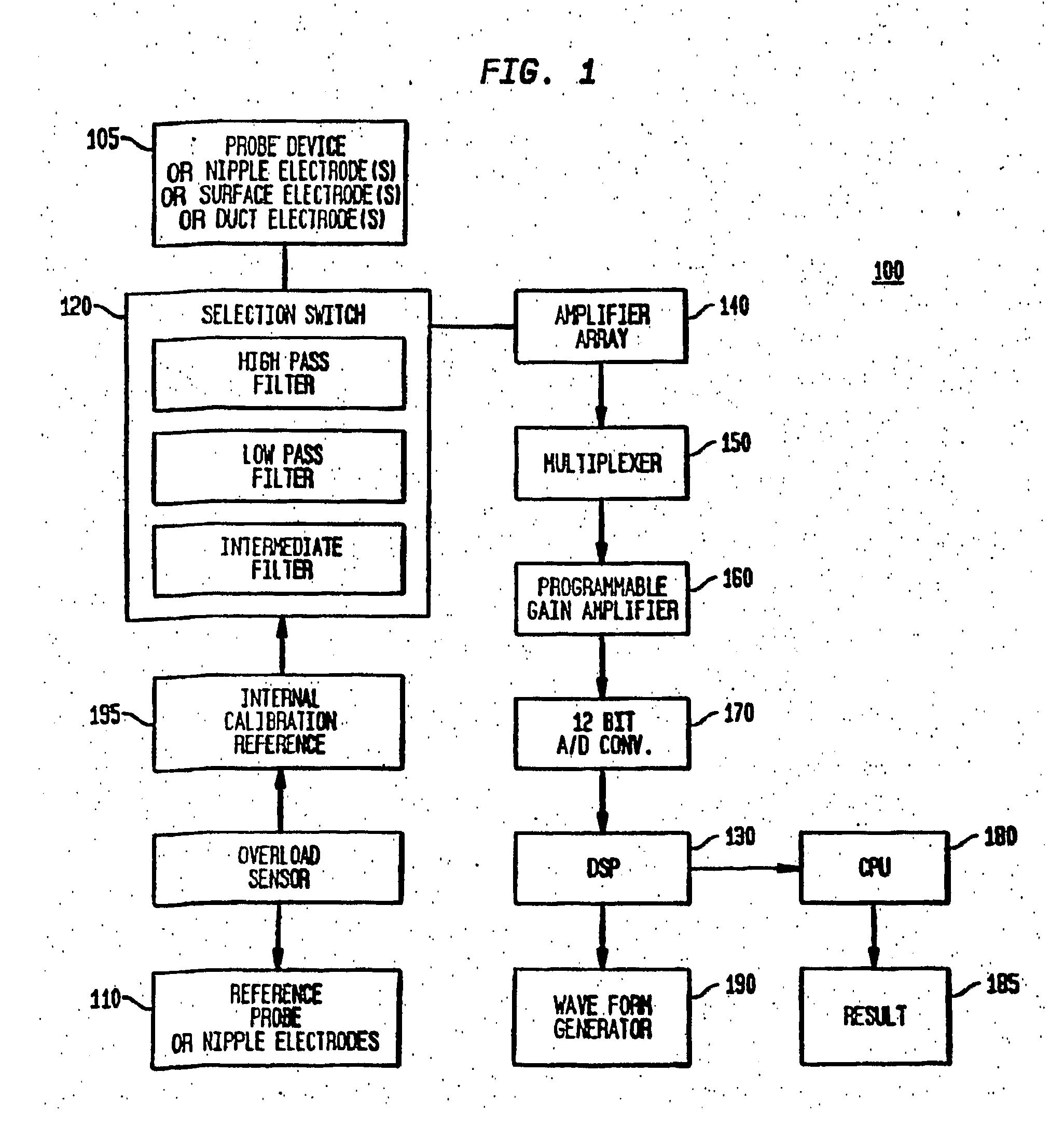
Method and System for Detecting Electrophysiological Changes in Pre-Cancerous and Cancerous Tissue and Epithelium
- Summary
- Abstract
- Description
- Claims
- Application Information
AI Technical Summary
Benefits of technology
Problems solved by technology
Method used
Image

Examples
example 3
Electrophysiological Changes in Other Epithelia
[0163]The examples illustrated by FIGS. 12 and 13 were performed in human colon specimen removed at the time of surgery. Based on in vitro studies in breast epithelial tissues, similar changes in human ductal epithelium that can be measured in vivo are expected.
[0164]FIG. 12 demonstrates the short circuit current (ISC) of human colonic epithelium ex-vivo. The figure demonstrates the time course along, the x-axis while varying the potassium gradient across the tissue. The potassium permeability of the apical membrane of human colonic mucosa (PKa) was determined in surgical specimens of controls and grossly normal-appearing mucosa obtained 10-30 cm proximal to colorectal adenocarcinomas. The mucosa was, mounted in Using chambers and the basolateral, membrane resistance and voltage were nullified by elevating the K+ in the serosal bathing solution. The apical sodium (Na+) conductance was blocked with 0.1 mM amiloride. This protocol reduces...
example 1
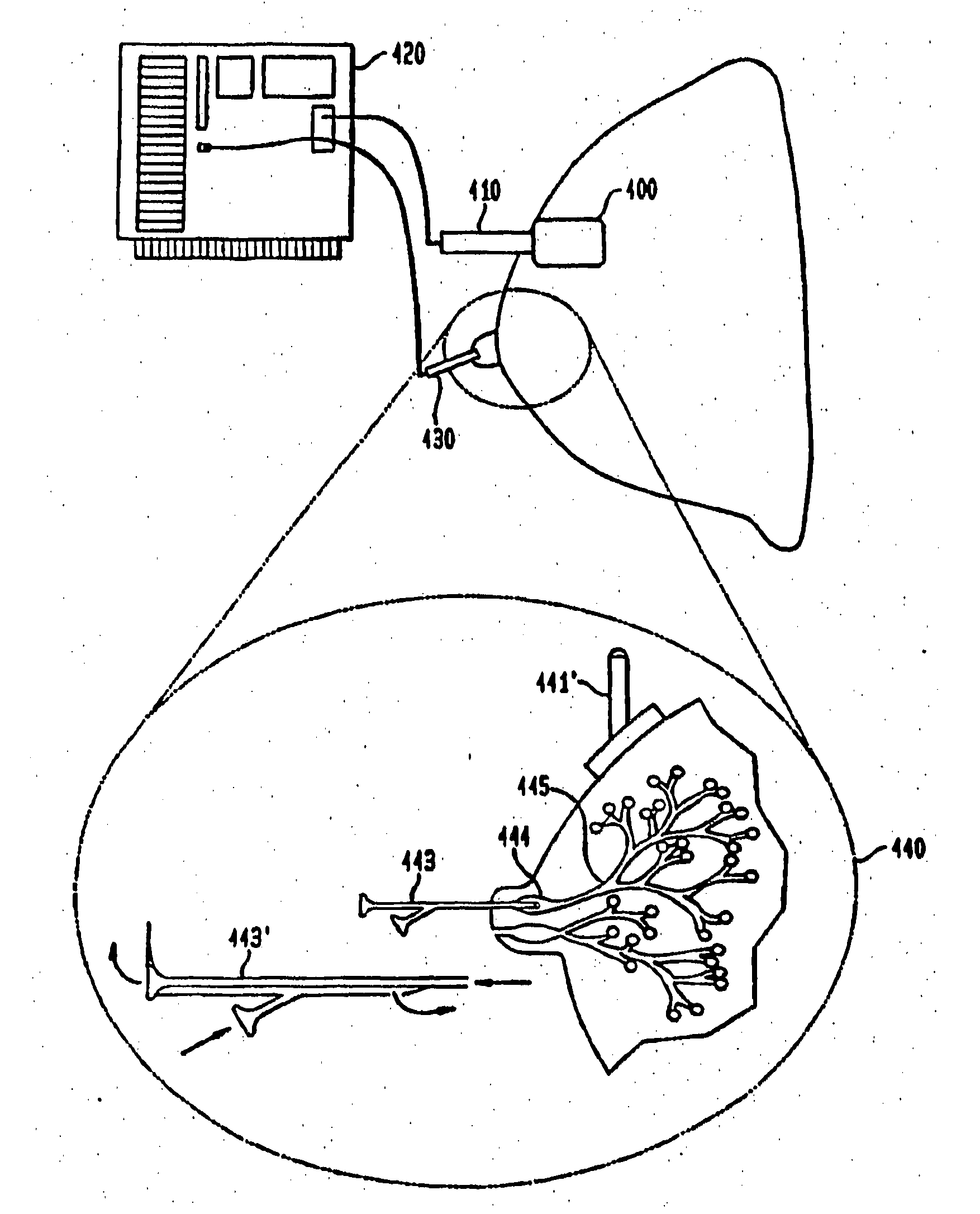
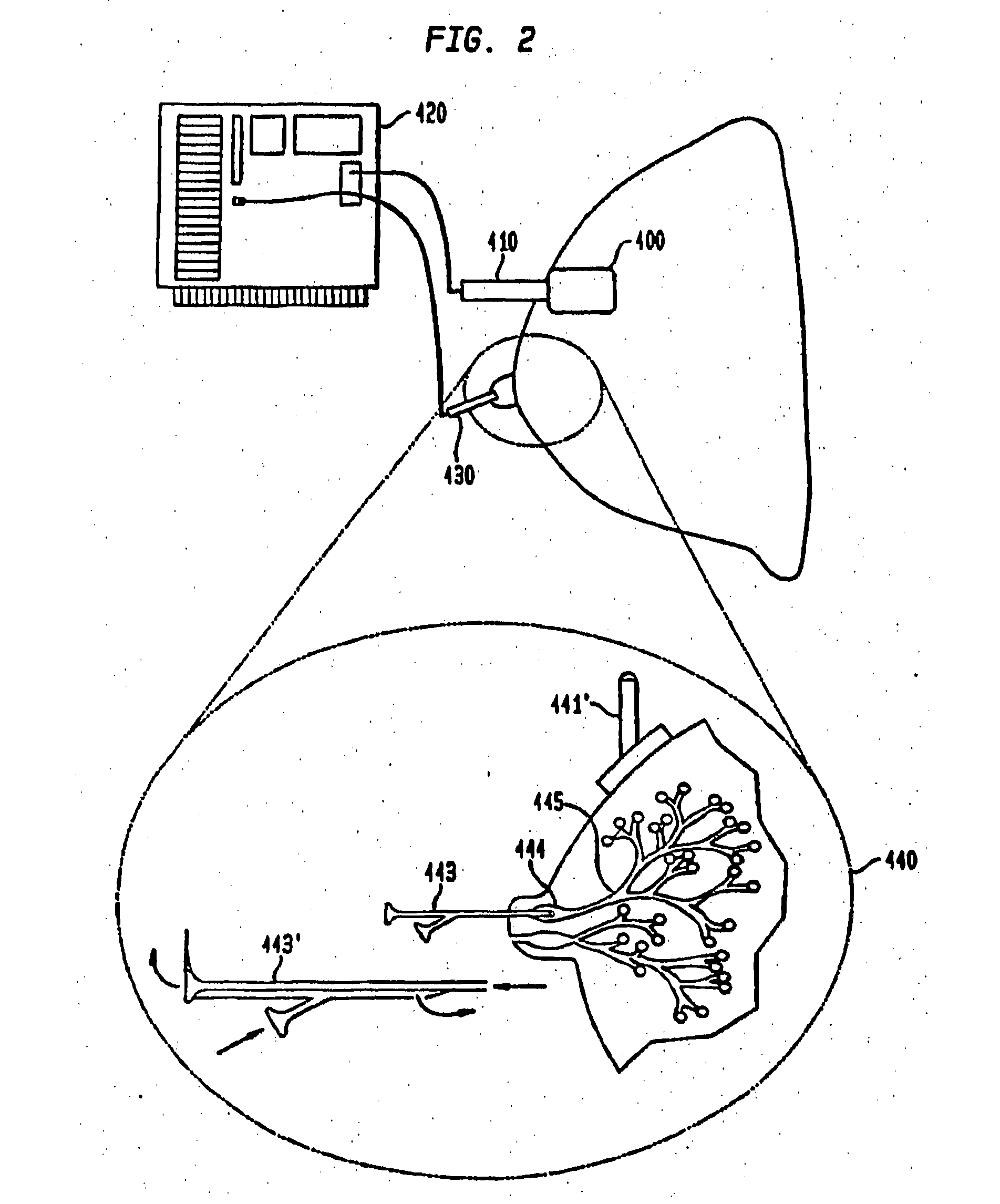
Breast Cancer Diagnosis Using Ductal Epithelial Impedance Spectroscopy
[0202]Alterations in ion conductance and transport [1]-(bracketed numbers identify references at the end of this example) represent early events in cellular proliferation and malignant transformation. Breast cancer develops within the ductal or lobular epithelium of the breast. Furthermore), functional changes occur in transgenic models of breast cancer, 2, 31, and it has been demonstrated that a down-regulation of specific ionic conductances occurs in human breast epithelia, during breast cancer development [4, 5]. Although electrical impedance scanning [6] or tomography, have been used to image mass lesions within the breast and for risk assessment, epithelial impedance spectroscopy has not been utilized to diagnose cancer in the epithelium, where breast cancer originates. One embodiment of the present invention illustrated in this example is an “Electronic Biopsy” to non-invasively identify breast cancer in the...
example 2
Expansion and Further Development of the Observations and Methods of Example 1
[0219]The diagnostic electrical characteristics described above have been applied to observations based on sample sizes varying between 110 and 150 patients as described below. The characteristics include: RSUB, Subepithelial Resistance—the resistance value at R∞, the intersection of the impedance curve with the X-axis at high frequency (for example, 60,000 Hz); REPI, Epithelial Resistance—the difference between R∞ and R0 (the high and the low intercept of the curve with the X-axis, or the difference in the resistance at low and high frequency); Capacitance (C)−derived from the curve using the formula Z (Impedance on the Y-axis)=1 / 2πfcC; Characteristic Frequency (fc)—the frequency at the highest point on the impedance curve; Depression Angle—the angle formed by a line drawn between the locus (center) of the impedance circle and the R0 or R∞ intercept on the x-axis; and open Circuit Potential or Transepithe...
PUM
 Abstract
Description
Claims
Application Information