Flocked medical instrument to be placed in the body, method of producing the medical instrument to be placed in the body and apparatus for producing the medical instrument to be placed in the body
- Summary
- Abstract
- Description
- Claims
- Application Information
AI Technical Summary
Benefits of technology
Problems solved by technology
Method used
Image

Examples
production example 1
Production Method for Hydroxyapatite Sinter
[0254]The following description deals with the production method for the hydroxyapatite sinter according to the present example.
[0255]A continuous oil layer of 40 ml including 0.5 g of a non-ionic detergent is prepared, by using dodecane as the continuous oil phase and pentaethylene glycol dodecyl ether whose cloudy sky atmosphere is 31° C. as the non-ionic detergent. Then, 10 ml of a Ca(OH)2 dispersing aqueous solution (2.5 mol %) is added to the prepared continuous oil layer. Next, after sufficiently stirring the fluid dispersion, 10 ml of KH2PO4 solution of 1.5% mol is added to the water / oil (W / O) emulsion, and was made to react in a reacting temperature of 50° for 24 hours. During the reaction, the solution was stirred. By thus separating the obtained reactants by centrifugation, hydroxyapatite is obtained. The hydroxyapatite is heated for one hour in the temperature of 800° C., which produced a hydroxyapatite sinter particle (hereafter...
example 1
[0266]The following description deals with a method for producing a percutaneous terminal by flocking the substrate with the KBE-cutSF-HAp.
[0267]The flocking device 1 as illustrated in FIG. 3 was used for the production of the percutaneous terminal. The steps were taken following the description of the section [2. Method and apparatus according to the present invention]. The conditions of the flocking were as follows:
Power source voltagedirect current (DC): 25 kVDistance between first electrodeapproximately 50 mmplate and second electrode plateDistance between second electrodeapproximately 15 mmplate and substrate ofpercutaneous terminalDistance between second electrodeapproximately 35 mmplate and substrate ofpercutaneous terminalHumidity controlrelative humidity of 95%Rotation rate of rotation3 rpmsupporting sectionProcessing time5 minutesTemperature inside apparatus25° C.Use of an atomizerYes
[0268]The KBE-cutSF-HAp was moistened with the atomizer before the KBE-cutSF-HAp was proce...
example 2
Evaluation of Flocking Density
[0277]The present Example performed an evaluation of flocking density of the percutaneous terminal produced by the operations and conditions described in Example 1.
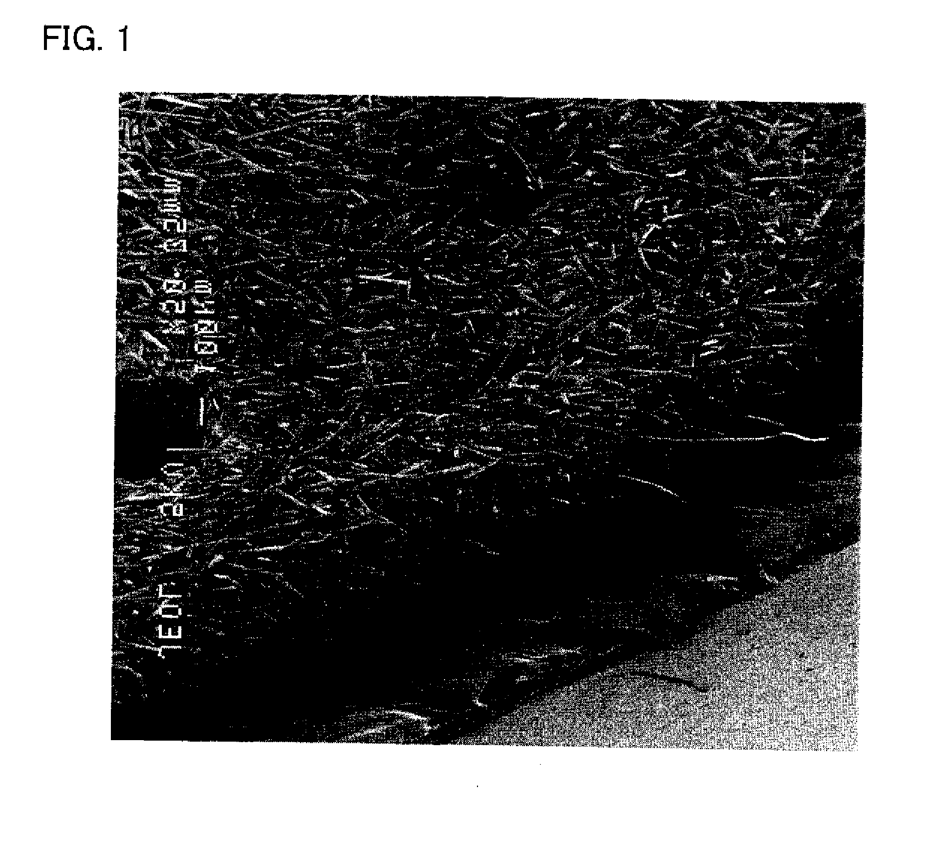
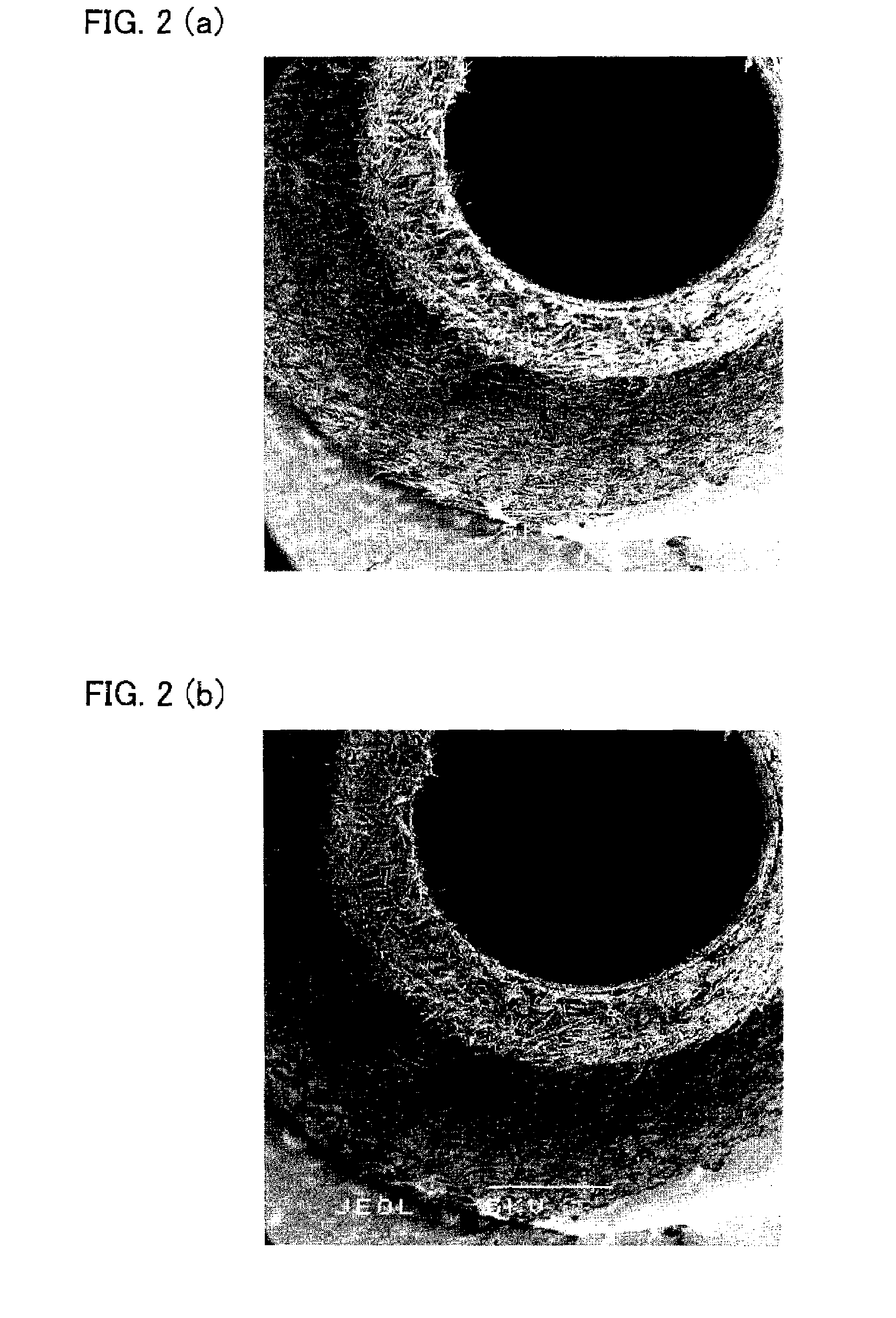
[0278]The evaluation of the flocking density was performed by cutting off one part of the flocked percutaneous terminal, and observing the surface of the part with a scanning electron microscope (SEM) (manufactured by Japan Electro Optical Laboratory Ltd. (JEOL Ltd.)). More specifically, the magnification was set as 100-power, and the number of flock existing within a square of a length of a side as 200 μm was counted. Five squares were randomly picked out for counting the number of flock, and the average value of the number of flock was evaluated as the flocking density.
[0279]The same operations were taken for the percutaneous terminal produced by the operations and conditions described in Comparative Example 1, and the flocking density thereof was evaluated.
[0280]A result of comparing (i) t...
PUM
| Property | Measurement | Unit |
|---|---|---|
| Length | aaaaa | aaaaa |
| Length | aaaaa | aaaaa |
| Length | aaaaa | aaaaa |
Abstract
Description
Claims
Application Information
 - R&D
- Intellectual Property
- Life Sciences
- Materials
- Tech Scout
- Unparalleled Data Quality
- Higher Quality Content
- 60% Fewer Hallucinations
Browse by: Latest US Patents, China's latest patents, Technical Efficacy Thesaurus, Application Domain, Technology Topic, Popular Technical Reports.
© 2025 PatSnap. All rights reserved.Legal|Privacy policy|Modern Slavery Act Transparency Statement|Sitemap|About US| Contact US: help@patsnap.com