Pre-angioplasty serration of atherosclerotic plaque enabling low-pressure balloon angioplasty and avoidance of stenting

a technology of atherosclerotic plaque and preangioplasty, which is applied in the field of preangioplasty serration of atherosclerotic plaque enabling low-pressure balloon angioplasty and avoiding stenting, which can solve the problems of only being prepared for blood vessels and not being fully dilated to their intended diameter

Active Publication Date: 2010-02-18
CAGENT VASCULAR INC
View PDF77 Cites 98 Cited by
  • Summary
  • Abstract
  • Description
  • Claims
  • Application Information

AI Technical Summary

Benefits of technology

[0012]To overcome the problems and disadvantages of prior practices of dilating plaque material in blood vessels through balloon angioplasty and with or without the use of post-angioplasty stent emplacement, the present invention employs an 
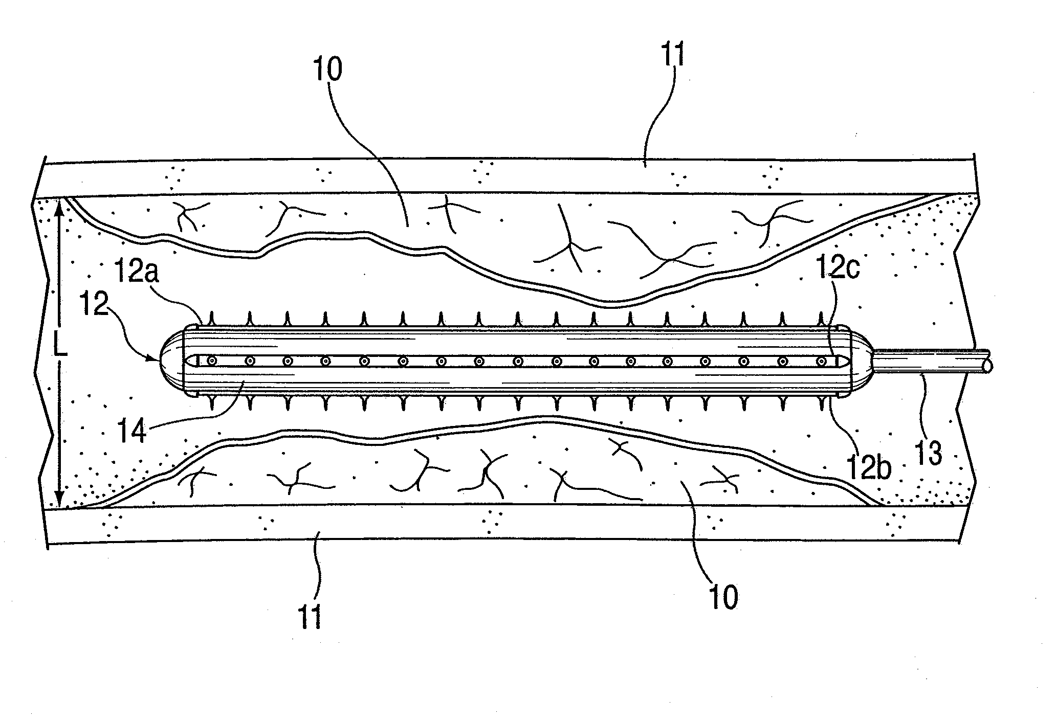
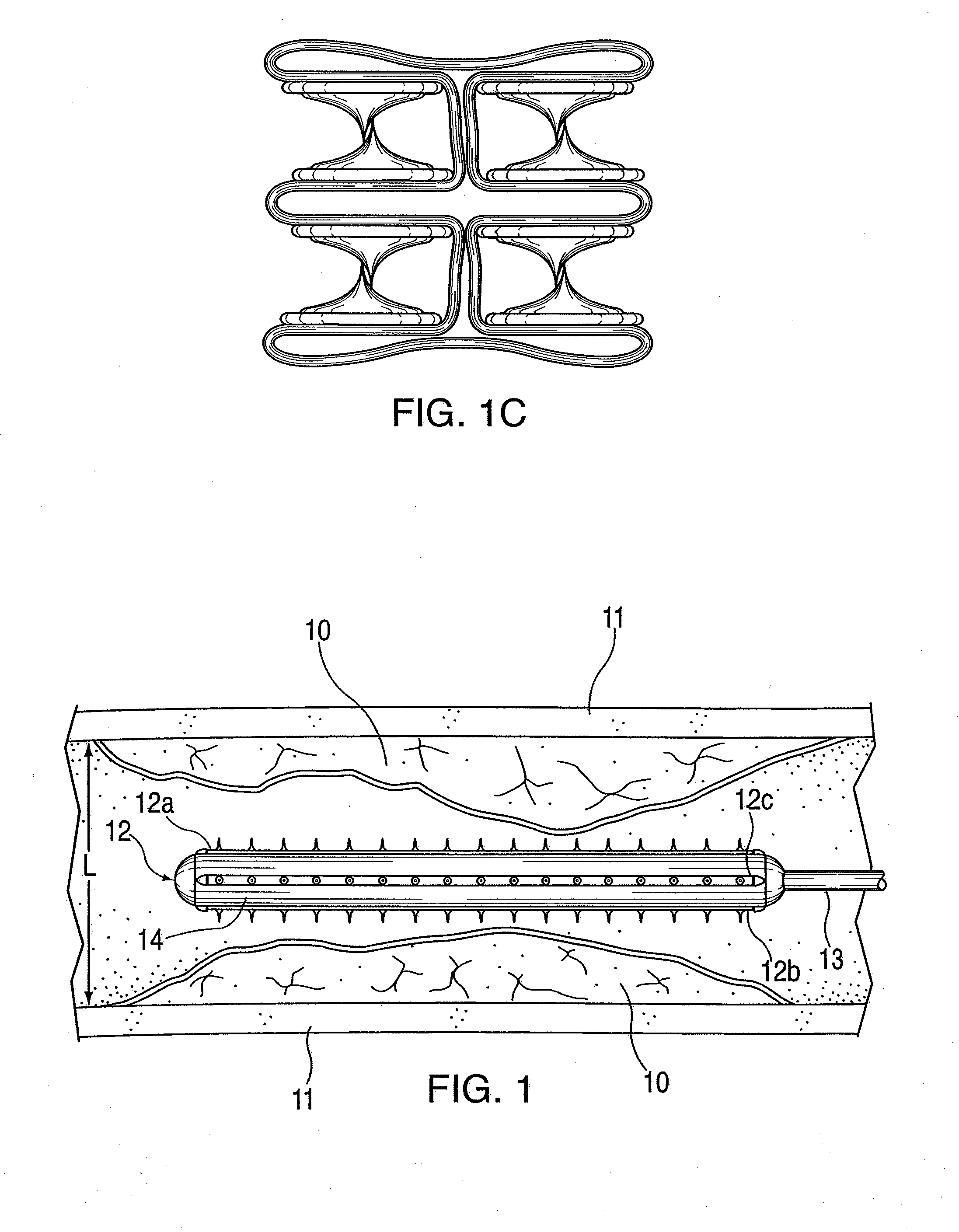
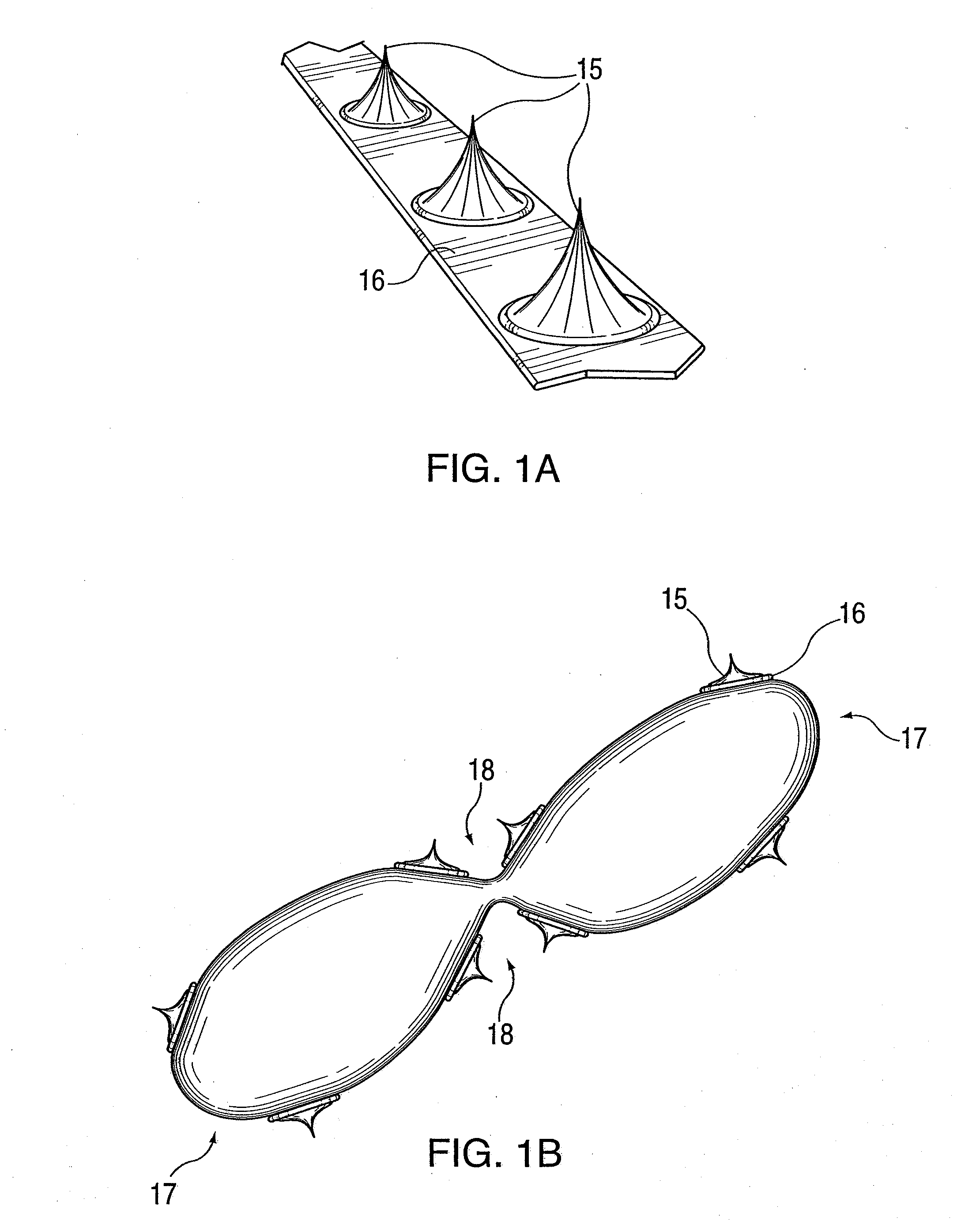
intravascular device for pre-angioplasty treatment carrying rows or patterns of small sharp spikes that are actuated by an expansion balloon or other apparatus to pierce the luminal surface of atherosclerotic plaque with lines or patterns of microperforations which act as serrations for forming cleavage lines, expansion lines, or planes in the plaque as a preparation prior to balloon angioplasty. When using a balloon actuated mechanism to press the spikes into the plaque to create the microperforations, expansion pressures of a full range may be used, from less than 2 atm to more than 10 atm. This pressure range may only be necessary for the purpose of introducing the spike elements into hardened calcified plaque. When a balloon actuated mechanism is used to create the microperforations, the blood vessel is only being prepared and is not being fully dilated to its intended diameter. The diameter of the artery is much larger than the fully expanded diameter of the spike device. Therefore the wall of the artery does not experience high pressure when the spike device is balloon actuated avoiding the danger identified in high pressure balloon angioplasty.
[0013]After preparation of the plaque with the microperforation and serration procedure, the plaque can be compressed and the artery lumen safely and accurately dilated and stretched, using low pressure balloon angioplasty, to its intended diameter without creating numerous and substantial dissections and elevated flaps. The microperforation and serration enable the plaque to be dilated more evenly and smoothly and avoid forming random cracks that may lead to dissection and residual stenosis. The plaque, after it has been pre-treated with microperforation and serration, may also be dilated with lower pressure than that which is used in standard balloon angioplasty. The lower intra-balloon pressure (e.g., less than or equal to 4 atm and very often less than or equal to 2 atm) causes less disruption of the plaque, fewer dissections, and less injury to the artery wall. This “low pressure” or “minimal injury” angioplasty is less likely to cause the biological reaction that often follows balloon angioplasty with neointimal hyperplasia or smooth muscle cell replication.
[0014]In addition, microperforation and serration permits the plaque to expand with less fracturing or disruption of the plaque during balloon angioplasty. By preparing the plaque using microperforations and then performing a balloon angioplasty at low pressure, the number and severity of dissections is reduced. This decreases the need for stent placement to be used to treat dissection or residual stenosis after balloon angioplasty. The subsequent balloon angioplasty may be performed at low balloon pressures of about 4 atmospheres or less due to preparation of the plaque with perforations, so as to avoid injury to the arterial wall. By performing plaque preparation and then low pressure angioplasty, there is less likelihood of a dissection occurring deeply and exposing the media layer of the artery. Exposure of this artery stimulates thrombus formation by collagen exposure and also stimulates smooth muscle cell growth which later causes neointimal hyperplastic occlusion of the artery. This decrease in number and also decrease in severity of dissection is a key differentiating factor in comparison to cutting or scoring devices.
[0017]The pre-angioplasty treatment of a plaque site may also be combined instead with drug-eluting balloon (DEB) angioplasty or drug-coated balloon (DCB) angioplasty. Due to the various applications of balloon angioplasty, there are a variety of medications that may be used, such as: plaque-reducing medication, thrombus inhibiting medication, inhibitors of cell growth, other biologically active treatments, and stem cell delivery. The intended effect is to have the medication taken up by or adhered to the plaque and / or wall of the diseased artery at the time of balloon angioplasty. The preparation of the plaque and the creation of microperforations enhances the uptake and biological activity of the medication. The creation of new plaque surface area in the depths of each of the microperforations, and also the location of the plaque that has been exposed, in the top layer of the plaque without exposing the medial layer, is further facilitative of the biological activity of the medication.
[0019]Another variation for the pre-angioplasty treatment is the use of a balloon-restricting mesh over the expansion balloon for restricting its maximum expansion diameter so that it is less than the blood vessel diameter. The mesh minimizes the potential of the balloon to expand beyond the stenosis site into an hour-glass shape, and also limits the amount of pressure that is delivered to the plaque by limiting the balloon to a defined radial expansion. The mesh structure can be include spike buttons or by milled (through grinding, laser cutting, metal extrusion, photolithography, or other means) to form sections with height variations that act as microperforations.

Problems solved by technology

When a balloon actuated mechanism is used to create the microperforations, the blood vessel is only being prepared and is not being fully dilated to its intended diameter.

Method used


Image

  • Pre-angioplasty serration of atherosclerotic plaque enabling low-pressure balloon angioplasty and avoidance of stenting
  • Pre-angioplasty serration of atherosclerotic plaque enabling low-pressure balloon angioplasty and avoidance of stenting
  • Pre-angioplasty serration of atherosclerotic plaque enabling low-pressure balloon angioplasty and avoidance of stenting

Examples


Embodiment Construction

[0035]The conventional practice of compression of plaque by expansion pressure during balloon angioplasty, i.e., by applying a high pressure expansion force equally in all directions radially from the inside to a heterogeneous, roughly circumferential plaque structure, can produce unpredictable and inconsistent results. In typical treatment of atherosclerotic plaques, the angioplasty balloon is inflated with 4 to 8 atmospheres of pressure, and pressures up to 22 atmospheres may be required in some cases. Such high pressures can cause injury to the intima and media in the artery at the treatment location. Arterial wall injury is one of the major stimulants to intimal hyperplasia, smooth muscle cell replication and intravascular scarring causing occlusion. Plaque is heterogeneous in nature composed of varying masses of soft and hard materials, calcium and highly variable topography, and can give way along paths of least resistance. Therefore, when standard balloon angioplasty is perfo...


PUM


Abstract

A device and method for intravascular treatment of atherosclerotic plaque prior to balloon angioplasty which microperforates the plaque with small sharp spikes acting as serrations for forming cleavage lines or planes in the plaque. The spikes may also be used to transport medication into the plaque. The plaque preparation treatment enables subsequent angioplasty to be performed at low balloon pressures of about 4 atmospheres or less, reduces dissections, and avoids injury to the arterial wall. The subsequent angioplasty may be performed with a drug-eluting balloon (DEB) or drug-coated balloon (DCB). The pre-angioplasty perforation procedure enables more drug to be absorbed during DEB or DCB angioplasty, and makes the need for a stent less likely. Alternatively, any local incidence of plaque dissection after balloon angioplasty may be treated by applying a thin, ring-shaped tack at the dissection site only, rather than applying a stent over the overall plaque site.

Description

[0001]This U.S. patent application is a continuation-in-part of co-pending U.S. patent application Ser. No. 12 / 408,035 of the same inventors, filed Mar. 20, 2009, which claimed priority to the filing date of U.S. Provisional Appln. 61 / 038,477, filed on Mar. 21, 2008.TECHNICAL FIELD[0002]The present invention is directed to a device and method for opening blood vessels in the body occluded by atherosclerotic plaque by pre-angioplasty serration and dilatation of the plaque.BACKGROUND[0003]Atherosclerotic occlusive disease is the primary cause of stroke, heart attack, limb loss, and death in the US and the industrialized world. Atherosclerotic plaque forms a hard layer along the wall of an artery and is comprised of calcium, cholesterol, compacted thrombus and cellular debris. As the atherosclerotic disease progresses, the blood supply intended to pass through a specific blood vessel is diminished or even prevented by the occlusive process. One of the most widely utilized methods of tr...

Claims


Application Information

Patent Timeline
Patent Type & Authority Applications(United States)
IPC IPC(8): A61B17/22
CPCA61B17/205A61B17/320725A61B2017/22061A61M25/1011A61M25/1027A61M25/104A61M2025/1084A61M2025/1013A61M2025/1031A61M2025/105A61M2025/1059A61M2025/1086A61M2025/109A61M2025/0096A61B2017/00004A61M2037/0023A61M2037/003A61M2037/0046
Inventor SCHNEIDER, PETERGIASOLLI, ROBERT
Owner CAGENT VASCULAR INC
Why Eureka
Try Eureka