
Pan-Kinase Activation and Evaluation of Signaling Pathways

a signaling pathway and activation technology, applied in the field of pankinase activation and evaluation of signaling pathways, can solve the problems of cumbersome assays and lack of sensitivity, and achieve the effect of small sample size and increased specificity and sensitivity

Inactive Publication Date: 2010-04-08
BECKMAN COULTER INC +1
View PDF15 Cites 52 Cited by
  • Summary
  • Abstract
  • Description
  • Claims
  • Application Information

AI Technical Summary

Benefits of technology

[0035]A significant advantage of the present invention is that the increased specificity and sensitivity allows for use of the methods with very small sample sizes. This is important where only a small patient sample is obtainable, for example, with neonates.
[0036]It should be noted that as used herein and in the appended claims, the singular forms “a,”“and,” and “the” include plural references unless the context clearly dictates otherwise.
[0037]Although any methods, devices, and materials similar or equivalent to those described herein can be used in the practice of the invention, the particular methods, devices, and materials are now described.Preparation of the Test Sample
[0038]The terms test sample and sample are used interchangeably herein. The test sample in the methods of the present invention can include any leukocyte-containing sample, for example, whole blood, blood plasma, bone marrow aspirates (or any cells obtained from bone marrow), urine, serum, saliva, cerebral spinal fluid, urine, amniotic fluid, interstitial fluid, feces, mucus, body tissue extracts or cellular extracts. In certain embodiments, the sample is a blood sample. In a preferred embodiment, the blood sample is whole blood. The whole blood can be obtained from the individual or test subject using standard clinical procedures.
[0039]Obtaining a blood sample encompasses obtaining the blood sample directly from the patient, for example by a phlebotomist. Obtaining a blood sample also encompasses obtaining a blood sample that was previously obtained from a patient, for example a laboratory technician obtaining a patient's blood sample for analysis using the methods of the present invention. Plasma can be obtained from whole blood samples by centrifugation of anti-coagulated blood. Such a process provides a bottom layer of packed red blood cells, an intermediate buffy coat layer and a supernatant plasma layer.
[0040]Leukocytes can be isolated from whole blood samples by any of various techniques including buoyant density centrifugation and others known in the art. The leukocytes, i.e., white blood cells, comprise monocytes, lymphocytes, neutrophils, basophils, and eosinophils. Monocytes are especially preferred for use in the methods of the present invention.

Problems solved by technology

Current assays rely on immunoblotting techniques that cannot identify the actual cell-type generating the response in a mixed cell population.
Also, by having to remove the drug from the samples, these assays are cumbersome and are subject to a lack of sensitivity.

Method used


Image

  • Pan-Kinase Activation and Evaluation of Signaling Pathways
  • Pan-Kinase Activation and Evaluation of Signaling Pathways
  • Pan-Kinase Activation and Evaluation of Signaling Pathways

Examples


example 1

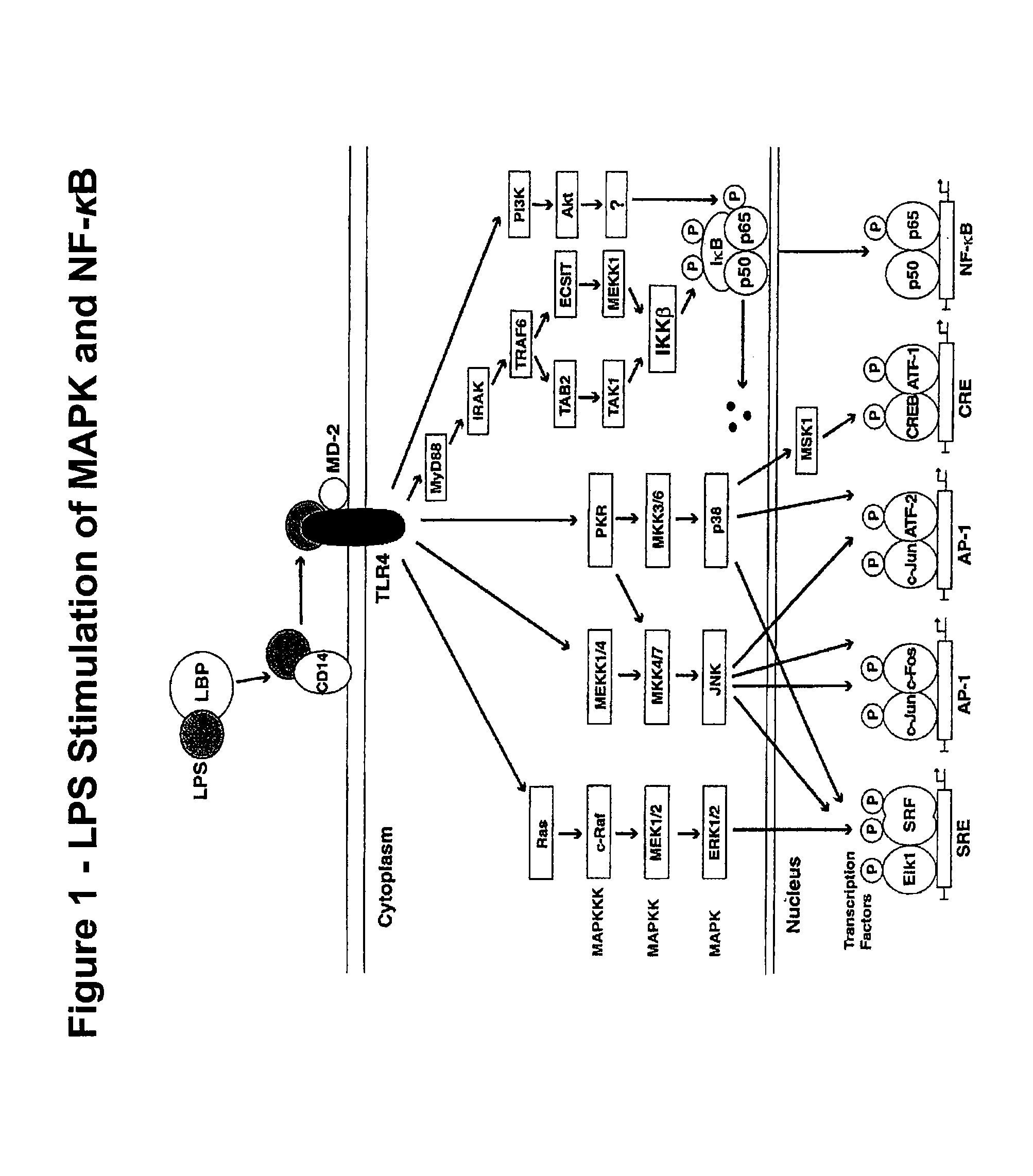
Protocol for LPS Activation of MAPK Signaling in Whole Blood Samples

[0178]100 μl of blood was inserted into the bottom of 12×75 mm tubes. Blood was removed from the side of tube with cotton swab to eliminate potential contamination of the sample with unfixed cells. 100 ng lipopolysaccharide (LPS) was added to activation tubes (or equal volume of phosphate buffered saline (PBS) to control tubes) and the tubes were placed in a 37° C. water bath.

[0179]After various incubation times (e.g. 1 minute to 60 minutes, the first tube was removed from the incubator and 65 ul 10% formaldehyde was added into tube. The tubes were mixed and placed in a rack and incubated at room temperature. After exactly a 10 minute incubation with formaldehyde, 1 ml Lyse / Permeabilization Buffer (585 μl Triton X-100 10% stock solution to 500 ml with PBS. 0.1165% Triton X-100 solution at room temperature in the dark and pre-heated to 37° C. immediately prior to use.) was added to each tube, vortexed vigorously and ...

example 2

Flow Cytometry Based Sepsis Assay

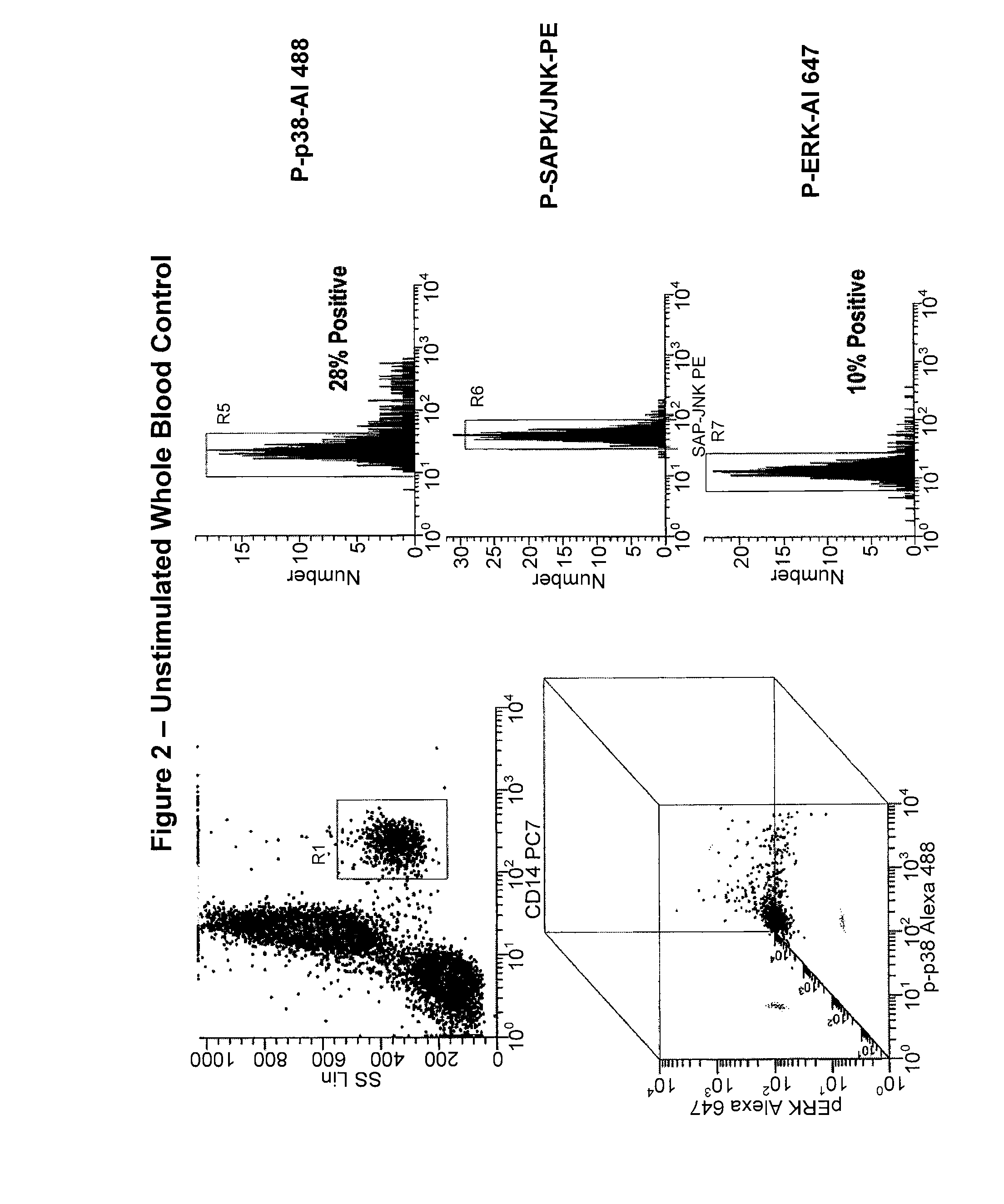
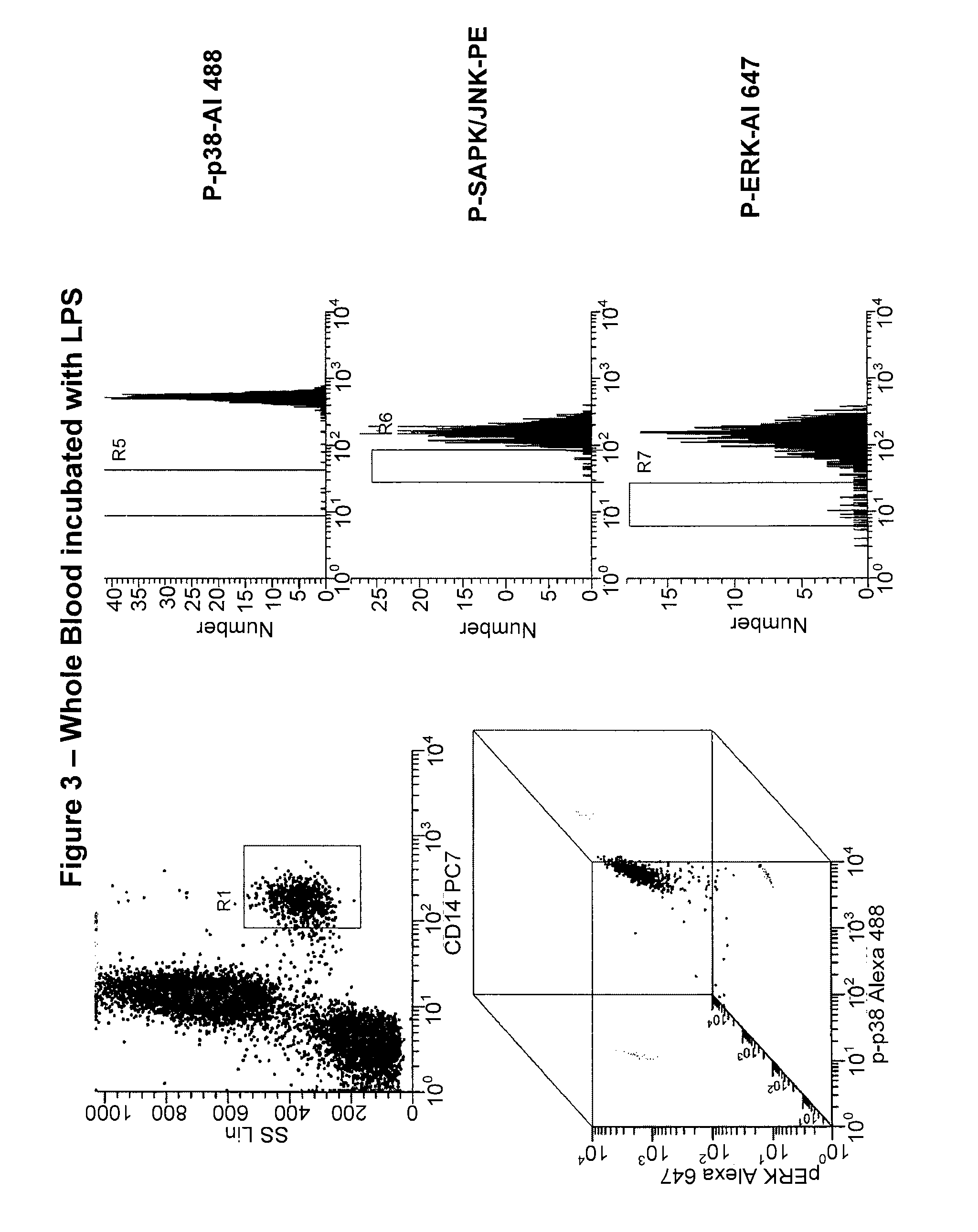
[0185]In this study, a simultaneous measurement of four different signaling phospho-epitopes, which include P-ERK, P-p38 MAP Kinase, P-SAPK (Stress-Activated Protein Kinase), and P-S6 ribosomal protein (a measurement of new polypeptide synthesis), was conducted. This assay employed a measurement of the time course of LPS activation, in both “naïve” whole blood, and whole blood samples previously exposed to LPS activation (“re-activated”). This assay also employed the fixation and permeabilization approach described above to measure both cell surface (CD14 to identify monocytes) and cytoplasmic or nuclear localized phospho-epitopes (P-ERK, P-p38, P-SAPK, and P-S6). This assay also employed an internal negative control to analyzing the data, relying on populations, rather than isotype controls, and measuring changes in MFI (mean fluorescence intensity) rather than percent positive.

[0186]The results from this study for each individual marker is presente...


PUM


Abstract

Methods and reagents are provided for determining the activation state of a signal transduction pathway signaling protein. There exists a need in the art for methods that can monitor the efficacy of a signal transduction inhibitor in a patient. Other needs exist for detecting and monitoring certain disease or disorders that are associated with aberrant activation of a signal transduction pathway signaling protein. The present assay provides a highly sensitive assay that is also useful in patient populations in which obtaining a large cellular sample is difficult, for example, neonates.

Description

CROSS-REFERENCE TO RELATED APPLICATIONS[0001]This application claims the benefit of U.S. Provisional Application 61 / 094,304, filed Sep. 4, 2008, which is incorporated herein in its entirety.BACKGROUND OF THE INVENTION[0002]1. Field of the Invention[0003]Certain embodiments of the invention relate to methods for determining the activation state of a signal transduction pathway signaling protein. In at least some embodiments, methods are provided for monitoring the efficacy of a signal transduction inhibitor in a patient. The present invention further provides highly sensitive assays useful in patient populations in which obtaining a large cellular sample is difficult, for example, neonates. Certain embodiments of the invention relate to methods of identifying novel signal transduction pathway protein inhibitors. Certain embodiments of the invention relate to methods for detecting sepsis in a patient sample. Kits for practicing the methods of the invention are also provided.[0004]2. B...

Claims


Application Information

Patent Timeline
IPC IPC(8): G01N33/53
CPCG01N33/5041G01N33/5047G01N33/573G01N33/5008G01N33/56972
Inventor HEDLEY, DAVIDCHOW, SUESHANKEY, T. VINCENT
Owner BECKMAN COULTER INC
Learn More