Inkjet gene printing

a gene printing and inkjet technology, applied in the field of inkjet gene printing, can solve the problems of unsatisfactory tissue engineering application of existing techniques, unsuitable for studies, and ineffectiveness,

Active Publication Date: 2010-06-24
WAKE FOREST UNIV HEALTH SCI INC
View PDF8 Cites 39 Cited by
  • Summary
  • Abstract
  • Description
  • Claims
  • Application Information

AI Technical Summary

Problems solved by technology
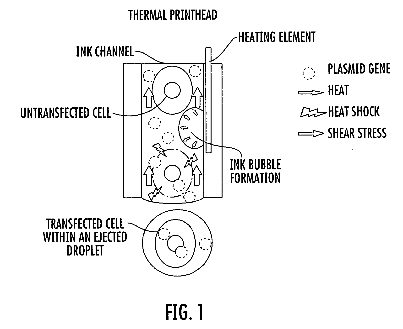
Although there are established methods in the art for delivering genes into cells, the application of existing techniques to tissue engineering is not ideal.
Although viral transfection has a high efficiency rate, it also has many drawbacks, such as residual pathogenicity, host immune response, and the potential induction of neoplastic growth following insertional mutagenesis.
These concerns have restricted its application in medicine and in the biomedical areas, especially in clinical gene therapy.
However, it is not efficient and not appropriate for studies and applications that require a significant number of cells to become transfected.
Low cell viability is a major limitation of transfection by electroporation.
However, this method is limited by a low transfection efficiency.

Method used


Image

  • Inkjet gene printing
  • Inkjet gene printing
  • Inkjet gene printing

Examples


example 1

[0070]In vitro transfection by coprinting. A porcine aortal endothelial cell line (PAEC) was used, which was established previously in our lab by enzymatic dispersion of adult pig aorta, and the passage 15, 16 and 17 of the cell line were used for gene transfection. PAEC cells were maintained in F12 medium (GIBCO, Invitrogen, Carlsbad, Calif.) supplemented with 10% fetal bovine serum (GIBCO), and 100 IU penicillin and 100 mg / ml streptomycin at 37° C. in a humidified 5% CO2 (95% air) atmosphere.

[0071]Cytomegalovirus (CMV) early immediate promoter driven plasmids encoding the cDNAs were used. The pmaxGFP™ (Amaxa GmbH, Germany, and Gaithersburg, Md.), pIRES-VEGF-GFP (BD Biosciences, Bedford, Mass.), and pIRES-lacZ (BCCM / LMBP, Belgium) plasmids were each amplified in DH5a strain of Escherichia coli, isolated by alkaline lysis, and purified by ion exchange column chromatography (Qiagen Inc., Valencia, Calif.). The pmaxGFP™ plasmid encodes the green fluorescent protein (GFP) from the cope...

example 2

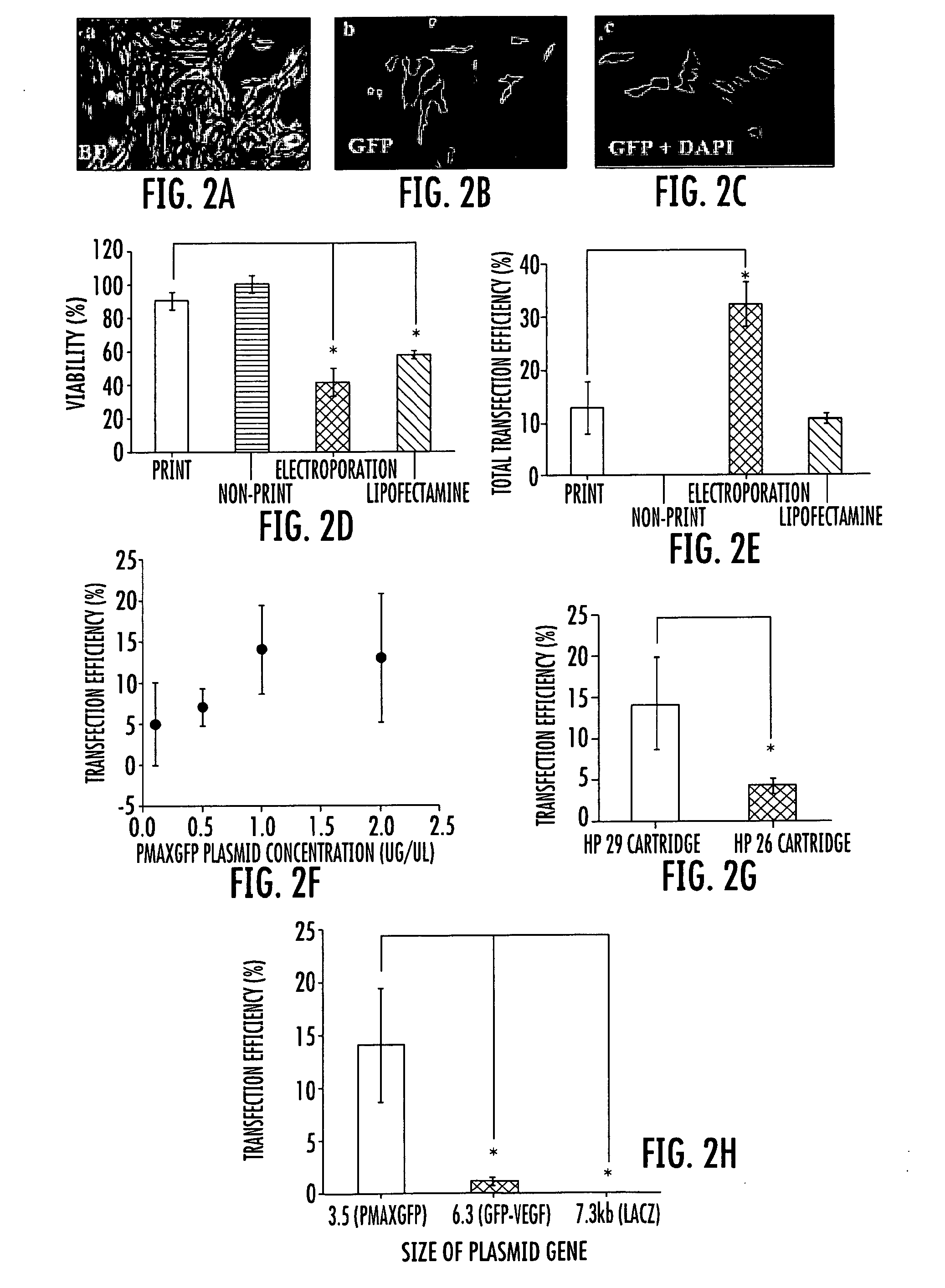
[0076]Comparison with common transfection methods. We further compared the cell viability and the transfection efficiency of the inkjet printing method with other chemical and physical transfection methods commonly used in tissue engineering. For this purpose, a common liposome based agent and an electroporation approach were performed. Lipofectamine™ 2000 (Invitrogen), a common liposome based agent, and Nucleofection, an electroporation approach, were compared to the printing method using the PAEC cell cultures.

[0077]For transfection with Lipofectamine™ reagent, PAEC cells were seeded in a 24-well plate at a density of 2×10⁵ cells per well the day before transfection. Transfection was performed according to the manufacturer's protocol. Briefly, 0.8 μg pmaxGFP™ (Amaxa) and the equivalent amount of Lipofectamine™ 2000 reagent were each added to 50 μl of serum free F12 medium. After incubation for 5 min at room temperature (RT), they were mixed and further incubated for 20 min at room...

example 3

[0086]Comparison of ink cartridges, concentration and size of plasmid. Different HP cartridges were also tested for inkjet gene transfection. The HP 51629a cartridge demonstrated higher gene transfection efficiency than the HP 51626a cartridge, as shown in FIG. 2g. The HP 51629a and HP 51626a cartridges share many similar printing parameters and mechanisms, but differ in the nozzle size. It is believed that the HP 51629a cartridge has a relatively smaller nozzle diameter than the HP 51626a cartridge, which may cause the higher stress, thus leading to higher transfection efficiency. After trypsin treatment, PAEC cells are about 10-20 μm. The nozzles of the HP 51629a and HP 51626a cartridges are about 40 and 50 micrometers, respectively (see Xu et al., Biomaterials (2006) 27(19):3580-88).

[0087]It was also found that several other factors could affect the transfection. Different concentrations of the plasmids in the print suspension were tested. As shown in FIG. 2f, the gene expression...


PUM

PropertyMeasurementUnit
timeaaaaaaaaaa
diameteraaaaaaaaaa
diameteraaaaaaaaaa

Abstract

Provided herein are methods and apparatuses for transfecting a cell with a compound of interest by stressing the cell, e.g. with shear stress. The compound of interest may be nucleic acids, proteins, molecules, nanoparticles, drugs, etc., or any combination thereof. Methods of printing cells with an inkjet printing device are also provided, wherein at least a portion of viable cells (preferably at least 1%) are transfected with a compound of interest. Preferably, at least 25% of the cells are viable after printing. In addition, methods of forming an array of viable cells are provided wherein at least a portion of the viable printed cells (preferably at least 1%) are transfected with at least one compound of interest.

Description

RELATED APPLICATIONS[0001]This application claims the benefit of U.S. Provisional Patent Application Ser. No. 60 / 942,549, filed Jun. 7, 2007, the disclosure of which is incorporated herein by reference in its entirety.FIELD OF THE INVENTION[0002]The present invention concerns the delivery of compounds of interest into living cells.BACKGROUND OF THE INVENTION[0003]In the interdisciplinary field of tissue engineering, powerful new therapies are being developed to address structural and functional disorders of human health by utilizing living cells as engineering materials. See Viola et al., “The Emergence of Tissue Engineering as a Research Field,” prepared for the National Science Foundation, Oct. 14, 2003, available at nsf.gov. In some areas of tissue engineering, researchers are creating two- and three-dimensional tissues and organs from combinations of cells in order to repair or replace diseased or damaged tissues.[0004]In instances where normal tissue cannot be engineered with t...

Claims


Application Information

Patent Timeline
Patent Type & Authority Applications(United States)
IPC IPC(8): C12N15/87C12N5/00C40B50/14C40B60/14
CPCC12N15/63C12N15/895C12N15/89C12N15/87A61K31/713
Inventor XU, TAOYOO, JAMES J.ATALA, ANTHONY
Owner WAKE FOREST UNIV HEALTH SCI INC
Why Eureka
Try Eureka