Methods for detecting nucleic acids in a sample

a nucleic acid and sample technology, applied in biochemical apparatus and processes, specific use bioreactors/fermenters, after-treatment of biomass, etc., can solve the problems of increasing the sample-to-answer time beyond what, not meeting the need for low-cost, easily manufactured devices, etc., and achieves the effect of simplifying the work flow

Inactive Publication Date: 2010-07-01
NEXUS DX
View PDF3 Cites 19 Cited by
  • Summary
  • Abstract
  • Description
  • Claims
  • Application Information

AI Technical Summary

Benefits of technology

[0018]Accordingly, in one instance, the present disclosure relates to a lateral flow test device that is feasible for point of care (POC) applications, which test device may be employed in a method for the detection of a nucleic acid. The methods and devices of the present disclosure have many advantages over other nucleic acid detection systems, such as the following advantages: (a) the methods and devices of the present disclosure utilize non-native capture units, such as pR...

Problems solved by technology

Hybridization assays, however, require specialized instrumentation and multiple pipetting, incubation, and washing steps.
However, typical DNA microarray technology suffers from several drawbacks, Long hybridization incubations are required for microarray assays, which increases the sample-to-answer times beyond what is acceptable for a rapid screening assay.
Additionally, microarray methods employ designs that remain reliant upon fluorescent detection and supporting instrumentation, and do not address the need for low-cost, easily manufactured devices that can be used in the absence of laboratory infrastructures.
Real-time PCR, however, requires highly specialized, expensive equipment along with costly reagents.
The process also involves expensive devices and instruments.
Flow cytometry, however, although suitable for the development of multiplex assays, also requires expensive instrumentation.
The disadvantage of these methods is that the sequence used for the immobilization can potentially hybridize with the sequence to be immobilized, forming intramolecular secondary structures, may hybridize with another sequence to be immobilized, forming intermolecular secondary structures, or may hybridize with nucleic acids from the sample.
The risk of such an unwanted or interfering interaction increases with the length of the nucleic acid(s) to be immobilized, as well as with the complexity of a sample (e.g., possible contaminating nucleic acids.
For instance, a significant economic and time disadvantage of using natural nucleic acids as immobilization agents is that a certain minimum sequen...

Method used


Image

  • Methods for detecting nucleic acids in a sample
  • Methods for detecting nucleic acids in a sample
  • Methods for detecting nucleic acids in a sample

Examples


example 1

Detection Using pRNA Based Synthetic Binding System and Europium Label

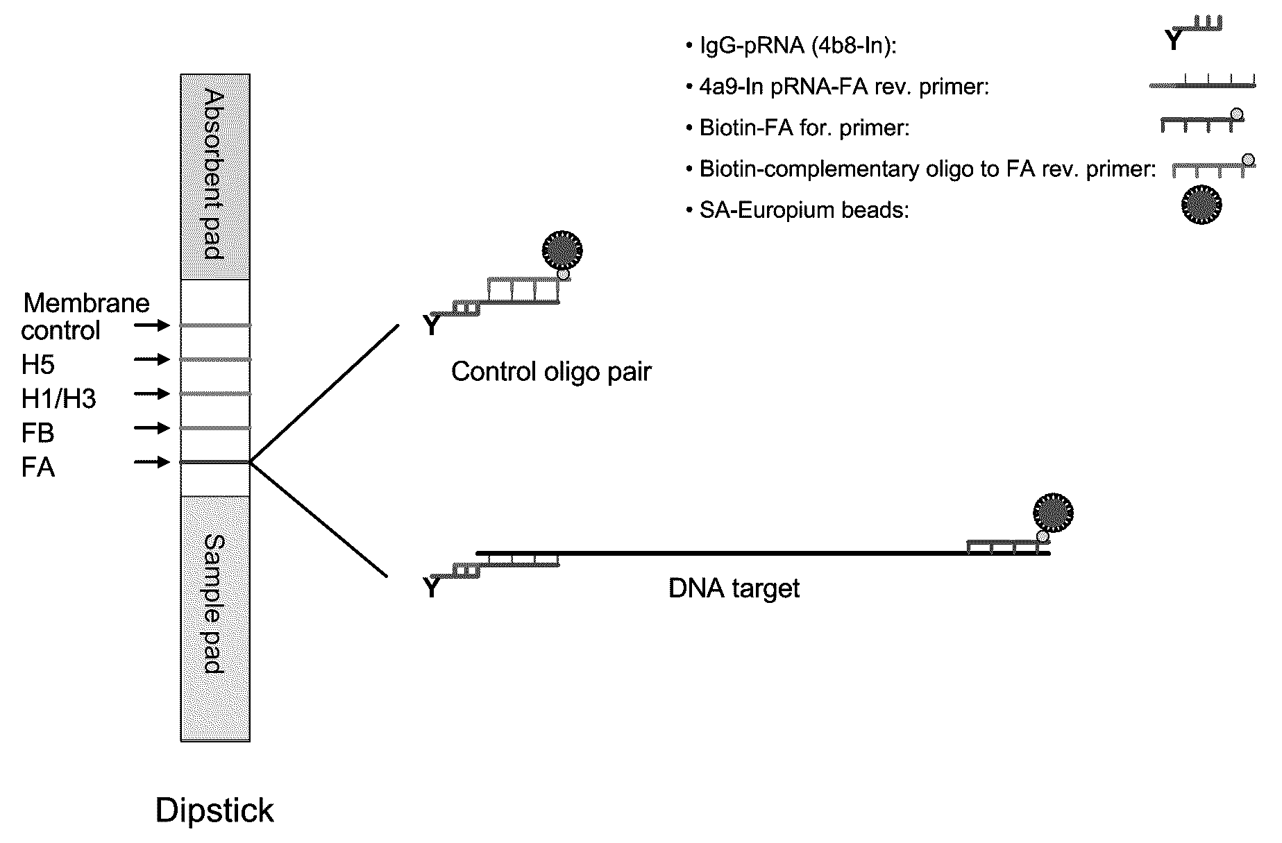
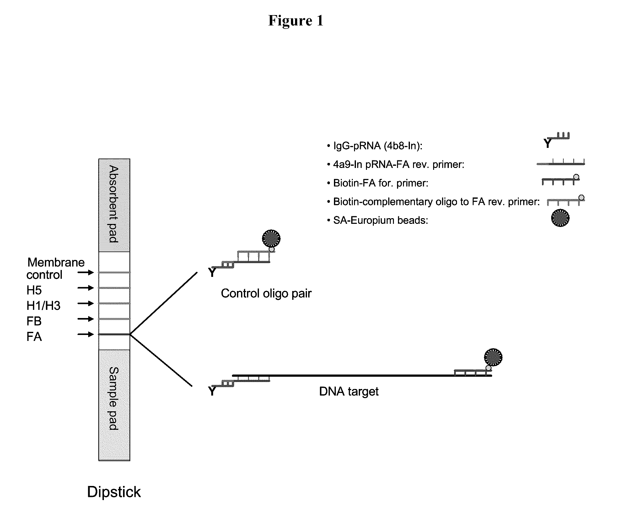
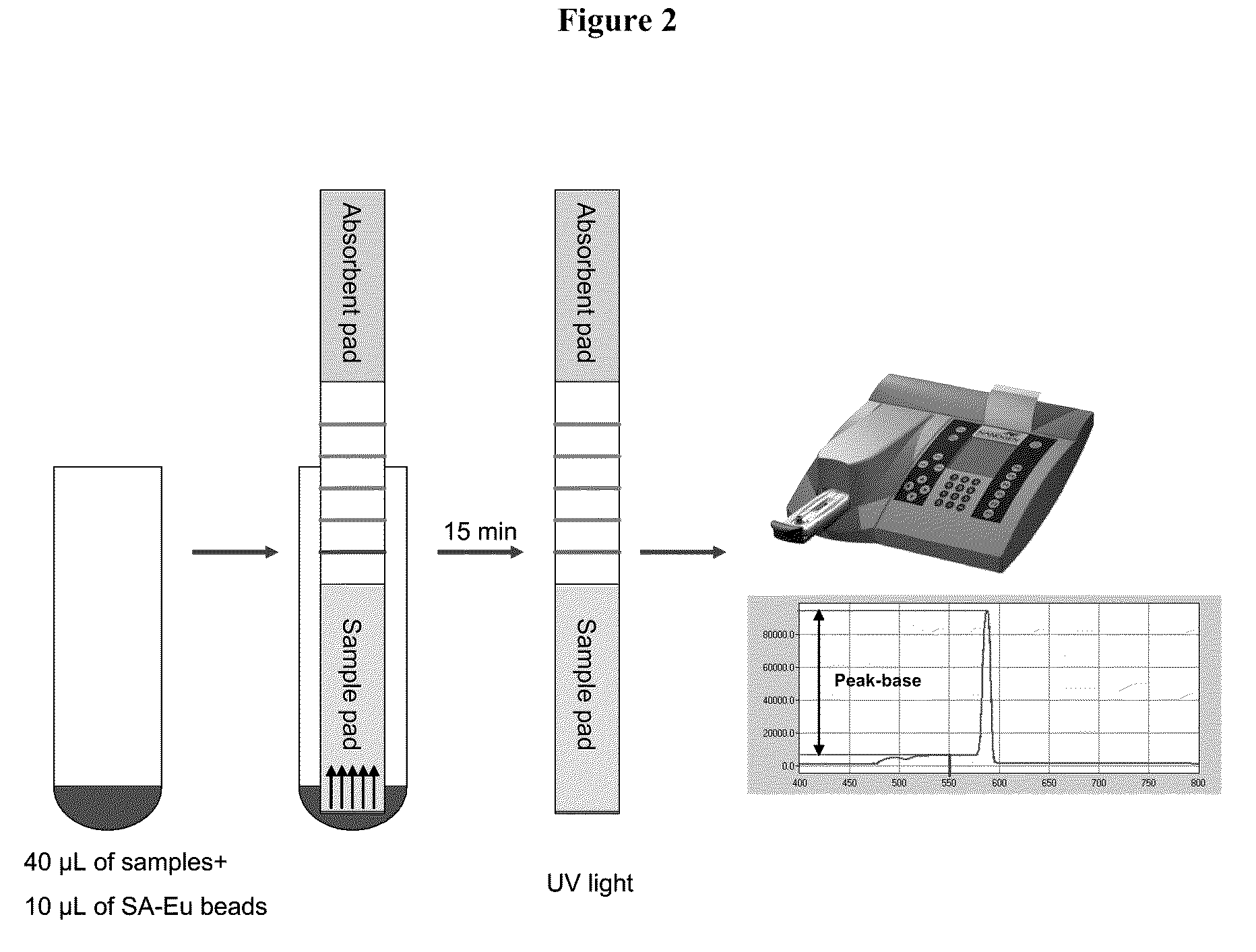
[0121]In a representative embodiment, the method utilizes pRNA technology and europium beads to detect nucleic acids on a lateral flow strip. The strip consists of three different materials including sample pad, nitrocellulose (NC) membrane and an absorbent pad. One or more unique synthetic nucleotide pRNA polymers (SCU) are coupled to a protein (e.g., immunoglobulin) and then spotted onto NC membranes forming specific test lines. PCR amplicons are generated using a pair of modified primer, of which one primer is conjugated with one type of pRNA (SBU) which is complementary to the pRNA on the NC membrane (SCU) and the other primer is biotinylated. Strips are dipped into samples containing PCR amplicons and streptavidin coated europium beads and incubated at room temperature for 15 min. The presence of amplicons are then detected under UV light on the membrane of the strip through bridging between pRNA and europium...

example 2

Detection Results for the Control Reaction Pair

[0130]The control oligonucleotide pair (no amplicon of influenza A DNA) comprising biotinylated complementary oligonucleotide and the 4a9-In pRNA-FA reverse primer which binds to IgG-pRNA were processed as follows:

[0131]25 μM of the control oligonucleotide pair was incubated in 1×PCR buffer II at room temperature for ˜45 minutes. The reaction was diluted in LF buffer 1 to 10, 1, 0.5, 0.25, 0.125 and 0.1 pmol / test (pmol / 40 μL). SA-Eu beads were diluted in LF buffer 1 to 2 μg / test (2 μg / 10 μL) and sonicated for 1 sec for 4 pulses. 40 μL of the sample and 10 μL of SA-Eu beads suspension were transferred to a sample tube. Test strip dipsticks were inserted into each tube and incubated for 15 minutes at room temperature. Each dipstick was checked under UV light for presence of Eu binding. Each dipstick was inserted into a cassette suitable for reading with a fluID™ reader and the scan data were analyzed.

[0132]The data from the scan is shown ...

example 3

Detection Results for FA Amplicons

[0133]FA amplicons were generated by reverse transcriptase PCR (RT-PCR) reaction with 10, 100 and 1000 copies of transcripts. The amplicon concentration was measured using a capillary electrophoresis system and listed in the following Table.

TABLE 6FA amplicon reactionsFA-transcriptsMix 2Mix 3copies / RT-PCRMix 1(s / s)(bio / pRNA)(s / pRNA)Mix 4 (bio / s)reactionng / μLng / μLng / μLng / μL100014.9410.274.757.214.315.093.988.0214.437.5157.081004.21.270.450.613.881.480.440.174.971.190.642.8910000000000000NTC0000

[0134]FA amplicons were diluted in LF buffer 1 at 1 / 10, 1 / 100, and 1 / 1000 fold. SA-Eu beads were diluted in LF buffer 1 to 2 μg / test (2 μg / 10 μL) and sonicated for 1 sec for 4 pulses. 40 μL of the sample and 10 μL of SA-Eu beads suspension were transferred to a sample tube. Test strip dipsticks were inserted into each tube and incubated for 15 minutes at room temperature. Each dipstick was checked under UV light for presence of Eu binding. Each dipstick was ins...


PUM

PropertyMeasurementUnit
Volumeaaaaaaaaaa
Molar densityaaaaaaaaaa

Abstract

Systems and methods are provided for immobilizing nucleic acid amplicons and protein antigens on a test device. Amplicons comprising a synthetic binding unit and a detectable label are generated and immobilized at predetermined locations on a test device by specific binding interactions between the synthetic binding unit and a synthetic capture unit located at the predetermined locations. The synthetic binding unit may include a unique design such that during amplification, a region of the synthetic binding unit is not subject to the amplification reaction, and thus the amplicon remains single stranded and available for binding to the synthetic capture unit during the capture process. In certain embodiments, the synthetic binding unit and a synthetic capture unit include synthetic nucleic acid analogs that do not interact with native nucleic acids or enzymes that act thereon. In one embodiment the synthetic binding unit and synthetic capture unit comprises puranosyl RNA (pRNA).

Description

TECHNICAL FIELD OF THE DISCLOSURE[0001]The present disclosure relates to a method and device for the identification of an analyte. In particular, the disclosure relates to a method, device, and system for the rapid detection of a nucleic acids and / or protein using non-native nucleic acid probes. More particularly, the disclosure relates to a nucleic acid detection system employing a lateral flow format.BACKGROUND OF THE DISCLOSURE[0002]There has been increasing interest and efforts devoted to developing biosensor technologies for identifying biological agents and / or markers, for instance, pathogens, particularly in areas such as biological weapons and emerging disease diagnostics. Rapid, accurate, and sensitive detection of biological agents typically employ a broad-spectrum assay that is capable of discriminating between closely related markers, such as closely related microbial or viral pathogens. Nucleic acid sequences traditionally provide the most robust and phylogenetically in...

Claims


Application Information

Patent Timeline
IPC IPC(8): C12Q1/68C12M1/00
CPCC12Q1/6837C12Q2565/514
Inventor HUANG, YINGLIGHT, II, JAMESMATHER, ELIZABETHWEISBURG, WILLIAM
Owner NEXUS DX
Why Eureka
Try Eureka